
- 📖 Geeky Medics OSCE Book
- ⚡ Geeky Medics Bundles
- ✨ 1300+ OSCE Stations
- ✅ OSCE Checklist PDF Booklet
- 🧠 UKMLA AKT Question Bank
- 💊 PSA Question Bank
- 💉 Clinical Skills App
- 🗂️ Flashcard Collections | OSCE, Medicine, Surgery, Anatomy
- 💬 SCA Cases for MRCGP
To be the first to know about our latest videos subscribe to our YouTube channel 🙌
Introduction
The cranial nerves are twelve pairs of nerves from the central nervous system. The cranial nerves are loosely based on their functions. In this summary, we discuss the nomenclature of the cranial nerves and supply some background information that might make it easier to understand the nerves and their function.
This summary should read alongside the complete articles for each of the cranial nerves:
- Olfactory nerve (CN I)
- Optic nerve (CN II)
- Oculomotor nerve (CN III)
- Trochlear nerve (CN IV)
- Trigeminal nerve (CN V)
- Abducens nerve (CN VI)
- Facial nerve (CN VII)
- Vestibulocochlear nerve (CN VIII)
- Glossopharyngeal nerve (CN IX)
- Vagus nerve (CN X)
- Accessory nerve (CN XI)
- Hypoglossal nerve (CN XII)
Cranial nerve nuclei
The cranial nerve nuclei will be covered in more detail in each cranial nerve article. A nucleus refers to a collection of neuronal cell bodies within the central nervous system and they give rise to one of seven major types of fibres (below):
- GSA (general somatic afferent): receive sensory information from the skin, skeletal muscles and joints
- GVA (general visceral afferent): receive sensory information from the viscera (organs)
- SSA (special somatic afferent): receive sensory information from the ectodermal retina, cochlear and vestibular apparatus
- SVA (special visceral afferent): receive sensory information from the endodermal nose and tongue
- GSE (general somatic efferent): provide motor innervation to skeletal muscles
- GVE (general visceral efferent): provide secretomotor function to smooth muscle and glands
- SVE (special visceral efferent): provide motor innervation to skeletal muscles of the pharyngeal arches
Afferent fibres carry sensory information back to the brain. Efferent fibres carry motor information away from the brain.
Olfactory nerve (CN I)
CN I is the olfactory nerve. It provides special visceral afferent fibres for the sense of smell.
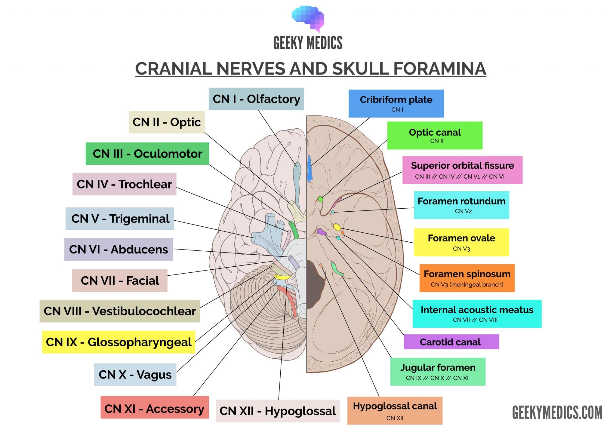
CN I connects to the brain (not the brainstem). It passes through the cribriform plate of the skull
Optic nerve (CN II)
CN II is the optic nerve. It provides special somatic afferent fibres for vision. It is the afferent limb for the pupillary light reflex.
CN II connects to the brain (not the brainstem). It passes through the optic canal of the skull
Oculomotor nerve (CNIII)
CN III is the oculomotor nerve. It provides general somatic efferent and general visceral efferent fibres to the extraocular muscles and pupillary constrictor muscles respectively. It is the efferent limb for the pupillary light reflex.
The muscles are the levator palpebrae superioris, inferior oblique, and superior, medial and inferior recti. CN III palsy causes a ‘down and out’ eye.
CN III connects to the midbrain. It passes through the superior orbital fissure of the skull
Trochlear nerve (CN IV)
CN IV is the trochlear nerve. It provides general somatic efferent to the extraocular superior oblique muscle. It assists in depressing and abducting the eye.
CN IV connects to the midbrain and is the only cranial nerve to leave the pontomesencephalic junction posteriorly. It passes through the superior orbital fissure of the skull.
Trigeminal nerve (CN V)
CN V is the trigeminal nerve. It has three sensory nuclei:
- Mesencephalic – proprioception
- Principal – light touch and discrimination
- Spinal – pain, temperature, crude touch
It is the afferent limb of the corneal reflex; CN VII is the bilateral efferent limb.
CN V emerges from the pons. It has three divisions (ophthalmic, maxillary and mandibular).
Table 1. The three divisions of the trigeminal nerve.
|
Division |
Modality |
Sensory function |
Motor function |
Foramen |
|
Ophthalmic V1 |
General somatic afferent |
Above the lower eyelid |
X |
Superior orbital fissure |
|
Maxillary V2 |
General somatic afferent |
Lower eyelid to the upper lip |
X |
Foramen rotundum |
|
Mandibular V3 |
General somatic afferent Special visceral efferent |
Below upper lip |
Muscles of mastication |
Foramen ovale |
Abducens nerve (CN VI)
CN VI is the abducens nerve. It provides general somatic efferent fibres for eye abduction. It innervates the lateral rectus muscle.
CN VI originates in the pontomedullary region. It passes through the superior orbital fissure of the skull.
Facial nerve (CN VII)
CN VII is the facial nerve. It originates in the pontomedullary region, passes through the internal auditory meatus and exits through the stylomastoid foramen.
The facial nerve loops around the abducens nucleus.
Lower motor neurone facial nerve lesions cause upper and lower facial paralysis
Upper motor neurone facial nerve lesions cause lower facial paralysis only
Table 2. Function of the facial nerve.
|
General somatic afferent |
Special visceral afferent |
General visceral efferent |
Special visceral efferent |
|
Skin behind the ear |
Taste to anterior 2/3 of the tongue |
Parasympathetic to the lacrimal, sublingual and submandibular glands |
Muscles of facial expression |
Vestibulocochlear nerve (CN VIII)
CN VIII is the vestibulocochlear nerve. It provides special somatic afferent fibres for hearing and balance.
CN VIII originates in the pontomedullary region. It passes through the internal auditory meatus and does not leave the skull.
The cochlea transmits sound waves to mechanical ossicle movements to electrochemical action potentials.
The vestibular apparatus detects changes in head motion.
Glossopharyngeal nerve (CN IX)
CN IX is the glossopharyngeal nerve. It originates in the medulla oblongata and passes through the jugular foramen.
It draws fibres from the solitary nucleus (taste) and nucleus ambiguus (motor).
It draws more fibres from the inferior salivatory nucleus (parotid gland) and dorsal motor nucleus (DMX; pharyngeal sensation).
Table 3. Functions of the glossopharyngeal nerve.
|
General somatic afferent |
Special visceral afferent |
General visceral efferent |
Special visceral efferent |
|
Sensation from the posterior 1/3 of the tongue, pharynx |
Taste to posterior 1/3 of the tongue |
Parasympathetic to parotid glands |
Motor to stylopharyngeus |
Vagus nerve (CN X)
CN X is the vagus nerve. It originates in the medulla oblongata and passes through the jugular foramen with CN IX and XI.
Its key role is parasympathetic innervation of the viscera
The recurrent laryngeal nerve loops under the right subclavian artery and (left) aortic arch.
Table 4. Functions of the vagus nerve.
|
General somatic afferent |
Special visceral afferent |
General visceral afferent |
General visceral efferent |
Special visceral efferent |
|
Skin around ear |
Taste and sensation to the epiglottis |
Sensory information to body viscera |
Parasympathetic to glands of GI tract |
Motor innervation to soft palate, pharynx and larynx |
Accessory nerve (CN XI)
CN XI is the accessory nerve. It originates in the medulla oblongata and superior cervical cord region. It exits the spinal cord in the neck and enters the skull through the foramen magnum.
It provides general somatic efferent fibres to the trapezius and sternocleidomastoid
Hypoglossal nerve (CN XII)
CN XII is the hypoglossal nerve. It provides general somatic efferent fibres for controlling tongue muscles.
It originates in the medulla oblongata and exits the skull through the hypoglossal canal.
Cranial nerve summary table
You can download our cranial nerve summary table in PDF format here.
|
Nerve |
Modality |
Fibre type |
Function |
Foramen |
|
I – Olfactory |
Sensory |
SVA |
Smell |
Cribriform plate |
|
II – Optic |
Sensory |
SSA |
Vision |
Optic canal |
|
III – Oculomotor |
Motor |
GSE, GVE |
Extraocular muscles and eyelid elevator |
Superior orbital fissure |
|
IV – Trochlear |
Motor |
GSE |
Superior oblique muscle |
Superior orbital fissure |
|
V – Trigeminal |
Both
|
GSA GSA GSA, SVE |
V1 – ophthalmic – face sensation V2 – maxillary – face sensation V3 – mandibular – face sensation |
V1 – superior orbital fissure V2 – foramen rotundum V3 – foramen ovale |
|
VI – Abducens |
Motor |
GSE |
Lateral rectus |
Superior orbital fissure |
|
VII – Facial |
Both |
GSA, SVA, GVE, SVE |
Muscles of facial expression + stapedius Taste to the anterior 2/3 of the tongue Tear and salivary ducts |
Internal acoustic meatus to stylomastoid foramen |
|
VIII – Vestibulocochlear |
Sensory |
SSA |
Balance – vestibular division Hearing – cochlear division |
Internal acoustic meatus |
|
IX – Glossopharyngeal |
Both |
GSA, SVA, GVE, SVE |
Taste for posterior 1/3 of the tongue Sensation to pharynx Innervates stylopharyngeus |
Jugular foramen |
|
X – Vagus |
Both |
GSA, SVA, GVA, GVE, SVE |
Parasympathetic innervation to viscera above splenic flexure Laryngeal muscles and palatoglossus |
Jugular foramen |
|
XI – Accessory |
Motor |
GSE |
Motor control to SCM and trapezius |
Jugular foramen |
|
XII – Hypoglossal |
Motor |
GSE |
Innervates tongue muscles except for palatoglossus |
Hypoglossal canal |
References
Reference texts
- Sinnatamby, C. S. (2011). Last’s Anatomy, International Edition: Regional and Applied. Elsevier Health Sciences.
- Moore, K. L., Dalley, A. F., & Agur, A. M. (2013). Clinically oriented anatomy. Lippincott Williams & Wilkins.
- Nolte, J. (2002). The human brain: an introduction to its functional anatomy.
- Snell, R. S. (2010). Clinical neuroanatomy. Lippincott Williams & Wilkins.
Reference images
- Patrick J. Lynch, medical illustrator. License: [CC-BY]. Modified by Dr Lewis Potter.
