
- 📖 Geeky Medics OSCE Book
- ⚡ Geeky Medics Bundles
- ✨ 1300+ OSCE Stations
- ✅ OSCE Checklist PDF Booklet
- 🧠 UKMLA AKT Question Bank
- 💊 PSA Question Bank
- 💉 Clinical Skills App
- 🗂️ Flashcard Collections | OSCE, Medicine, Surgery, Anatomy
- 💬 SCA Cases for MRCGP
To be the first to know about our latest videos subscribe to our YouTube channel 🙌
Introduction
The movements produced at joints by muscles are given specific anatomical names, often referred to as “anatomical terms of motion”. We usually make the assumption that the body is in normal resting anatomical position, and that joint movement occurs from this resting position.
In this article, we explore the difference between an axis and a plane, before describing different joint movements in the context of the larger joints in the body.
Planes
There are several different planes that we use to describe the body and movements.
There are three major planes.
Sagittal plane
- Divides the body into left and right halves
- Median plane refers to the midline
- Paramedian plane refers to subdividing one half
- Example: flexion and extension of the hip occurs in the sagittal plane
Coronal plane
- Divides the body into anterior and posterior halves
- Example: abduction and adduction of the shoulder occurs in the coronal plane
Transverse/axial plane
- Divides the body into superior and inferior halves
- Example: left and right rotation of the atlantoaxial joint occurs in the transverse plane
Axes
There are also several different axes we use to describe the movement of a joint. It is useful to think of an axis as a metal pole, and the joint rotating around this pole.
Sagittal axis
- Passes horizontally from anterior to posterior
- Formed by the intersection of the sagittal and transverse planes
- Example: abduction and adduction of the shoulder about the sagittal axis
Coronal axis
- Passes horizontally from left to right
- Formed by the intersection of the coronal and transverse planes
- Example: flexion and extension of the hip occurs about the coronal axis
Vertical axis
- Passes vertically from superior to inferior
- Formed by the intersection of the sagittal and coronal flames
- Example: left and right rotation of the atlantoaxial joint occurs about the vertical axis
Movements in an axis and along a plane
Bringing together planes and axes, we will describe the movements we used above.
Flexion and extension of the hip occur in the sagittal plane, and about the coronal axis.
Abduction and adduction of the shoulder occur in the coronal plane, and about the sagittal axis.
Left and right rotation of the atlantoaxial joint occurs in the transverse plane, and about the vertical axis.
Now, let’s make some more sense of what these movements actually are!
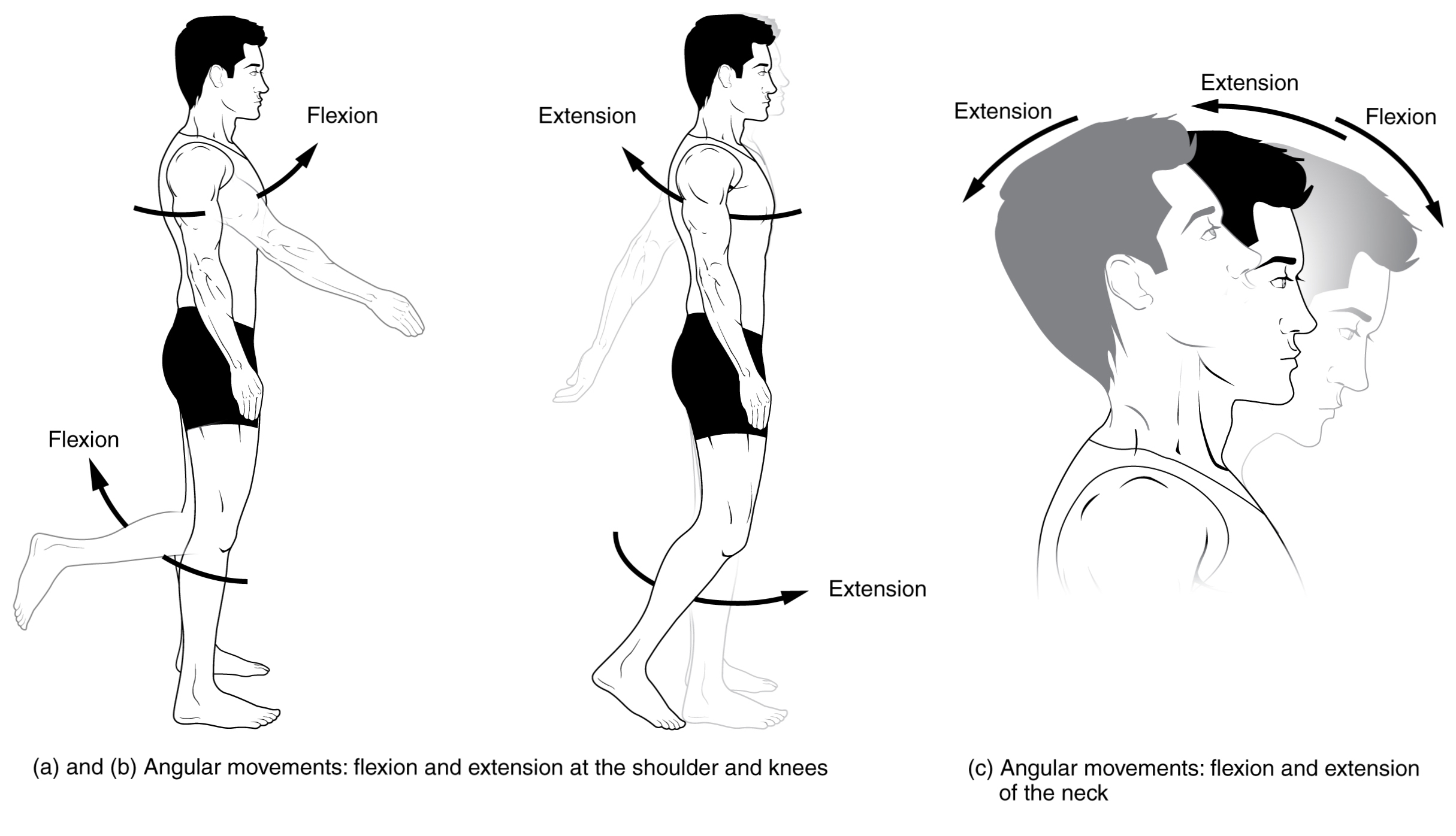
Flexion and extension
When talking about flexion and extension, we are usually referring to these movements as they occur about the coronal axis, and along the sagittal plane.
Flexion refers to decreasing a joint angle, and extension to increasing the joint angle back to resting anatomical position.
Examples of flexion
Examples of flexion include:
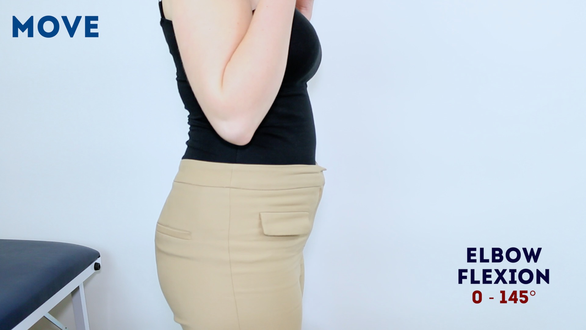
- Flexing the elbow to bring the radius and ulna closer to the humerus (Figure 2)
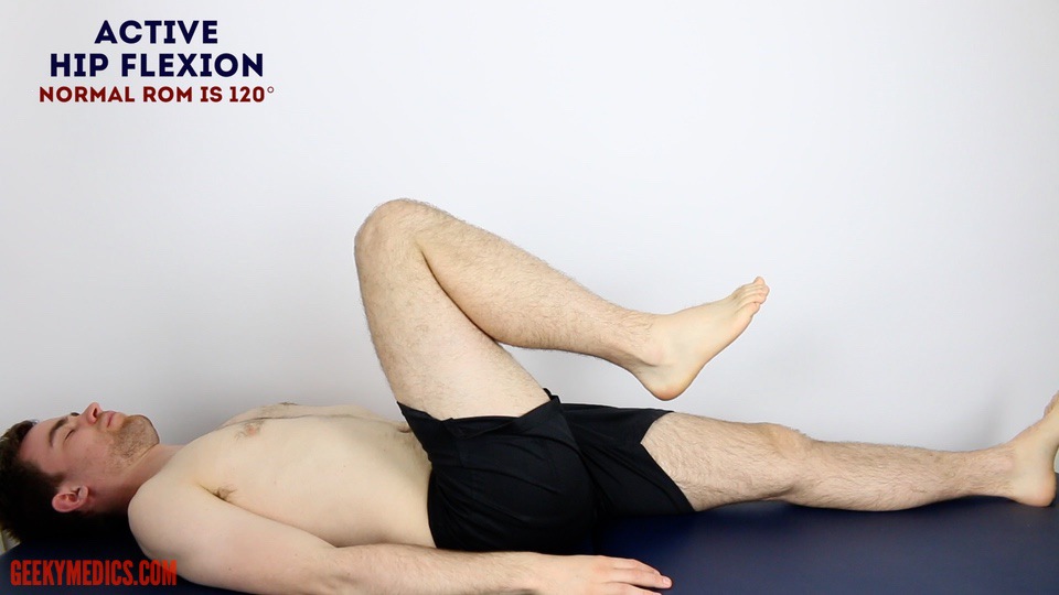
- Flexing the hip to bring the femur closer to the abdomen (Figure 3)
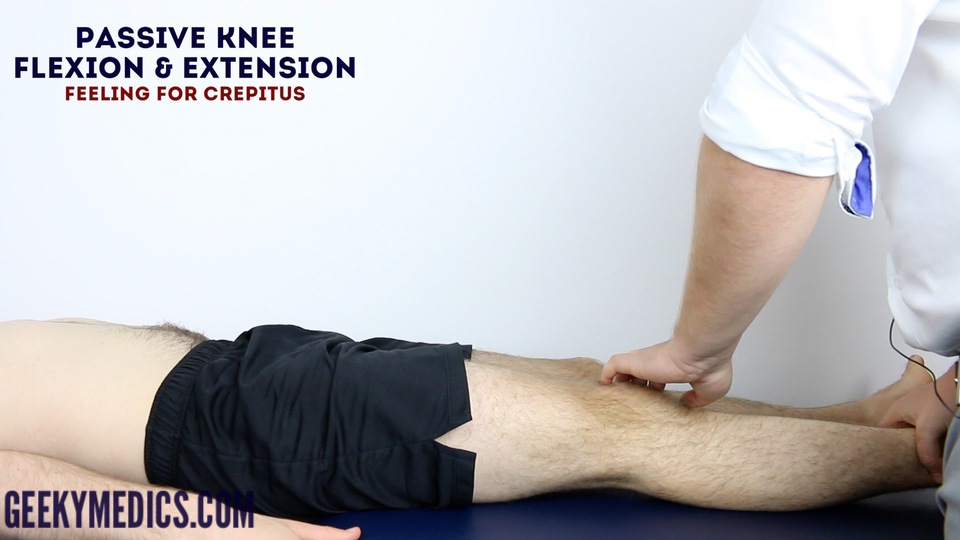
- Flexing the knee to bring the tibia and fibula closer to the femur (Figure 4)
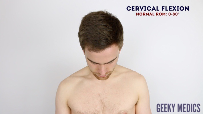
- Flexing the neck (atlanto-occipital joint) to bring the head closer to the chest (Figure 5)

Examples of extension
Examples of extension include:
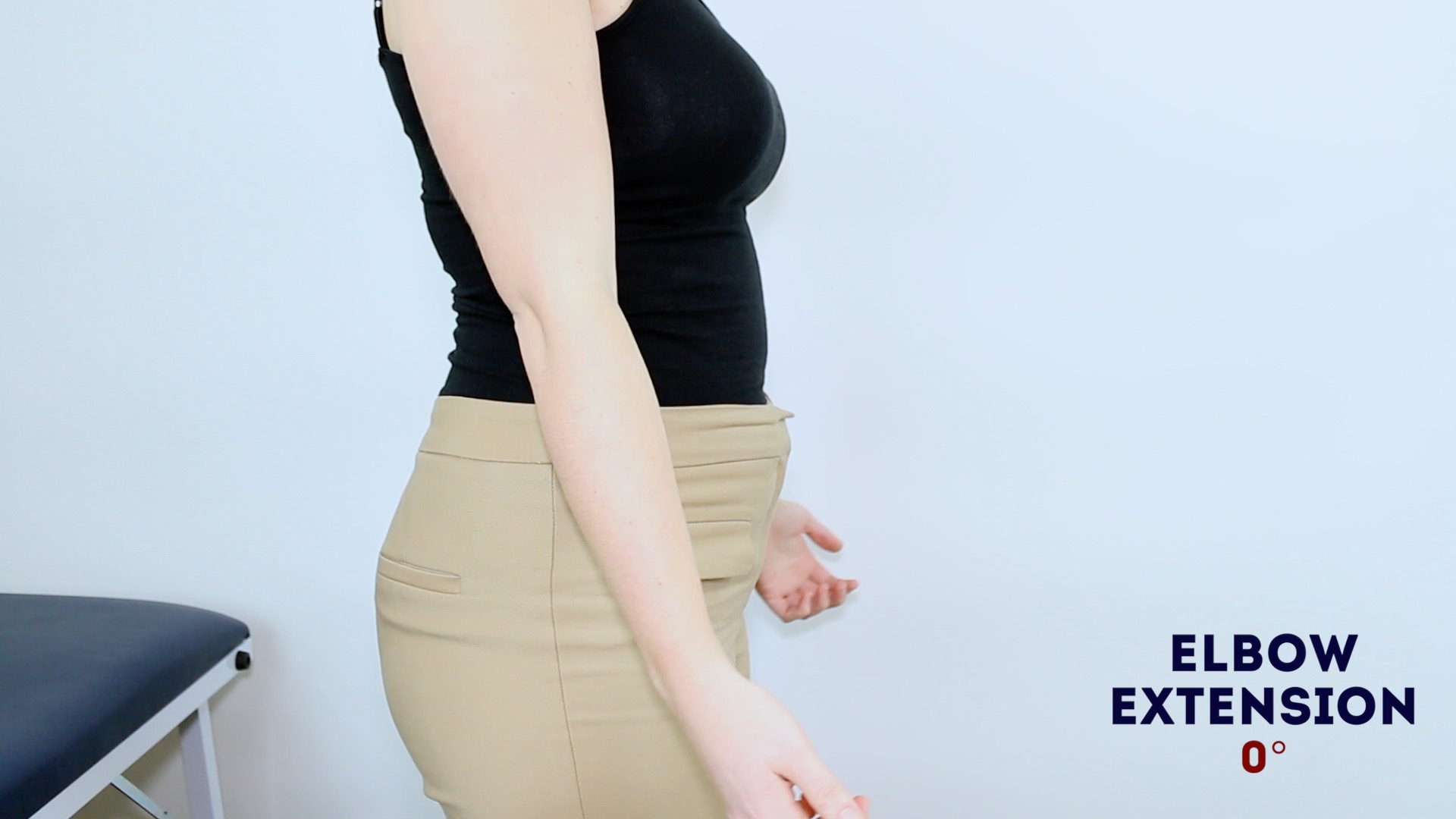
- Extending the elbow to return the radius and ulna back to resting anatomical position (Figure 6)
- Extending the hip to return the femur back to resting anatomical position;
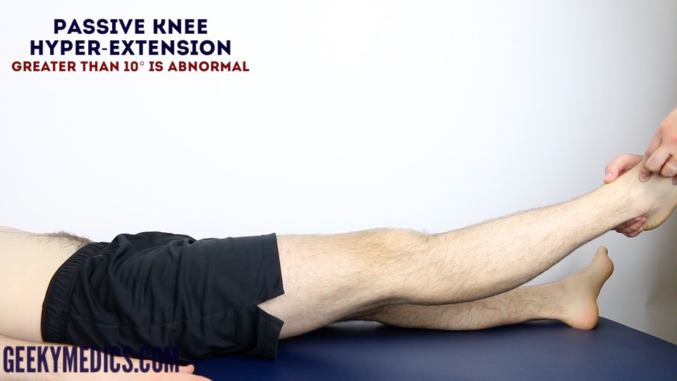
- Extending the knee to return the tibia and fibular back to resting anatomical position (Figure 7)
- Extending the atlanto-occipital joint to return the head back to resting anatomical position.
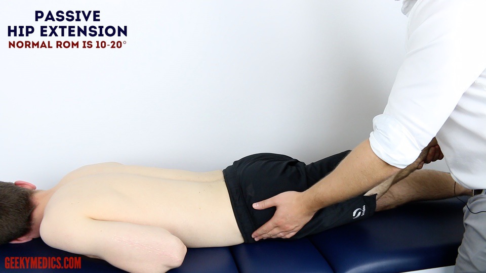
Examples of hyperextension
Examples of hyperextension include:
- Hyperextension of the shoulder refers to extending the shoulder past its resting anatomical position so that the arm is now behind the back;
- Hyperextension of the hip refers to extending the hip past its resting anatomical position, contracting the gluteus maximus to bring the femur closer to the vertebral column (Figure 9)
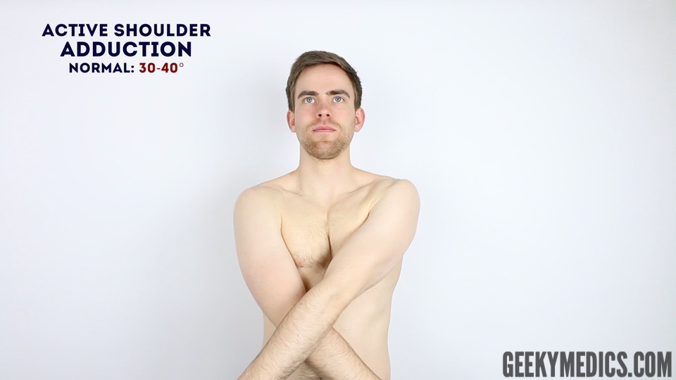
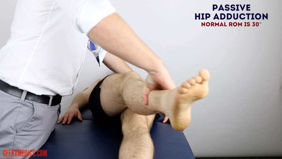
Abduction and adduction
Abduction and adduction refer to movements made about a sagittal axis and along the coronal plane.
Abduction is moving a body part away from its resting anatomical position in the coronal plane; adduction is returning it to its normal resting position (includes ‘hyperadduction’).
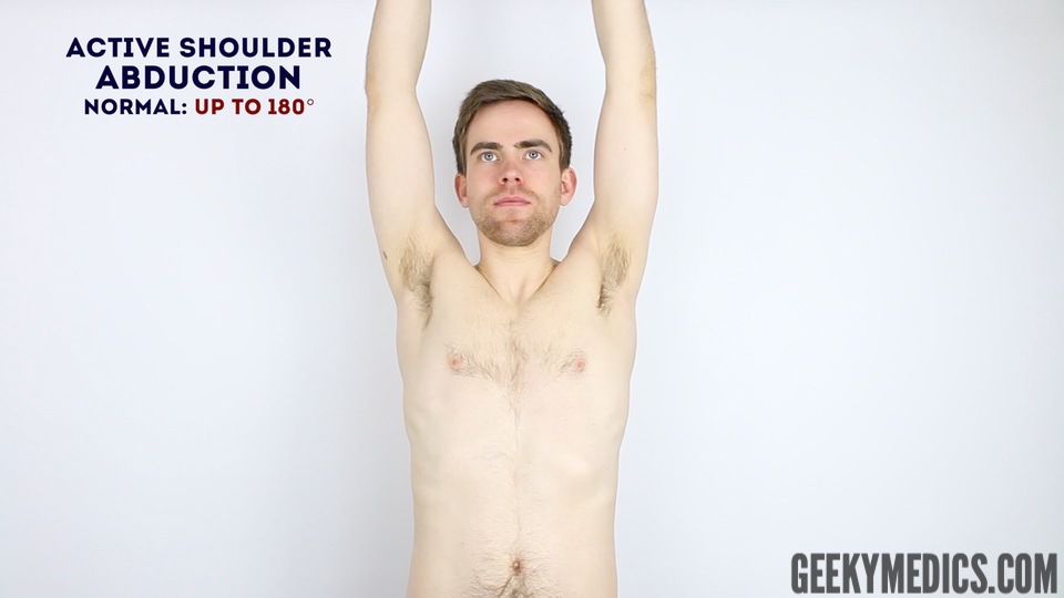
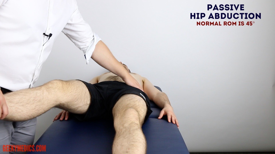
Examples of abduction
Examples of abduction include:
- Abducting the shoulder joint to lift the upper limb closer to the ears (Figure 11)
- Abducting the hip joint to lift move the lower limbs away from each other (Figure 12)
Examples of adduction
Examples of adduction include:
- Returning the shoulder joint back to resting anatomical position
- Returning the hip joint back to resting anatomical position (bring the lower limbs together again)
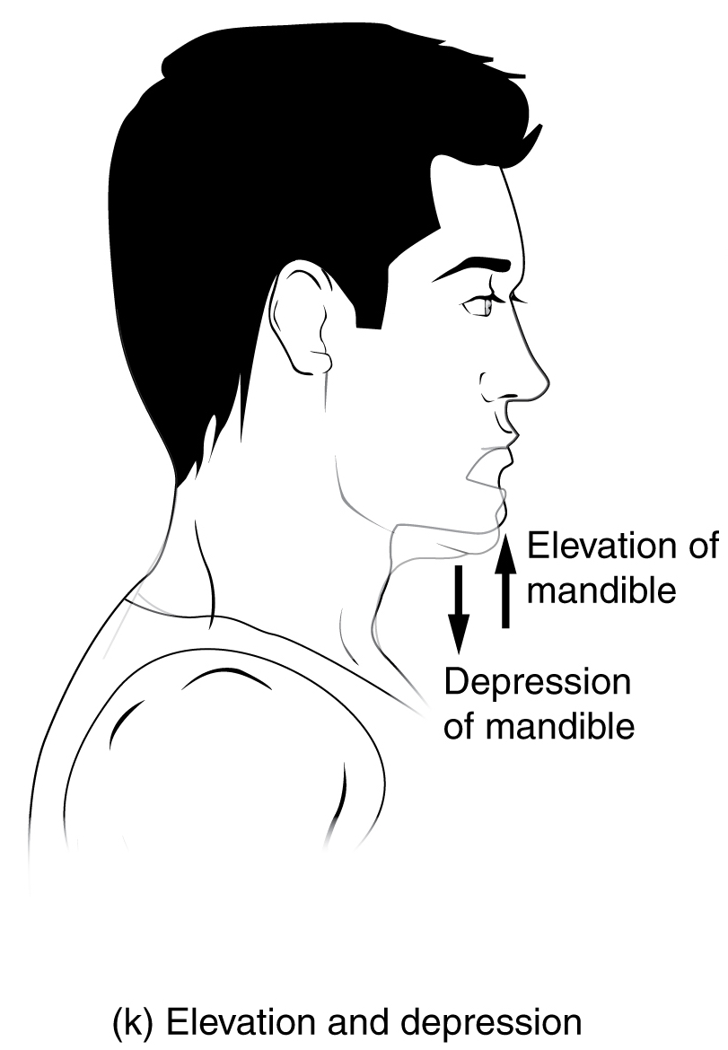
Elevation and depression
Elevation refers to lifting, and depression refers to lowering.
These movements only occur in several regions of the body and are a result of movement along the coronal plane.
Examples of elevation
Examples of elevation include:
- Elevating the shoulders to shrug
- Elevating the mandible during mastication to close the mouth

Examples of depression
Examples of depression include:
- Returning the shoulders to resting anatomical position after shrugging
- Depressing the mandible during mastication or phonation to open the mouth

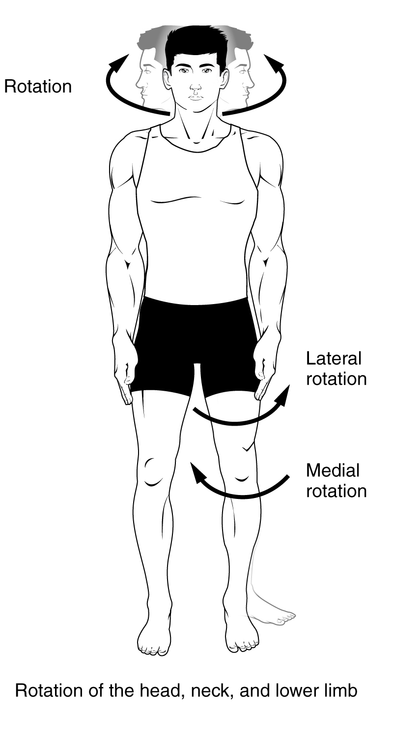
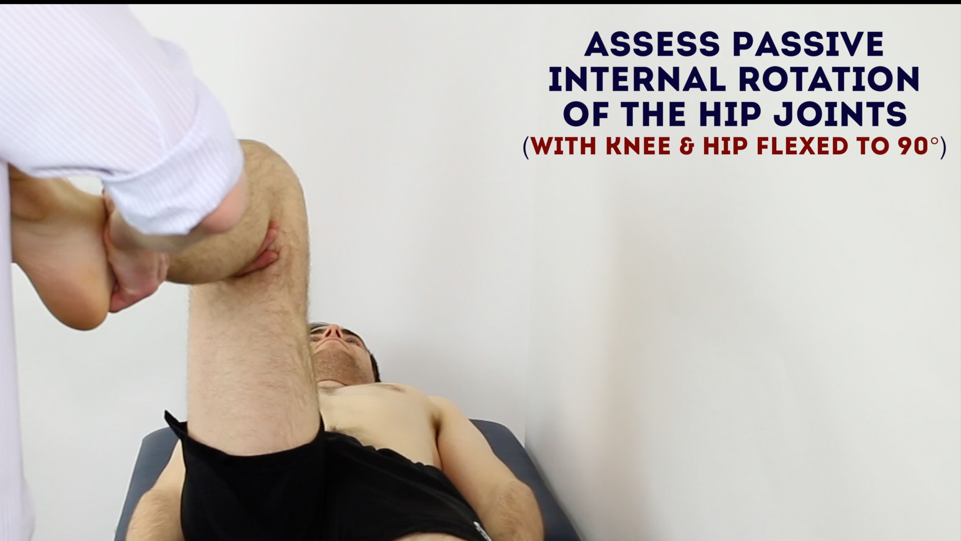
Internal and external rotation (medial and lateral rotation)
Rotation refers to movements made about the longitudinal axis and in the transverse plane.
Internal rotation is rotating a joint towards the midline and external rotation is rotating a joint away from the midline.
Examples of internal rotation
Examples of internal rotation include:
- With the elbow at 90 degrees of flexion, internally rotating the shoulder brings the forearm and hand toward the body (Figure 19)
- Rotating the lower limb at the hip joint to point the feet away from each other (Figure 20)

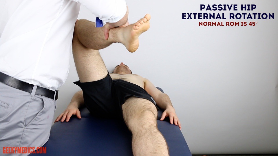
Examples of external rotation
Examples of external rotation include:
- With the elbows at 90 degrees of flexion, externally rotating the shoulder brings the forearm and hand away from the body (Figure 21)
- Rotating the lower limb at the hip joint to point the feet towards each other (Figure 22)

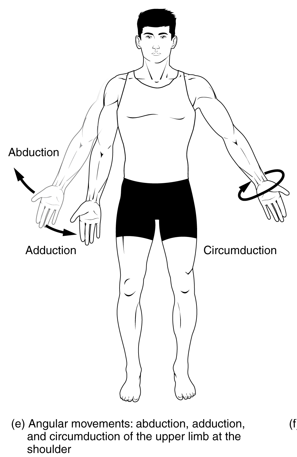
Circumduction
Circumduction is a compound movement that occurs only at ball and socket joints, which can perform multiple movement types.
As Figure 23, it is a combination of:
- Flexion and extension
- Abduction and adduction
- Rotation
Circumduction is described as a circular motion utilising each of these movements at different parts of the circular motion. Given that the shoulder and hip are the only joints to use circumduction and they have a relatively ‘fixed’ joint, the movement is more conical.
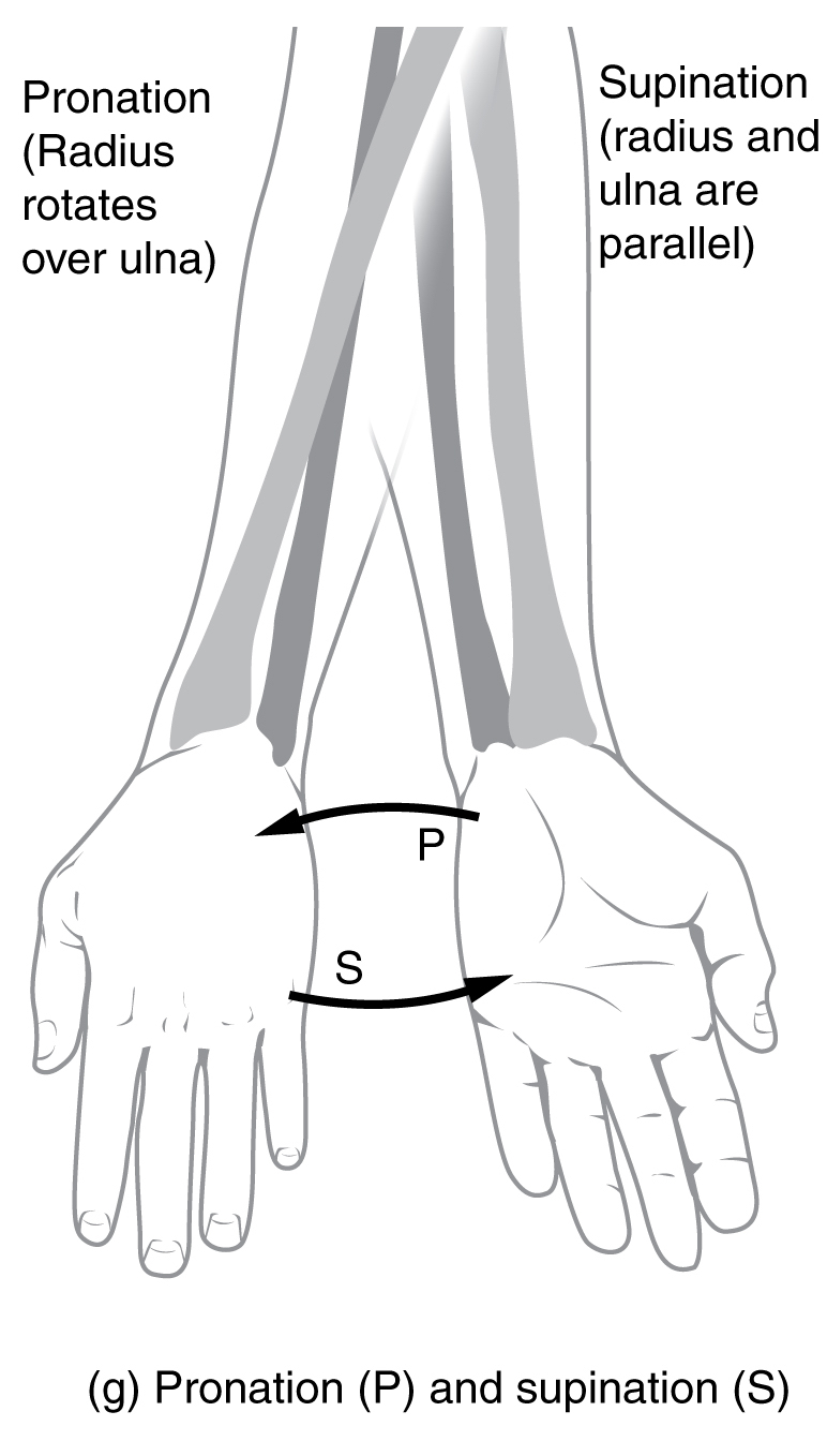
Pronation and supination
Pronation and supination occur at pivot joints. The most important example of this is the radiohumeral joint and the union of the radius and ulna through the interosseous membrane.
To remember pronation, think of lying prone (on your belly). Thus, pronation of the radiohumeral joint refers to the palm of the hand facing the ground.
To remember supination, think of holding a bowl of soup. You hold a bowl of soup with your palms facing up, this is the position of supination.
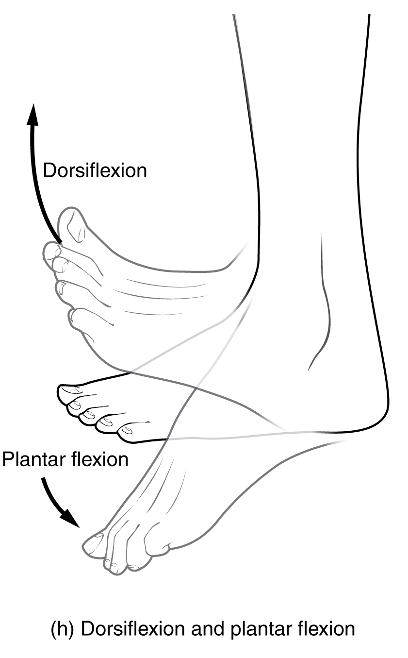
Dorsiflexion and plantarflexion
The foot is a little special and has four movements specific to it. The first two, dorsiflexion and plantarflexion, refer to the way the foot moves about the coronal axis and along the sagittal plane.
Dorsiflexion brings the dorsum (back) of the foot back toward the tibia, so the toes are beginning to point towards the sky. This is a position of high ankle stability.
Plantarflexion refers to pointing the foot away from the tibia and down into the ground. This is a position of low ankle stability, and most ligamentous ankle sprains occur in a position of plantarflexion.
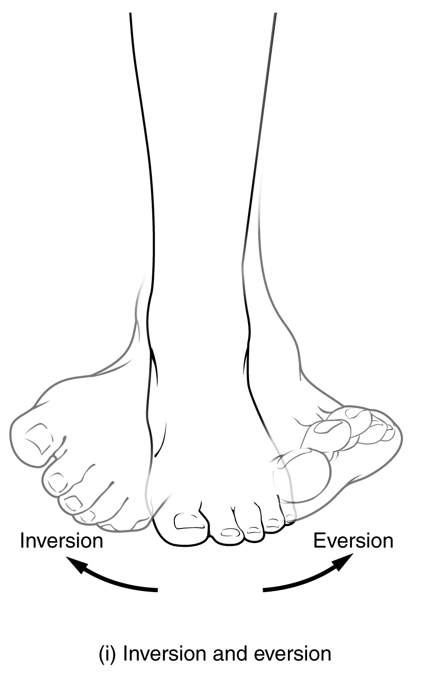
Eversion and inversion
The second set of movements specific to the foot are eversion and inversion. These movements occur about the sagittal axis and along the coronal plane.
Eversion refers to bringing the soles of the feet out, so they are facing away from the midline of the body.
Inversion refers to bringing the soles of the feet in, so they are facing towards the midline of the body (and each other).
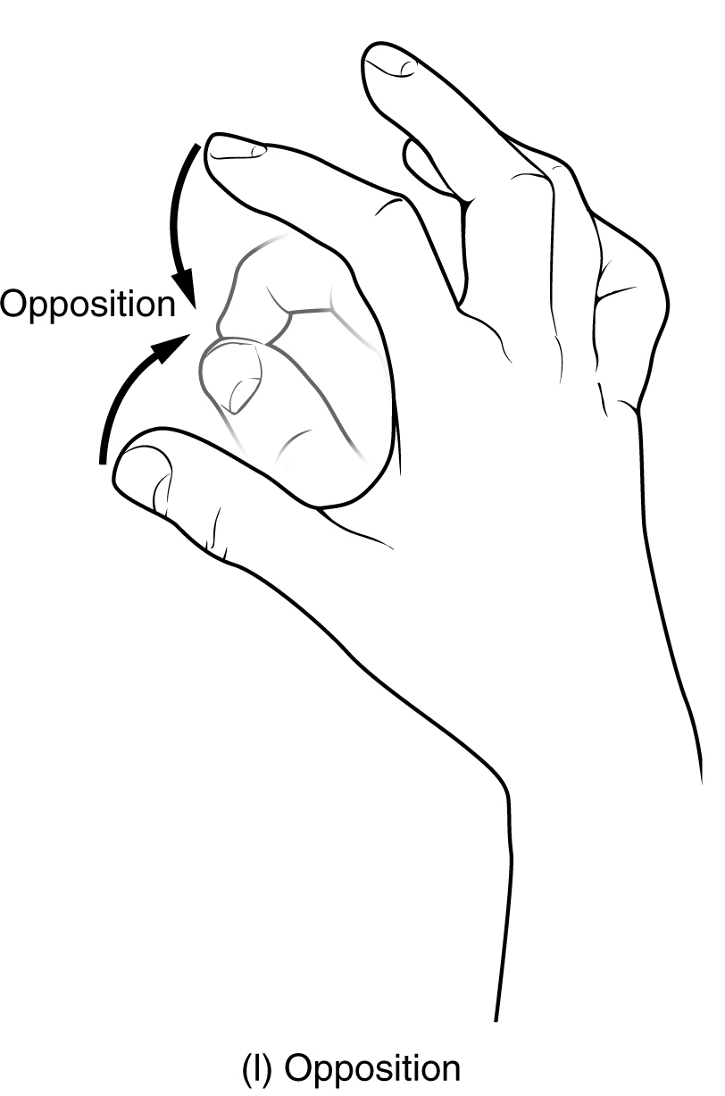
Opposition and re-position
Opposition and re-position are special movements unique to the human hand. They are movements that allow us fine dexterity to hold objects.
Opposition and re-position occur at the thumb and little finger (digits 1 and 5, respectively). They even have their own separate muscles for this movement!
Opposition of the thumb and little finger refers to the movement of bringing the pads of the thumb and little finger together in the midline of the hand. Technically, the thumb can be opposed to each of the other fingers on the same hand.
Re-position refers to returning the thumb and little (or one of the others!) finger back to resting anatomical position.
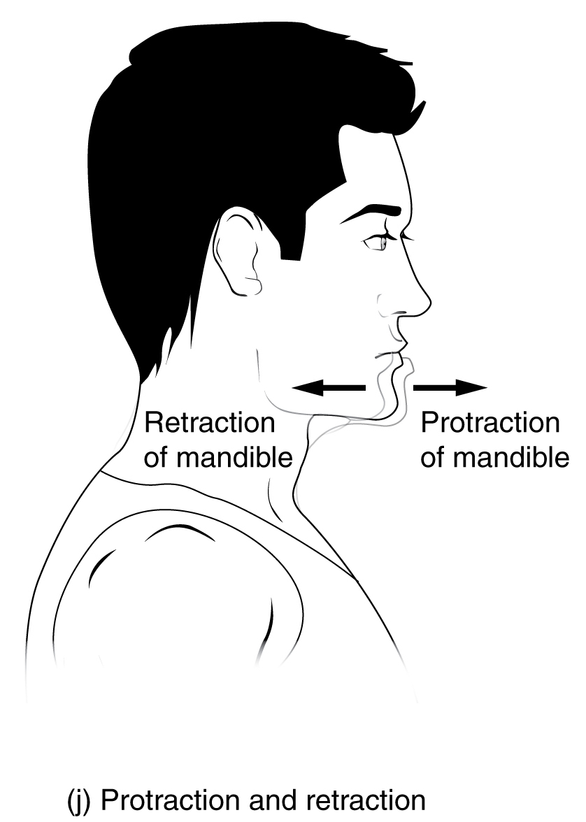
Protraction and retraction (and protrusion and retrusion)
Protraction and retraction occur in two major regions of the body – the scapula and the mandible.
Protraction refers to protruding or sticking out. Retraction refers to bringing together.
Examples of protraction
Examples of protraction include:
- With the shoulder in 90o of abduction, brings the arms forward as if to hug someone. The scapulae begin to slide laterally and then anteriorly along the thorax, this is protraction
- Protrusion (similar to protraction) of the jaw allows for anterior movement of the jaw, as if to move it out from the rest of the head
Examples of retraction
Examples of retraction include:
- Returning the scapulae from hugging someone with abducted shoulders in front of us to the resting anatomical position of the scapulae
- Retrusion (similar to retraction) of the jaw brings the jaw back to its resting anatomical position
Sliding
Sliding is a vital movement in the human body but is seldom discussed. It isn’t a movement that is easily visible as it occurs on a relatively small level – nonetheless, it is very important! Sliding occurs in synovial joints with two flat surfaces opposing each other. Examples include the carpal bones of the hand and wrist complex.
Sliding allows these bones to move slightly during movements such as flexion or extension of the wrist, or abduction and adduction of the wrist. Without sliding joints in the carpus, we’d find it difficult to move our wrists as well as we do!
References
Texts
- Sinnatamby, C. S. (2011). Last’s Anatomy, International Edition: Regional and Applied. Elsevier Health Sciences.
- Moore, K. L., Dalley, A. F., & Agur, A. M. (2013). Clinically oriented anatomy. Lippincott Williams & Wilkins.
Images
- Tonye Ogele CNX. License: [CC BY-SA]
- Connexions. License: [CC BY]
- Connexions. License: [CC BY]
- Tonye Ogele CNX. License: [CC BY-SA]
- Connexions. License: [CC BY]
- Connexions. License: [CC BY]
- Connexions. License: [CC BY]
- Osteomyoamare. License: [CC BY]
