

- 📖 Geeky Medics OSCE Book
- ⚡ Geeky Medics Bundles
- ✨ 1300+ OSCE Stations
- ✅ OSCE Checklist PDF Booklet
- 🧠 UKMLA AKT Question Bank
- 💊 PSA Question Bank
- 💉 Clinical Skills App
- 🗂️ Flashcard Collections | OSCE, Medicine, Surgery, Anatomy
- 💬 SCA Cases for MRCGP
To be the first to know about our latest videos subscribe to our YouTube channel 🙌
This article explains the dermatomes and myotomes of the human body, with an included dermatome map. Examining myotomes and dermatomes is a vital part of a thorough neurological examination, particularly when a patient has a spinal cord injury.
Embryology
Dermatomes and myotomes both arise from somites, which are divisions of the body of an embryo. Somites are paired structures which run cephalocaudally (along the head to toe axis of the body).
In the developing human embryo, somites form dermatomes and myotomes, as well as tendons, cartilage and sclerotomes (which form bone).
Dermatomes
A dermatome is an area of skin supplied by a single spinal nerve.
If you imagine the human body as a map, each dermatome represents the area of skin supplied with sensation by a specific nerve root.
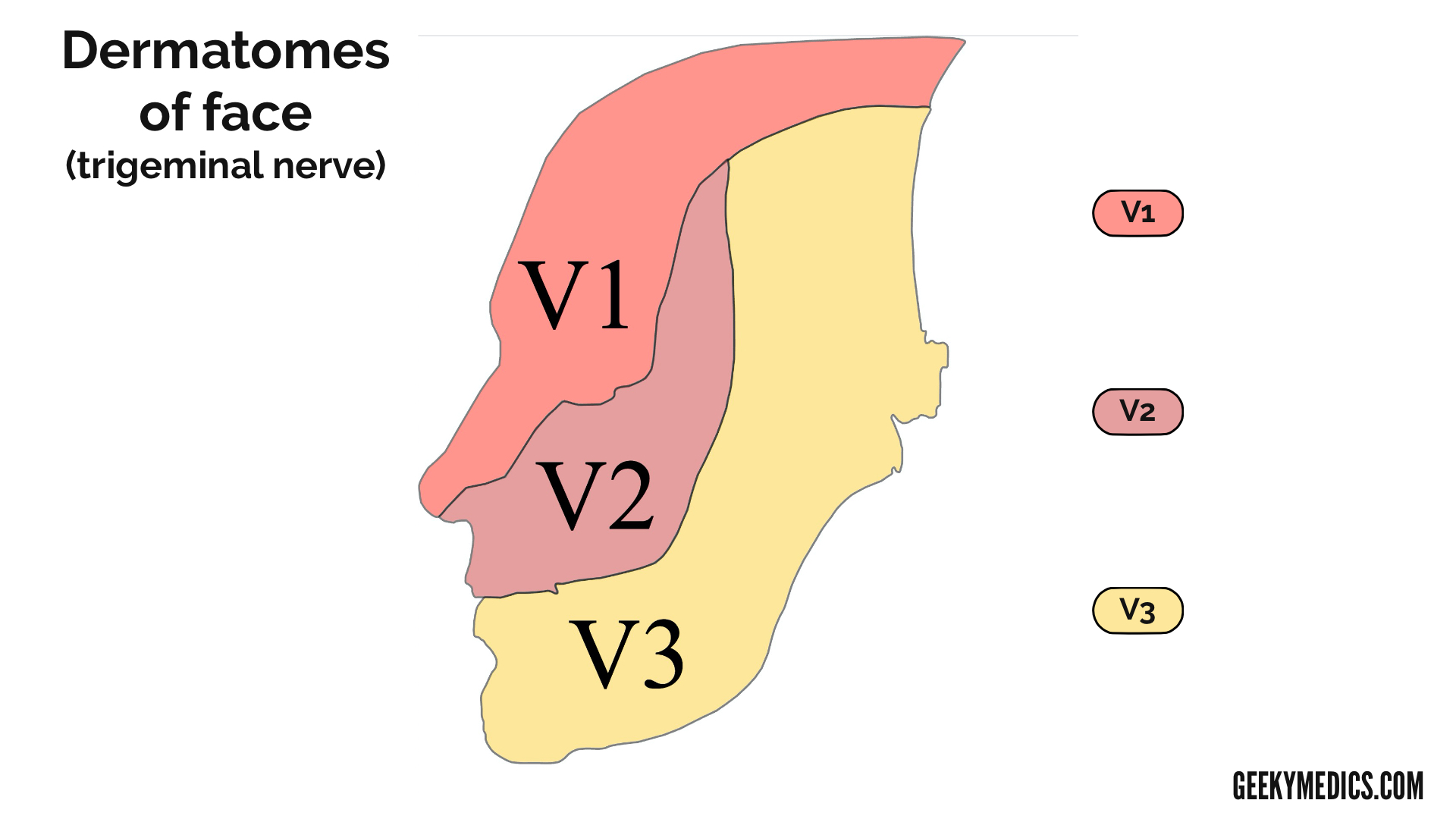
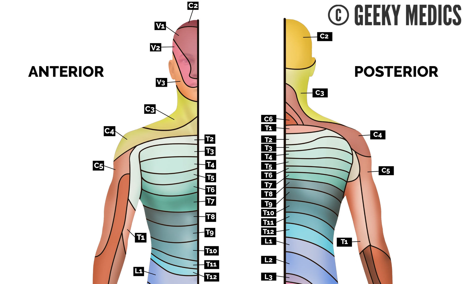
It is important to bear in mind that the dermatomes of the head are supplied by branches V1, V2 and V3 of the trigeminal nerve.
When assessing sensation, areas close to dermatomal boundaries should be avoided to minimise the risk of misinterpretation. The lists below describe locations that can be used to assess the dermatomes of the head, upper limb, torso and lower limbs.1 We have also included a selection of dermatomal maps to demonstrate the region of the skin each dermatome covers.
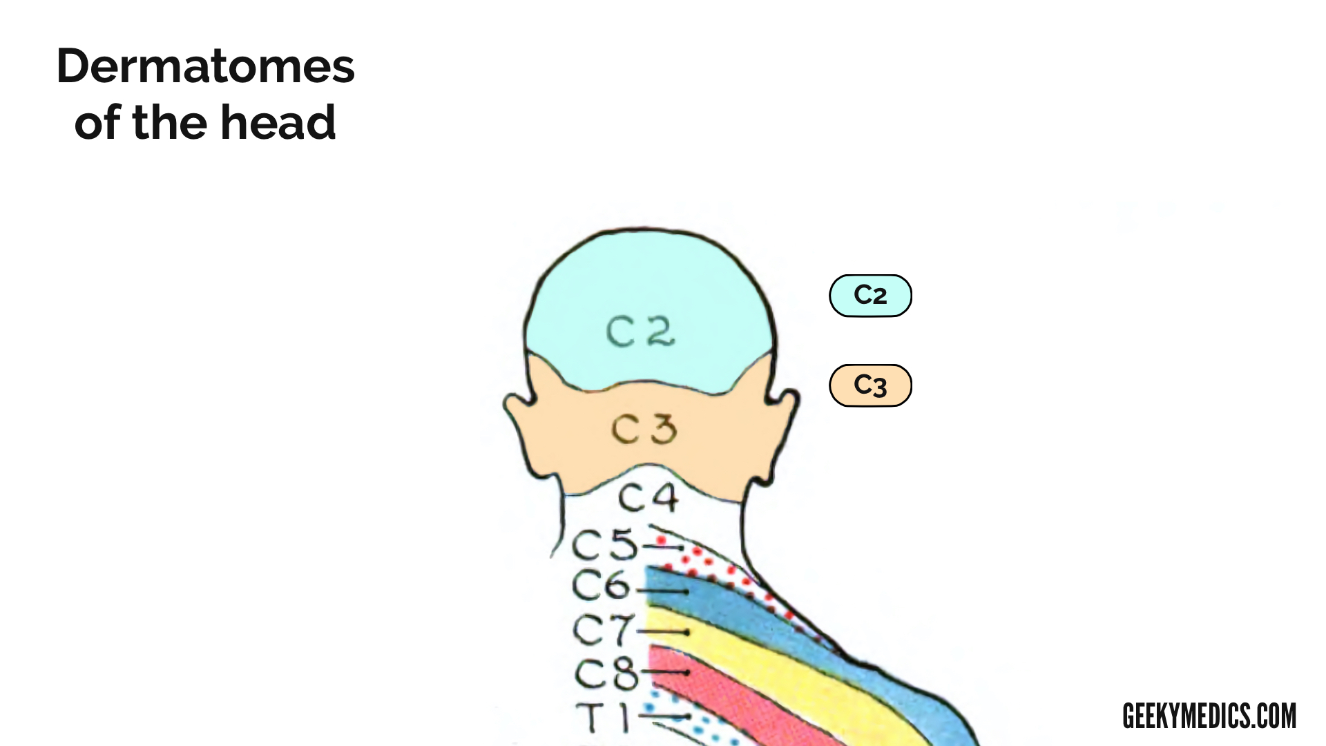
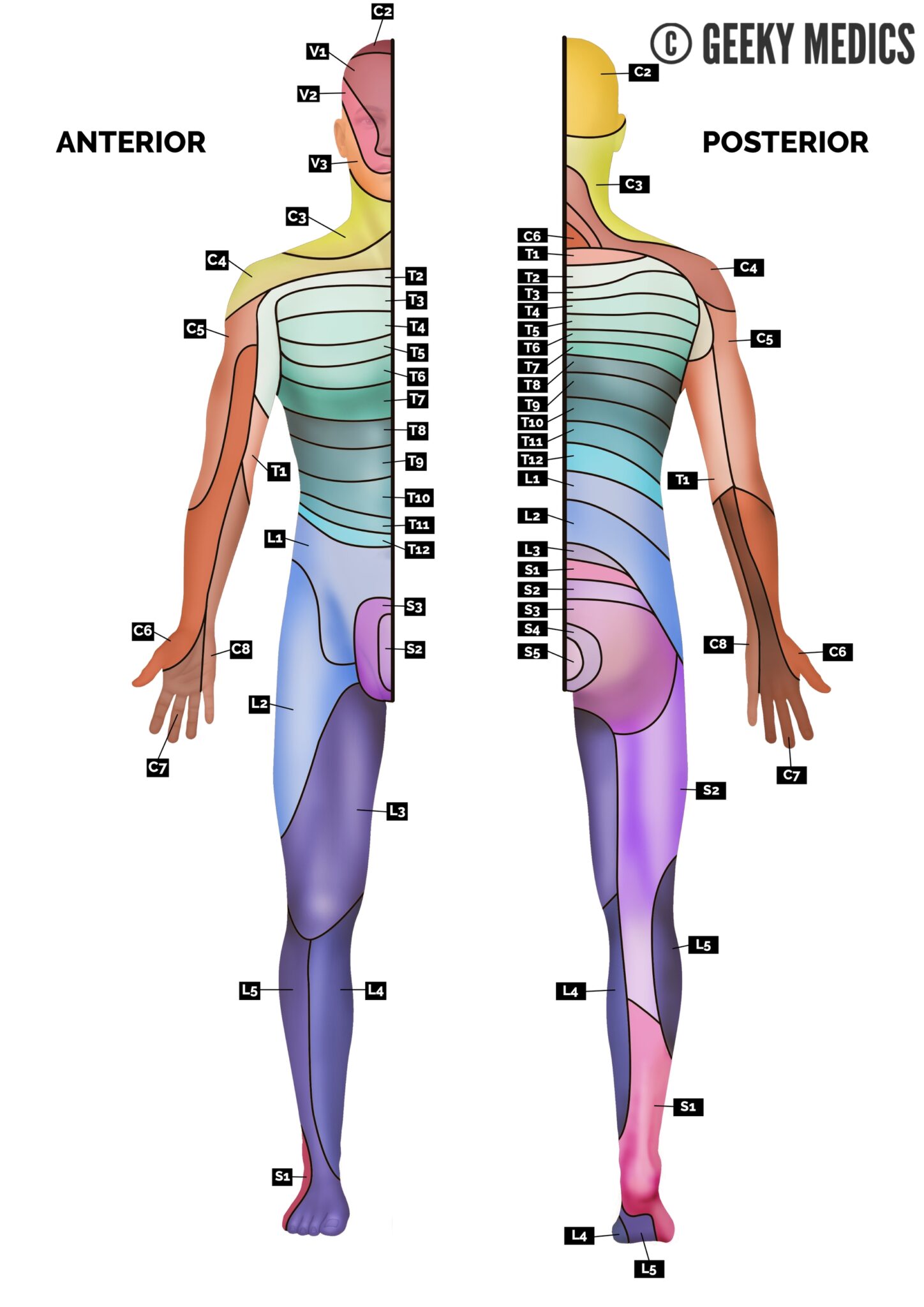
Dermatomes of the head
Trigeminal nerve (CN V)
- V1: ophthalmic branch – the lateral aspect of the forehead
- V2: maxillary branch – the cheek
- V3: mandibular branch – the lower jaw (avoid the angle of the mandible as it is supplied by C2/C3)
Other
- C2: 1-2 cm lateral to the occipital protuberance
- C3: the supraclavicular fossa in the midclavicular line.
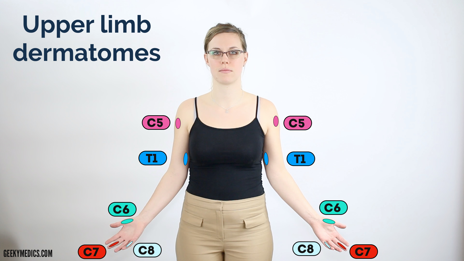
Dermatomes of the upper limb
- C4: over the acromioclavicular joint.
- C5: the lateral aspect of the lower edge of the deltoid muscle (known as the “regimental badge”).
- C6: the palmar side of the thumb.
- C7: the palmar side of the middle finger.
- C8: the palmar side of the little finger.
- T1: the medial aspect antecubital fossa, proximal to the medial epicondyle of the humerus.

Dermatomes of the torso
- T2: the apex of the axilla.
- T3: the intersection of the midclavicular line and third intercostal space.
- T4: the intersection of the midclavicular line and the fourth intercostal space at the level of the nipples.
- T5: the intersection of the midclavicular line and the fifth intercostal space, horizontally located midway between the level of the nipples and the level of the xiphoid process.
- T6: the intersection of the midclavicular line and the horizontal level of the xiphoid process.
- T7: the intersection of the midclavicular line and the horizontal level at one quarter the distance between the level of the xiphoid process and the level of the umbilicus.
- T8: the intersection of the midclavicular line and the horizontal level at one half the distance between the level of the xiphoid process and the level of the umbilicus.
- T9: the intersection of the midclavicular line and the horizontal level at three-quarters of the distance between the level of the xiphoid process and the level of the umbilicus.
- T10: the intersection of the midclavicular line, at the horizontal level of the umbilicus.
- T11: the intersection of the midclavicular line, at the horizontal level midway between the level of the umbilicus and the inguinal ligament.
- T12: the intersection of the midclavicular line and the midpoint of the inguinal ligament.
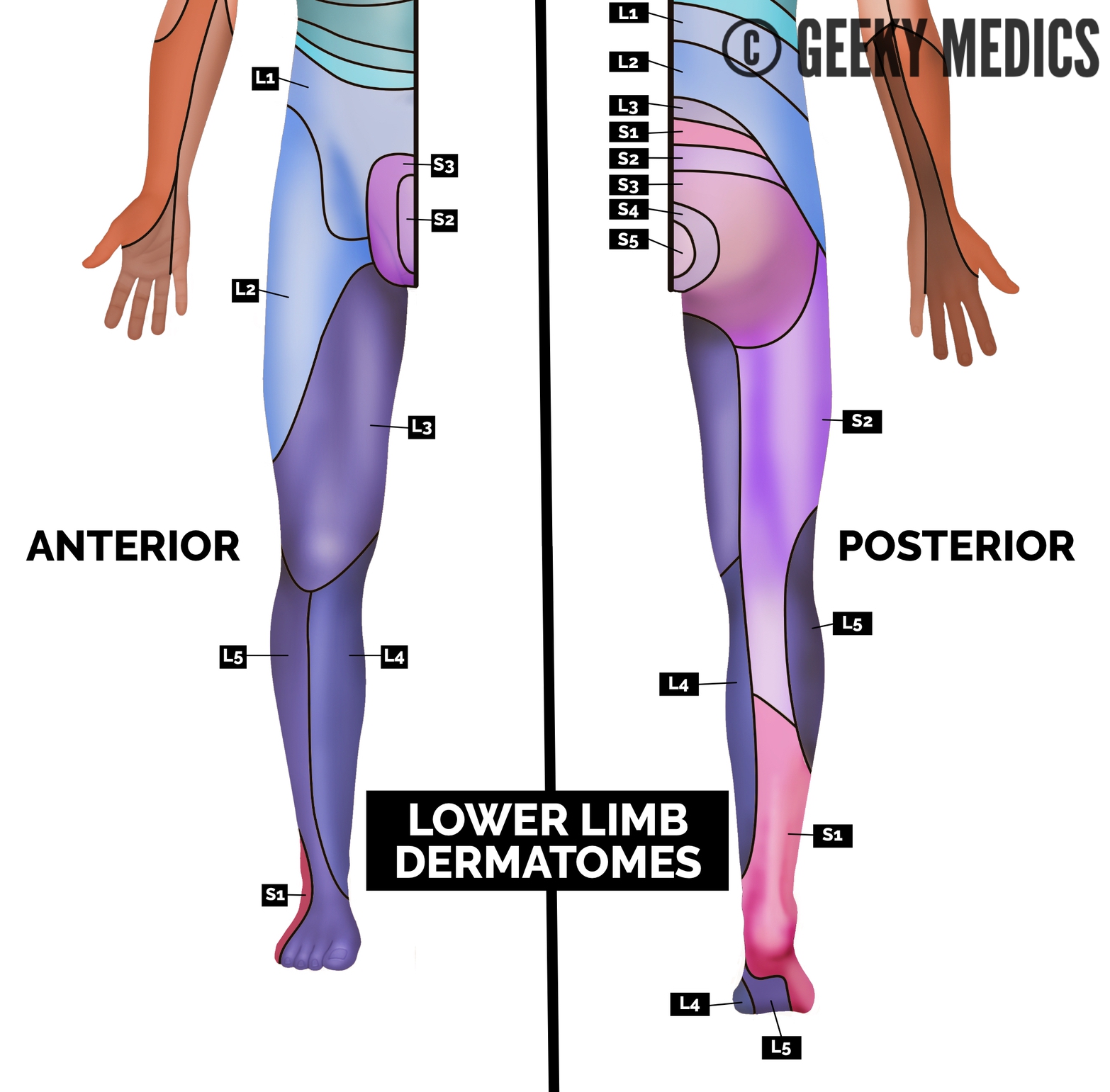
Dermatomes of the lower limb
- L1: the inguinal region and the very top of the medial thigh.
- L2: the middle and lateral aspect of the anterior thigh.
- L3: the medial epicondyle of the femur.
- L4: the medial malleolus.
- L5: the dorsum of the foot at the third metatarsophalangeal joint.
- S1: the lateral aspect of the calcaneus.
- S2: at the midpoint of the popliteal fossa.
- S3: at the horizontal gluteal crease (the horizontal crease formed by the inferior aspect of the buttocks and the posterior upper thigh).
- S4/5: the perianal area.
To learn how to assess sensation as part of a neurological examination, see our upper and lower neurological examination guides.
Dermatomal map of the whole body
Myotomes
A myotome is a group of muscles innervated by a single spinal nerve.
This list details some important myotome nerve roots and the actions that their associated muscles produce:
- C4: shoulder shrugs
- C5: shoulder abduction and external rotation; elbow flexion
- C6: wrist extension
- C7: elbow extension and wrist flexion
- C8: thumb extension and finger flexion
- T1: finger abduction
- L2: hip flexion
- L3: knee extension
- L4: ankle dorsiflexion
- L5: big toe extension
- S1: ankle plantarflexion
- S4: bladder and rectum motor supply
For information about examining myotomes, see the motor sections of the Geeky Medics upper and lower limb neurological examination guides.
Plexuses
We can classify groups of nerves into plexuses:
- Cervical plexus (C1 – C4): innervates the diaphragm, shoulders and neck.
- Brachial plexus (C5 – T1): innervates the upper limbs.
- Lumbosacral plexus (L2 – S4): innervates the lower extremities.
References
- American Spinal Injury Association (ASIA). Key Sensory Points (PDF). June 2008.
