

- 📖 Geeky Medics OSCE Book
- ⚡ Geeky Medics Bundles
- ✨ 1300+ OSCE Stations
- ✅ OSCE Checklist PDF Booklet
- 🧠 UKMLA AKT Question Bank
- 💊 PSA Question Bank
- 💉 Clinical Skills App
- 🗂️ Flashcard Collections | OSCE, Medicine, Surgery, Anatomy
- 💬 SCA Cases for MRCGP
To be the first to know about our latest videos subscribe to our YouTube channel 🙌
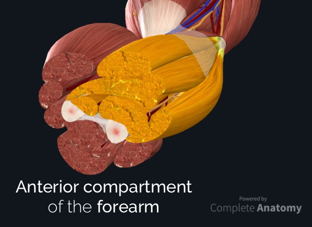
Overview
The forearm is the portion of the arm distal to the elbow and proximal to the wrist. There are 20 muscles separated into two compartments.
In this article, we will discuss the anterior compartment of the forearm in the setting of their attachment points, function, innervation and vascular supply, while providing clinical examples to reinforce this information. For a discussion of the posterior forearm compartment, check out this article.
Muscles of the anterior compartment of the forearm
- Produce wrist and/or finger flexion
- Anterior to the interosseous membrane
- Eight muscles arranged in three layers:
- Superficial
- Intermediate
- Deep
- Supplied by either the median or ulnar nerves
- Proximal blood supply:
- Brachial, ulnar or ulnar recurrent arteries
- Distal blood supply:
- Radial or ulnar arteries
- Deep veins mimic the arteries
When identifying the function of the forearm muscles, it is important to note that any forearm compartment muscle that crosses the elbow joint will act at this joint. Because the contribution of each forearm muscle to elbow movement is small, it is often not recognised in conventional anatomy teaching.
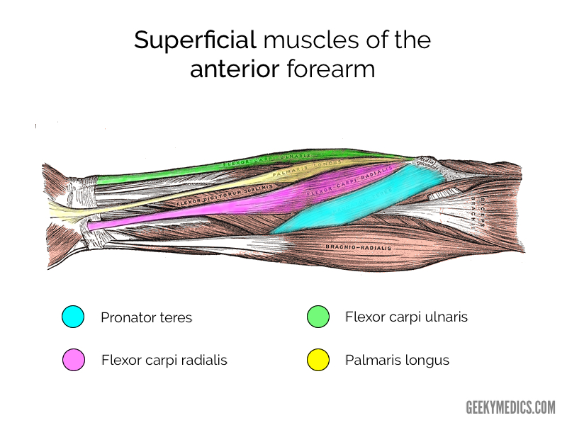
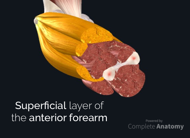
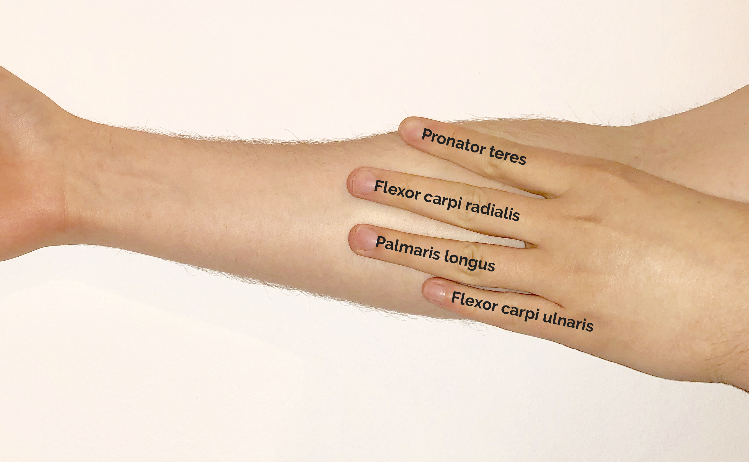
Superficial layer of the anterior compartment
The superficial layer contains 4 muscles: flexor carpi ulnaris, palmaris longus, flexor carpi radialis, and pronator teres.
All 4 muscles have a common origin at the medial epicondyle of the humerus, known as the common flexor tendon.
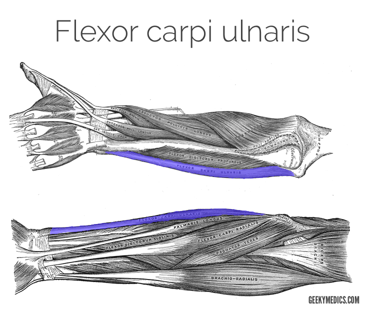
Flexor carpi ulnaris
The flexor carpi ulnaris is the most medial muscle in the superficial layer of the forearm. It is a relatively broad, strap-like muscle that plays a powerful role in movements at the wrist.
- Function: wrist flexion and adduction
- Origin: medial epicondyle, olecranon process and posterior border of the ulna
- Insertion: pisiform bone of the hand
- Innervation: ulnar nerve
- Arterial supply: ulnar artery
Palmaris longus
Palmaris longus is absent in ~15% of the population. When it is present, it lies between the flexor carpi ulnaris and flexor carpi radialis muscles. It is the most superficial forearm muscle and has a small functional role. Thus, it is often used in tendon transfers.
- Function: accessory wrist flexion
- Origin: medial epicondyle
- Insertion: into flexor retinaculum and palmar aponeurosis via a long, thin tendon
- Innervation: median nerve
- Arterial supply: ulnar artery

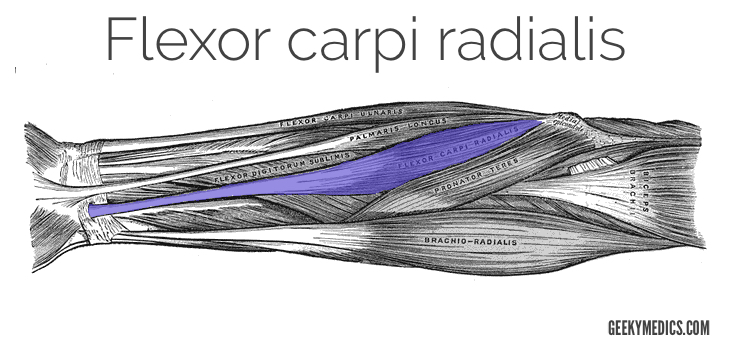
Flexor carpi radialis
When the palmaris longus is present, the flexor carpi radialis lies immediately lateral to the palmaris longus. As we move more laterally in the forearm, the radial artery can be palpated lateral to the tendon of flexor carpi radialis.
- Function: wrist flexion and abduction
- Origin: medial epicondyle
- Insertion: base of the metacarpals II and III
- Innervation: median nerve
- Arterial supply: radial artery
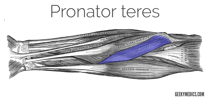
Pronator teres
The pronator teres muscle forms the medial border of the cubital fossa in the anterior elbow. It is a functionally important muscle that contains two heads.
The median nerve passes into the forearm between the two heads of the pronator teres and is separated from the ulnar artery by the ulnar head of pronator teres.
- Function: pronation of the forearm
- Origin:
- Humeral head: medial supracondylar ridge of the humerus
- Ulnar head: ulnar coronoid process
- Insertion: lateral surface of the distal radius
- Innervation: median nerve
- Arterial supply: brachial artery
Intermediate layer of the anterior compartment
The intermediate layer contains just one muscle, flexor digitorum superficialis.
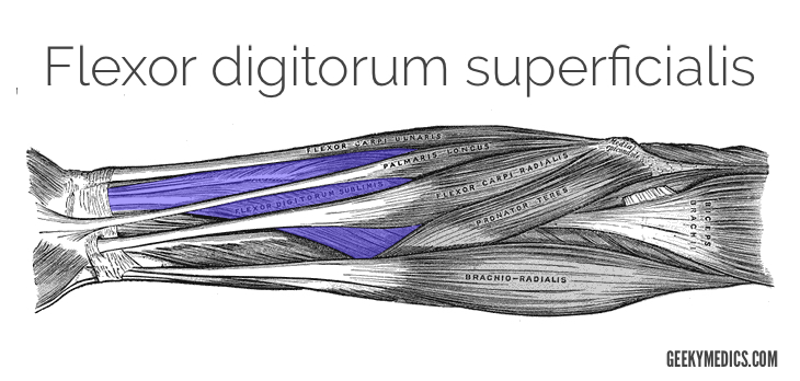
Flexor digitorum superficialis
There are several important things to know about the flexor digitorum superficialis.
Proximally, flexor digitorum superficialis contains two heads (humero-ulnar and radial), which lie either side of the median nerve and ulnar artery.
Distally, flexor digitorum superficialis divides into four tendons that pass through the carpal tunnel to digits II, III, IV and V. At the proximal phalangeal base of digits II-V, the tendon splits into two to pass laterally and then posteriorly around the tendon of flexor digitorum profundus before attaching to the lateral margins of the middle phalanx of digits II-V.
- Function: flexion at the proximal interphalangeal, metacarpophalangeal and wrist joints
- Origin:
- Humeroulnar head: medial epicondyle
- Radial head: anterior oblique line of the radius
- Insertion: middle phalanx of digits II-V
- Innervation: median nerve
- Arterial supply:
- Medial aspect: ulnar artery
- Lateral aspect: radial and median arteries

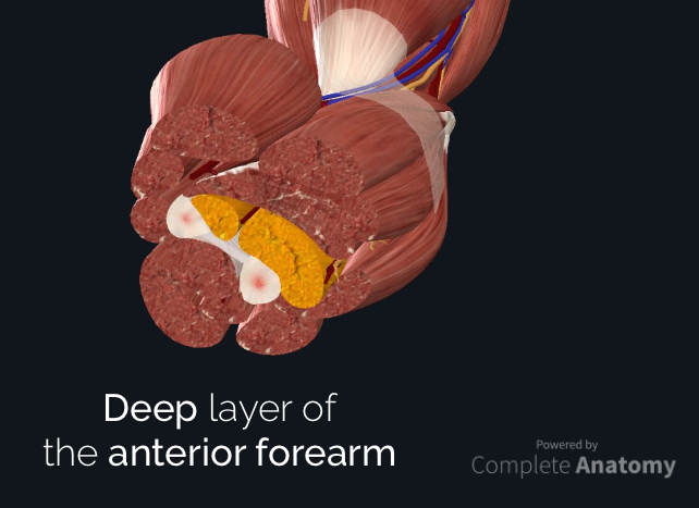
Deep layer of the anterior compartment
The deep layer contains three muscles:
- Flexor pollicis longus
- Flexor digitorum profundus
- Pronator quadratus
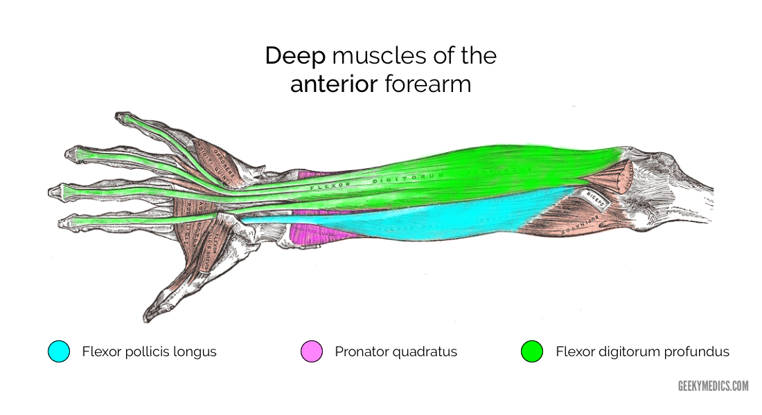
Flexor pollicis longus
The flexor pollicis longus is a powerful thumb flexor that sends a long, single tendon through the lateral region of the carpal tunnel.
- Function: flexion of the thumb
- Origin: anterior surface of the radius and interosseous membrane
- Insertion: base of the distal phalanx of the thumb
- Innervation: anterior interosseous nerve (from the median nerve)
- Arterial supply:
- Proximal aspect: anterior interosseous artery
- Distal aspect: radial artery

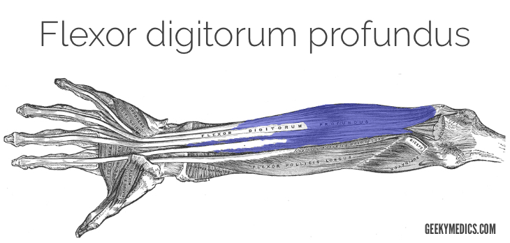
Flexor digitorum profundus
The flexor digitorum profundus is a powerful finger and wrist flexor that is one of several crucial elements in both grip strength and performing a pincer grip. It has four tendons that pass through the inferior region of the carpal tunnel before diverging to digits II-V, where it passes through the tunnel created by the splitting of the tendons of flexor digitorum superficialis.
- Function: flexion of the distal and proximal interphalangeal, metacarpophalangeal and wrist joints
- Origin: anterior and medial surface of the ulna and adjacent interosseous membrane
- Insertion: anterior surface of the base of the distal phalanx of digits II-V
- Innervation:
- Medial aspect: ulnar nerve
- Lateral aspect: anterior interosseous nerve (from the median nerve)
- Arterial supply: ulnar and anterior interosseous arteries
Pronator quadratus
The pronator quadratus is a flat square-shaped muscle in the distal forearm that assists and stabilises the forearm pronation produced by pronator teres.
- Function: pronation and stabilisation of the forearm
- Origin: antero-medial surface of the distal ulna
- Insertion: antero-lateral surface of the radius
- Innervation: anterior interosseous nerve (from the median nerve)
- Arterial supply: anterior interosseous artery

Clinical relevance – epicondylitis
Epicondylitis is a painful chronic inflammation of the tendons at either the medial or lateral epicondyles of the elbow. These injuries are often referred to as golfer’s (medial) elbow and tennis (lateral) elbow although several recreational and occupational can cause these injuries.
Risk factors
- Repetitive movements of the elbow
- Forceful activity (managing physical loads >20 kg)
Clinical presentation
- Extraarticular medial or lateral elbow pain, typically exacerbated by repetitive movements
Medial epicondylitis
- Localised tenderness over the medial epicondyle and proximal wrist flexors
- Pain with resisted wrist flexion with the elbow extended
- Pain with passive wrist extension with the elbow extended
Lateral epicondylitis
- Localised tenderness over lateral epicondyle and proximal wrist extensors
- Pain with resisted wrist extension with the elbow in full extension
- Pain with passive wrist flexion with the elbow in full extension
Diagnosis
- Clinical diagnosis based on history and exam findings
- See our elbow examination guide
Initial management
- Rest and activity modification
- Counterforce bracing
- NSAIDs (corticosteroid injection in severe cases)
- Physical therapy
Tips and tricks for the forearm muscles
1. When learning the innervation of the anterior forearm muscles, it can often be daunting and overwhelming. There are four things (underlined below) that you absolutely must know:
- Everything except for two muscles is innervated by the median nerve
- The flexor carpi ulnaris and ulnar half of flexor digitorum profundus are innervated by the ulnar nerve
2. If you get stuck on an exam and forget the name of a muscle, break down the name into logical components:
-
- Are you in the flexor or extensor compartment?
- Does this muscle move the wrist (carpi), fingers (digitorum) or thumb (pollicis)?
- Does this muscle run down the radial (radialis) or ulnar (ulnaris) aspect of the forearm?
- Does this muscle have a longer (longus) or shorter (brevis) sibling?
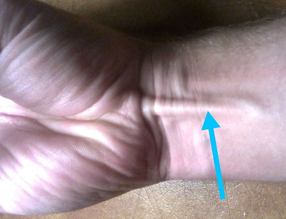
3. A neat trick to learn the superficial layer of the anterior forearm is to use your fingers as the models!
References
Images
- Complete Anatomy 2019. 3D4 Medical [LINK]
- Henry Gray. Modified by Geeky Medics [Published 2019]
- Photo created by Geeky Medics [2019]
Texts
- Richard L. Drake, A. Wayne Vogyl, Adam W.M. Mitchell. Gray’s Anatomy for Students. s.l. : Elsevier, 2015.
- Steven F Morris, G. Ian Taylor. Vascular territories. [book auth.] Peter C. Neligan Geoffrey C. Gurtner. Plastic Surgery: Volume 1. s.l. : Elsevier, 2018.
Edited by
William Maish
Geeky Medics Anatomy Lead
