
- 📖 Geeky Medics OSCE Book
- ⚡ Geeky Medics Bundles
- ✨ 1300+ OSCE Stations
- ✅ OSCE Checklist PDF Booklet
- 🧠 UKMLA AKT Question Bank
- 💊 PSA Question Bank
- 💉 Clinical Skills App
- 🗂️ Flashcard Collections | OSCE, Medicine, Surgery, Anatomy
- 💬 SCA Cases for MRCGP
To be the first to know about our latest videos subscribe to our YouTube channel 🙌
Introduction
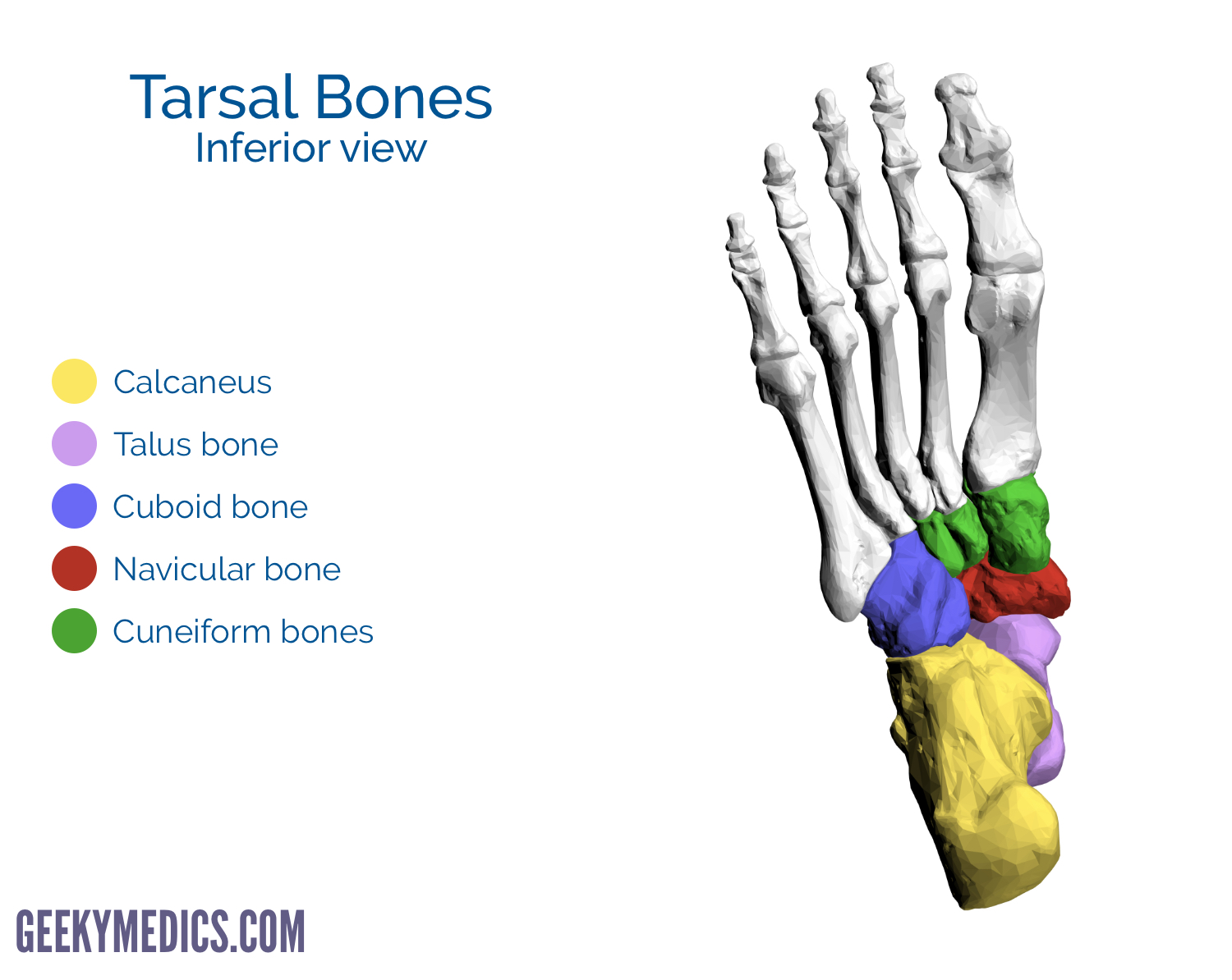
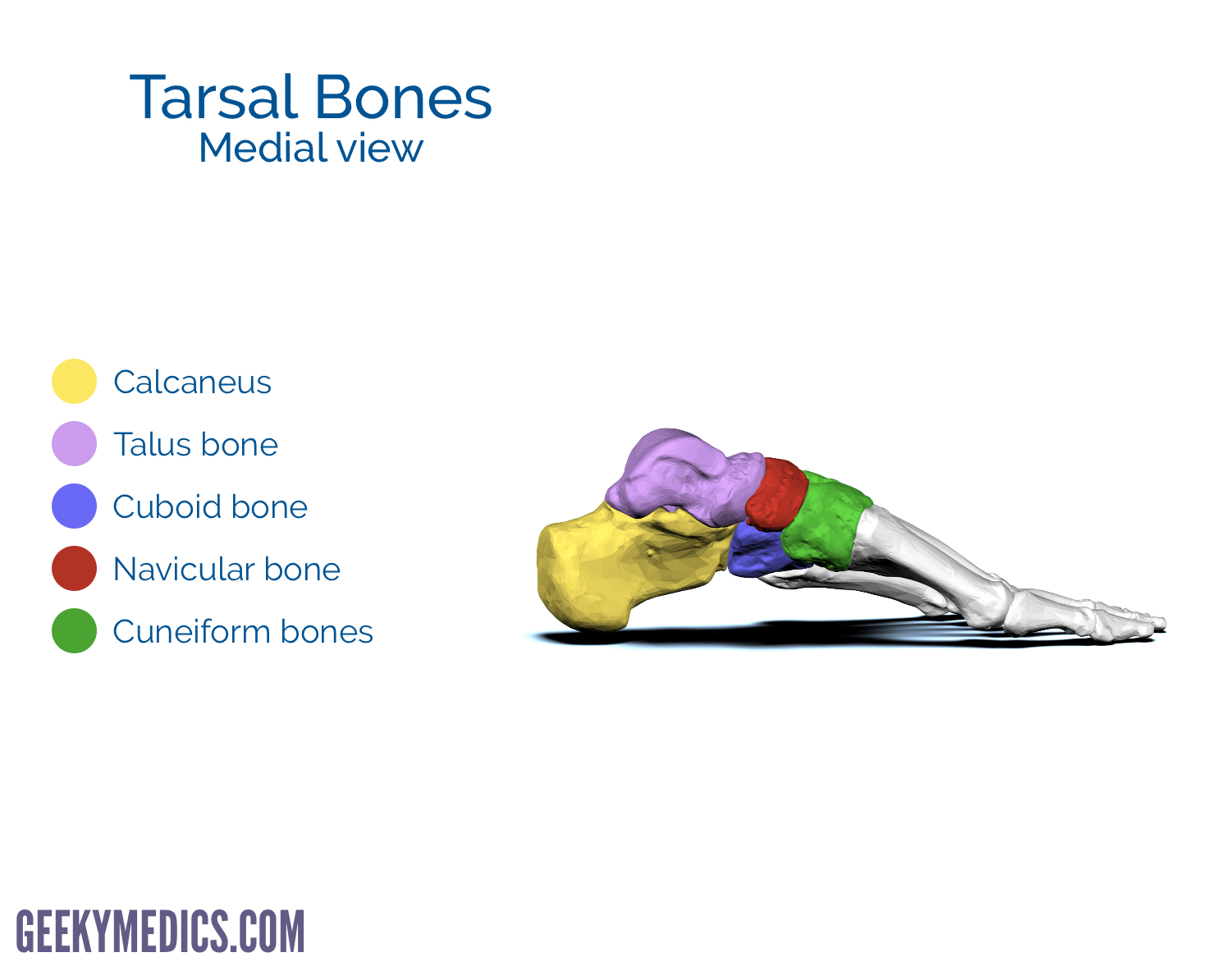
The skeleton of the foot consists of 26 bones and these can be grouped into three groups:
- The tarsus (ankle joint)
- The metatarsus
- The phalanges (bones of the toes)
There are 7 tarsal bones, 5 metatarsal bones and 14 phalanges.
Anatomically the foot can be divided into the forefoot (metatarsals and phalanges), the midfoot (cuboid, navicular and cuneiforms) and the hindfoot (calcaneus and talus).
Injuries to the bones of the foot commonly occur in athletes and active individuals. Most injuries are the consequence of acute trauma during sports.
Adolescents are most vulnerable to these injuries due to the incompletion of endochondral ossification and the existence of their cartilaginous epiphyseal plates. As such, it is important to have a basic anatomical knowledge regarding the bones of the foot, including their articulations and sites of attachment.
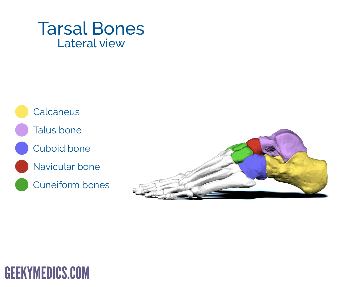
The tarsus
The tarsus consists of 7 bones which make the posterior aspect of the foot: talus, calcaneus, cuboid, navicular and three cuneiforms.
The tarsal bones can be divided into the hindfoot (talus and calcaneus) and midfoot (cuboid and cuneiforms). The navicular is the intermediate bone between these two groups.
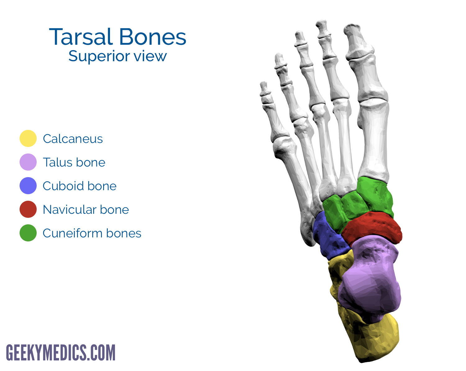
Talus
Talus (Latin for ankle) talus is the most superior bone of the tarsus and rests on top of the calcaneus. Three areas of articulation form the ankle joint:
- The superomedial aspect of the talus articulates with the medial malleolus of the tibia
- The superior aspect of the talus articulates with the distal portion of the tibia
- The lateral aspect of the talus articulates with the lateral malleolus of the fibula
Anteriorly the talus articulates with the navicular bone. Medially the talus articulates with the intermediate bone of the talus.
The talus has a body, head and neck. The superior surface, known as the trochlea, has the two malleoli attached which help with the stability of the joint. The trochlea is wider anteriorly, leading to a difference in stability depending on joint position. Dorsiflexion of the joint stretches the ligaments resulting in joint stability. Plantarflexion narrows the trochlea width resulting in decreased stability.
Calcaneus
The calcaneus is often referred to as the heel bone and is the largest and strongest bone of the foot. The weight of the body passes through the tibia, into the talus and then to the calcaneus. The sustentaculum tali is a bony prominence on the medial aspect of the calcaneus which supports the medial aspect of the talus bone.
The superior aspect of the calcaneus has three articular surfaces for articulation with the talus. The anterior surface articulates with the cuboid. The posterior aspect of the calcaneus has a large weight-bearing region called the calcaneal tubercle.
A hard fall on the heel (e.g. falling from a ladder) may result in a calcaneal fracture. Diagnosis is based on symptoms (pain, bruising, difficulty walking) and then can be confirmed by X-ray or CT. 3
Cuboid
The cuboid bone posteriorly articulates with the end of the calcaneus. It is the most lateral bone in distal row of tarsal bones. Anterior to the cuboid tuberosity, on the lateral and inferior surfaces of the bone is a groove which provides passage for a muscle tendon.
Navicular
The navicular bone’s name is derived from its resemblance to a small boat. The navicular is located between the head of the talus posteriorly and the three cuneiforms anteriorly. The navicular bone is on the medial aspect of the foot and articulates with the talus proximally, the three cuneiforms distally and the cuboid laterally.
Cuneiforms
The cuneiform bones are wedge-shaped. They are named medial, intermediate and lateral based on their relative position. Each cuneiform has a narrow inferior surface and a broad superior surface resulting in the transverse curvature of the foot. Each cuneiform articulates with the navicular posteriorly and their respective metatarsal anteriorly.
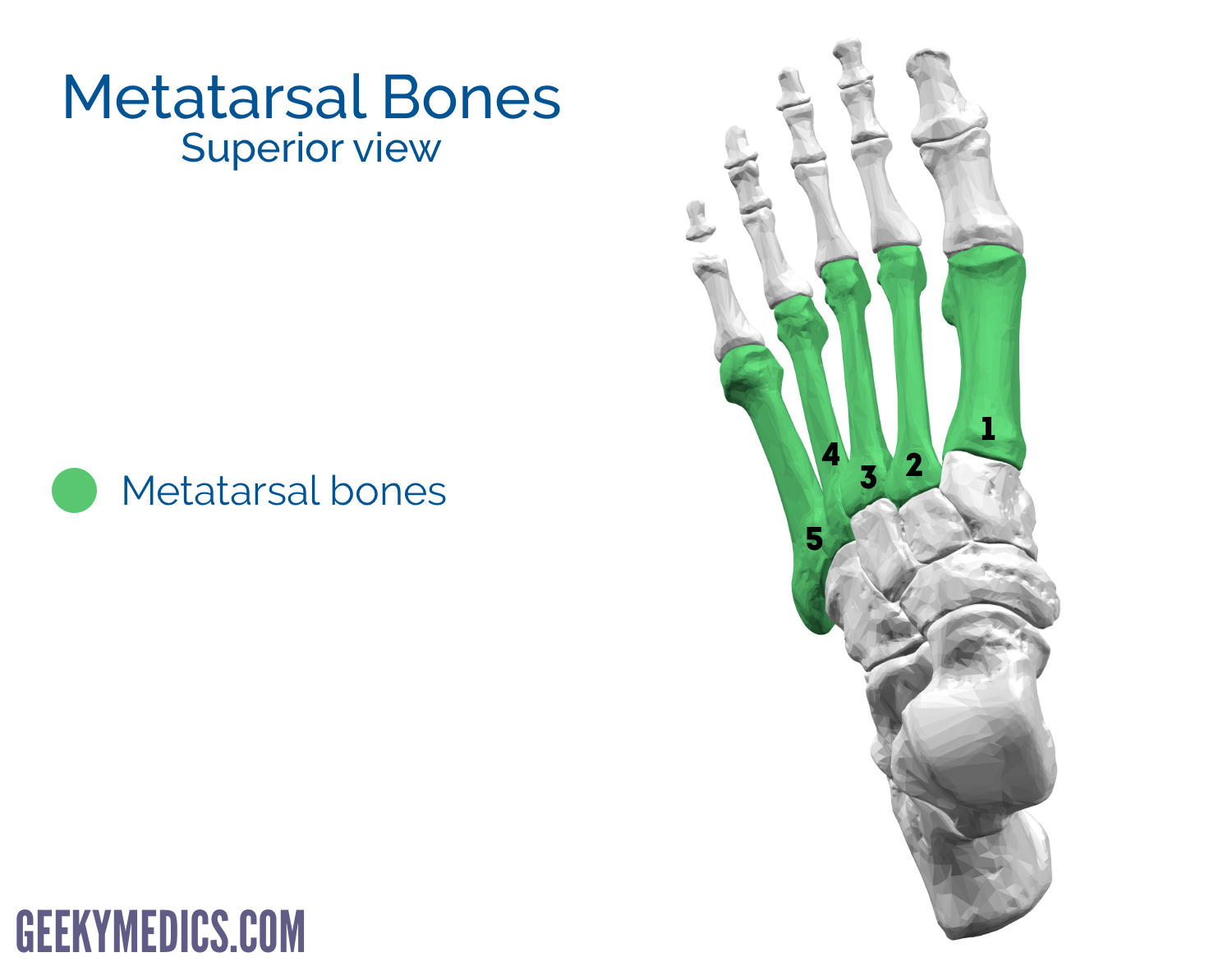
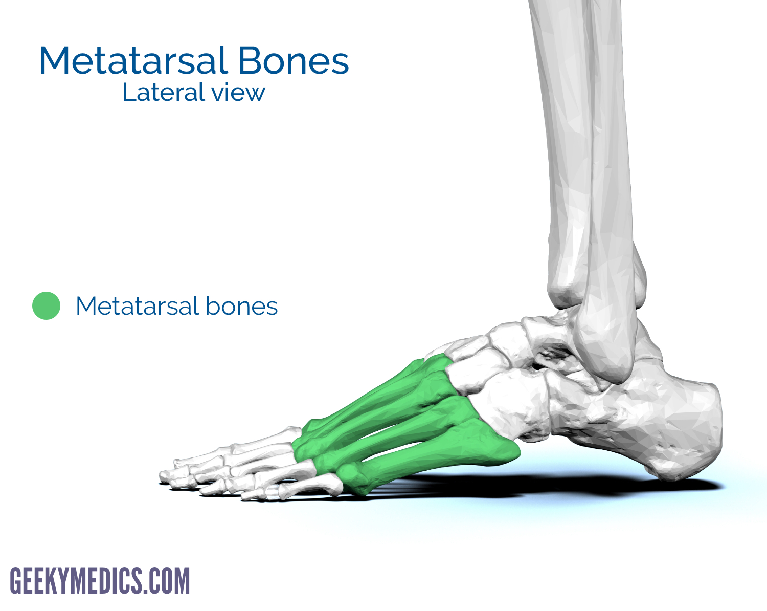
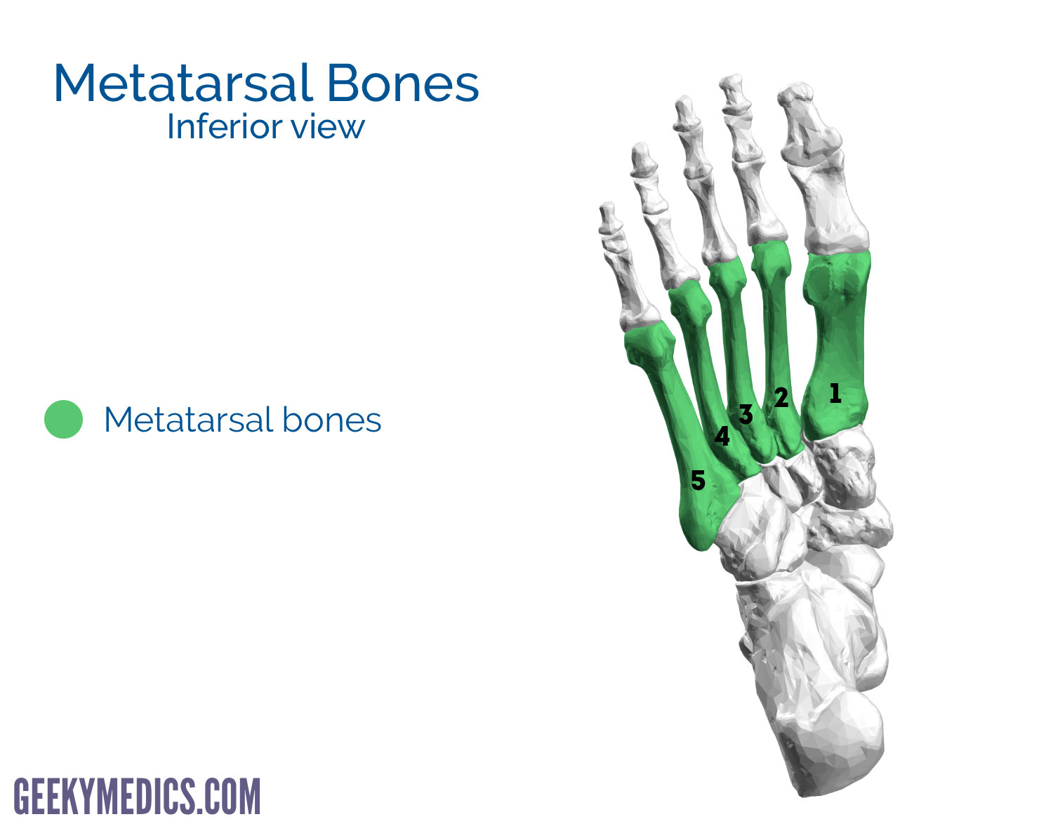
Metatarsals
The anterior portion of the foot is formed by the 5 metatarsal bones which are numbered 1-5 starting from the medial aspect of the foot. The first metatarsal is shorter and thicker than the others.
Each metatarsal has a base (proximally), a shaft, and a head (distally). The base of metatarsal articulates with the cuneiform or cuboid bones. The head of a metatarsal articulates with the proximal phalanges to form the metatarsophalangeal joint (MTP). The heads of the metatarsals lie in contact with the ground forming the ball of the foot. The bases of 5th metatarsals have a large tuberosity providing an area for tendon attachment.
Fractures of the metatarsals typically occur when a heavy object falls on the foot. These fractures are common in football players either as a result of direct trauma or as stress fractures (“march fractures”).
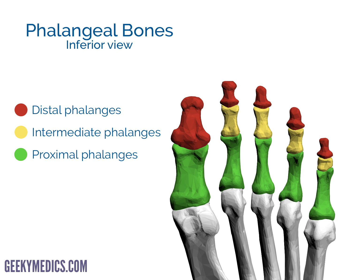
Phalanges
Finally, there are 14 phalanges in the foot which are arranged in a similar manner as in the hand. The 1st digit has two phalanges (proximal and distal) whereas the remaining four digits have three phalanges (proximal, intermediate and distal).
Like the metatarsals, each phalanx has a base (proximally), a shaft, and a head (distally). Like the 1st metatarsal, the first phalanges of the first digit are short and broad.
Reviewer
Mr Saleem Mastan
Trauma & Orthopaedic Registrar
References
- By BodyParts3D is made by DBCLS. (Polygon data is from BodyParts3D). License: [CC BY-SA 2.1]
- Chinn L, Hertel J. Rehabilitation of Foot and Ankle Injuries in Athletes. Clin Sport Med [Internet]. 2010;29(1):157–67. Available from: [LINK]
- Palmersheim K, Hines B, Olsen BL. Calcaneal fractures: update on current treatments. Clin Podiatr Med Surg [Internet]. 2012 Apr;29(2):205–20, vii. Available from: [LINK]
