

- 📖 Geeky Medics OSCE Book
- ⚡ Geeky Medics Bundles
- ✨ 1300+ OSCE Stations
- ✅ OSCE Checklist PDF Booklet
- 🧠 UKMLA AKT Question Bank
- 💊 PSA Question Bank
- 💉 Clinical Skills App
- 🗂️ Flashcard Collections | OSCE, Medicine, Surgery, Anatomy
- 💬 SCA Cases for MRCGP
To be the first to know about our latest videos subscribe to our YouTube channel 🙌
Introduction
The human skull serves the vital function of protecting the brain from the outside world, as well as supplying a rigid base for muscles and soft tissue structures to attach to.
The bones of the skull are held rigidly in place by fibrous sutures. In this article, we explore the bones of the skull during development before discussing their important features in the context of function and pathology.
Embryology of the skull
The skull begins to form prior to week 12 of embryogenesis. A cartilaginous mould begins to grow and is slowly replaced by bone in a process called intramembranous ossification.
This process occurs at different rates in two different regions of the skull:
- Calvarium (roof): the frontal, occipital and parietal bones tend to grow slightly earlier
- Base: the sphenoid, ethmoid, occipital and temporal bones tend to grow slightly later
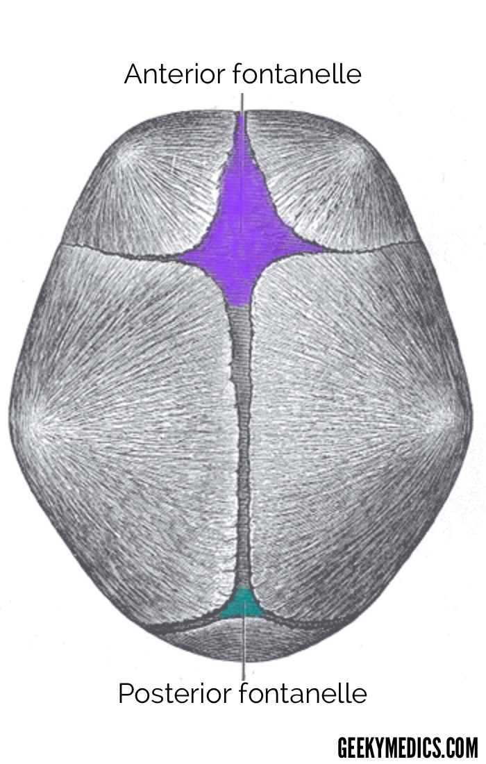
By week 37, the cartilaginous frame for the skull is almost entirely replaced by bone, and only two relatively small cartilaginous structures remain:
- Anterior fontanelle: the soft region on the front of the infant’s head
- Posterior fontanelle: the soft region on the back of the infant’s head
The fontanelles undergo complete ossification within 9 – 18 months from birth.
Clinical relevance: Fontanelle abnormalities
An enlarged anterior fontanelle can be due to endocrine pathologies like congenital hypothyroidism.
A bulging anterior or posterior fontanelle may suggest raised intracranial pressure, which may occur in hydrocephalus.
A sunken anterior or posterior fontanelle may suggest dehydration.
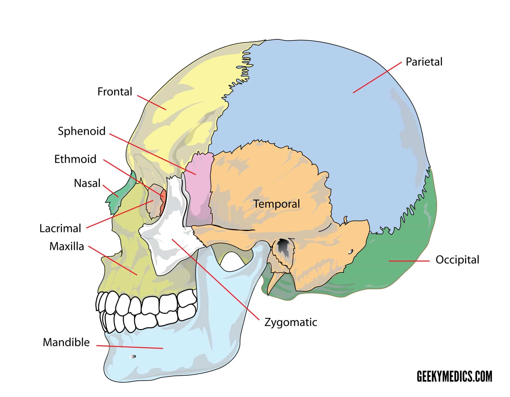
Bones of the calvarium
The calvarium, also known as the roof or skull cap, consists of three bones:
- Frontal bones
- Parietal bones
- Occipital bones
These bones protect the brain superiorly, but also provide an anchor for important muscles of facial expression and eye movement. The parts of these bones that lie inferior to the brain are considered to be a part of the skull base (mentioned below).
Of particular importance is the strength of these bones relative to how thin they are. These bones of the skull cap contain two thin layers of cortical bone separated by diploë, which is synonymous with the spongy bone seen in long bones.
Diploë provides structural integrity and transmits forces along the length of the bones of the skull cap.
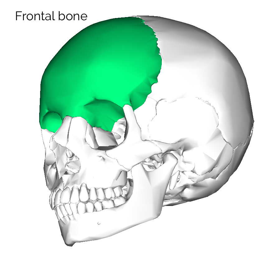
Frontal bone
The frontal bones start as two paired bones. During childhood and adolescence, these two bones fuse into one.
The fusion of these frontal bones forms a metopic suture, which may be visible in some skull specimens.
There are three important parts of the frontal bone to know:
- Orbital plate of the frontal bone: forms the roof of the orbital cavity and separates the frontal lobe of the brain from the eye
- Glabella: the brow ridge that serves as an attachment point for the frontalis muscle and superior portion of the orbicularis oculi
- Supraorbital notch: nerves supplying the forehead to emerge through this aperture
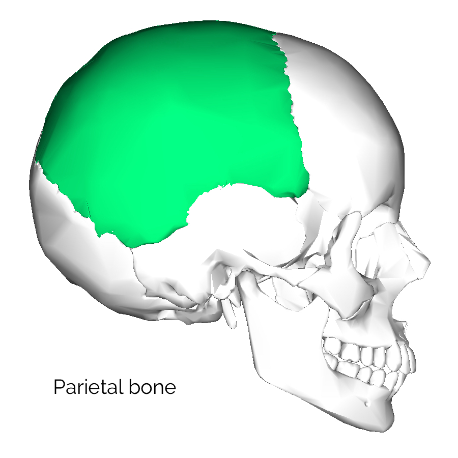
Parietal bone
The two parietal bones join in the sagittal line to form the sagittal suture. They are each joined anteriorly to the frontal bone by the coronal suture.
Overlying the parietal bones is a thick connective tissue layer known as the galea aponeurotica. The galea joins the frontalis muscle belly anteriorly to the occipitalis muscle belly posteriorly. This is why raising your eyebrows can create the appearance that the back of the head is moving.
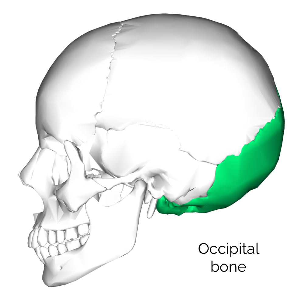
Occipital bone
The occipital bone forms a cup for the occipital lobe of the brain to sit in.
The occipital bone is joined superiorly to the parietal bones by the lambdoidal suture. It contains an external occipital protuberance that can be felt on the back of your head.
The foramen magnum, housing the brainstem, is also a part of the occipital bone. Either side of the foramen magnum are the hypoglossal canals for cranial nerve XII.
The clivus is an incline in the occipital bone that the ventral pons and medulla rest on. Anteriorly, the occipital bone joins to the sphenoid and temporal bones.
Bones of the skull base
The bones of the skull base include:
- Part of the frontal bone
- Part of the occipital bone
- Temporal bone
- Sphenoid bone
- Ethmoid bone
We will first briefly revisit the bones of the calvarium that contribute to the skull base (frontal and occipital) and then discuss the temporal, sphenoid and ethmoid bones in some more detail.
Frontal bone
The orbital plate of the frontal bone is technically part of the skull base, as it lies below the brain. It forms the anterior cranial fossa for the frontal lobe.
Occipital bone
The part of the occipital bone that is horizontal and creates the posterior cranial fossa for the occipital lobe is part of the skull base. The clivus is technically part of the skull base.
The foramen magnum is considered part of the skull base
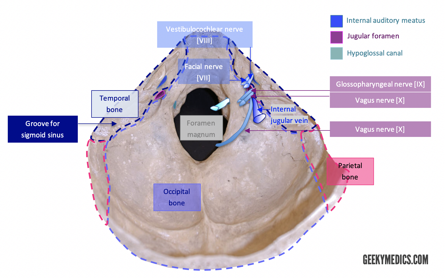
Temporal bone
The temporal bone is a complex anatomical structure that transmits many veins, arteries and nerves into and out of the skull.
The temporal bone consists of four main parts: squamous, petrous, tympanic and mastoid.
Squamous
- Shaped like a fish scale (for which it is named)
- Contributes to the temporal fossa
Petrous
- Pyramid or wedge-shaped
- Densest bone in the body
- Contains the internal auditory meatus for cranial nerves VII and VIII
Tympanic
- Curved plate below the squamous portion
- Originates as a separate bone (the tympanic bone) which fuses with the temporal bone
- Contains the external auditory meatus
Mastoid
- Named after its breast-like appearance
- Contains the mastoid and styloid processes
Sphenoid bone
Literally translating to “wedge-like,” the sphenoid bone is named after its appearance. However, it is classically referred to as being shaped like a butterfly, moth or bat with its wings spread.
There are several important structures to know:
- Greater wings: form the floor of the middle cranial fossa for the temporal lobe to sit in
- Sella turcica: the “Turkish chair” that contains the pituitary gland
- Orbital surface: forming the posterior wall of the orbit
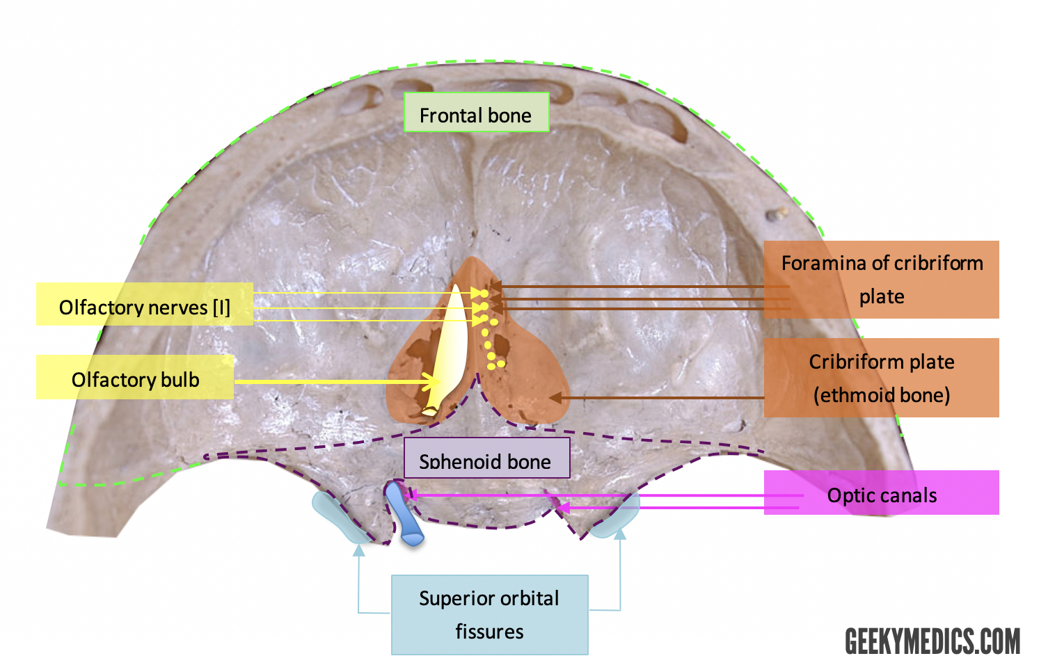
Ethmoid bone
The ethmoid bone separates the nasal cavity from the brain.
It forms part the perpendicular plate of the osseous nasal septum.
It also contains small foramen that allow the olfactory nerve to move from within the skull to the top of the nasal cavity to sample air particles. This part of the ethmoid bone is called the cribriform plate.
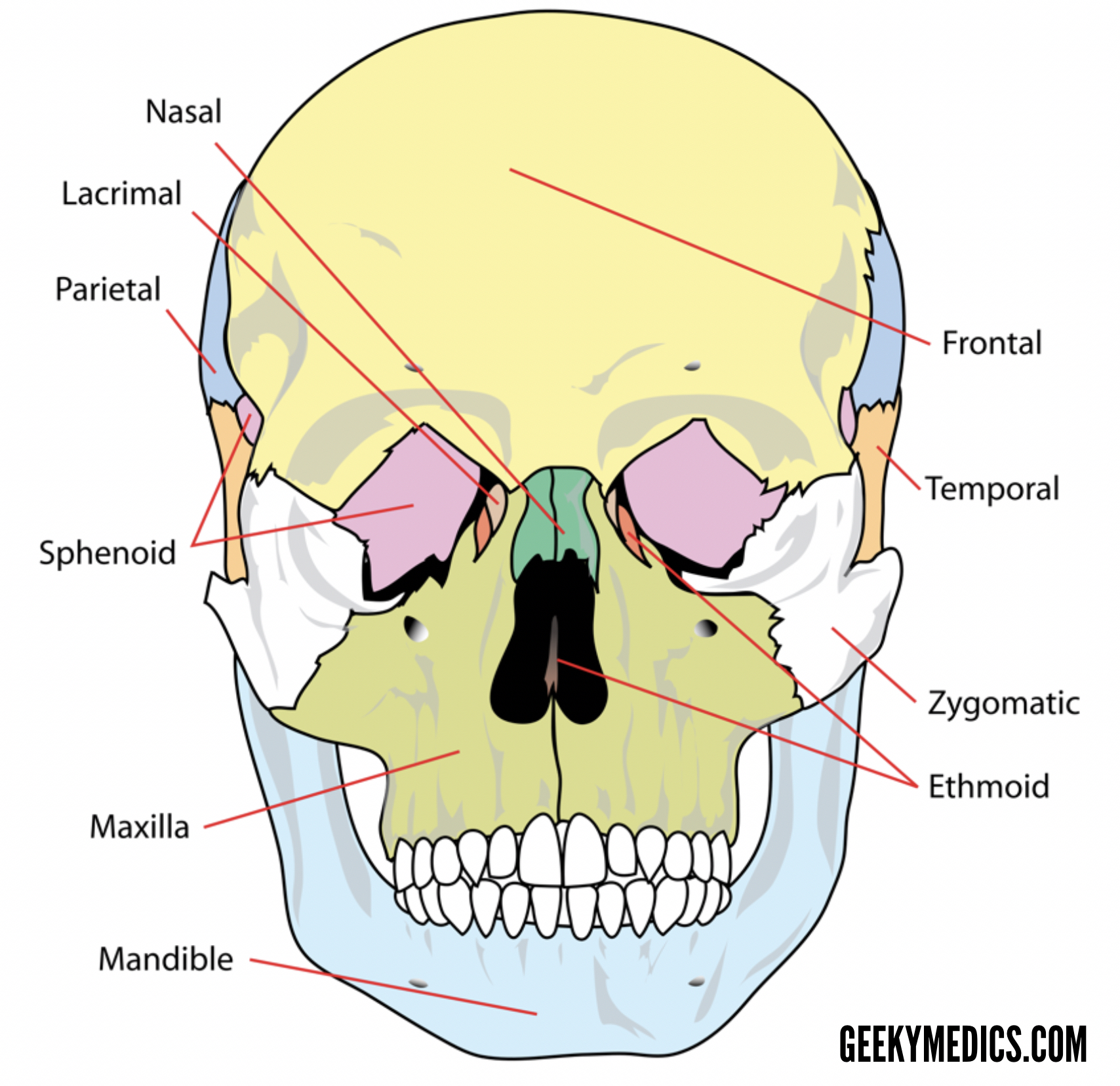
Bones of the face
There are 14 individual bones of the face that support soft tissue structures to determine the appearance of the face.
These bones fuse to form part of the orbital, nasal and oral cavities, as well as some of the sinuses of the face.
Nasal bones
- Lie between the maxillae
- Form the top, osseous part of the nose
Inferior nasal conchae
- Two bones in the nasal cavity that increase air turbulence and surface area for humidification
Maxillae
- Form the upper jaw
Lacrimal bones
- Forming the medial wall of the orbit
- House the lacrimal sac
Zygomatic bone
- Form the inferolateral floor of the orbital cavity
- Form the cheekbones and zygomatic arch
Palatine bones
- Posteriorly-located in the oral cavity
- The soft palate and uvula hang from the palatine bones
Vomer
- Form the lower part of the osseous nasal septum
Mandible
- Form the lower jaw
- Joined to the temporal bone at the temporomandibular joint (TMJ)
Foramina of the skull
The foramina (singular: foramen) of the skull are numerous and it is easy to get lost identifying them. We will revisit each of these foramina again in articles on the cranial nerves and cranial vasculature.
Foramen to know are briefly discussed below, to learn more, you can check out our guide to the foramina of the skull.
Olfactory foramina
- Located in the ethmoid bone
- Transmits the olfactory nerves
Optic canal
- Located in the sphenoid bone
- Transmits the optic nerve
Superior orbital fissure
- Located in the sphenoid bone
- Transmits cranial nerves III, IV, V1, VI, and several vascular structures
Foramen rotundum
- Located in the sphenoid bone
- Transmits V2
Foramen ovale
- Located in the sphenoid bone
- Transmits V3 and portio minor (and several vascular structures)
Foramen spinosum
- Located in the sphenoid bone
- Transmits the middle meningeal vessels
Internal acoustic meatus
- Located in the temporal bone
- Transmits cranial nerves VII (within the skull bone) and VIII
Stylomastoid foramen
- Located in the temporal bone
- Transmits cranial nerve VII (exit)
Carotid canal
- Located in the temporal bone
- Transmits the internal carotid artery and plexus
Jugular foramen
- Located in the temporal bone anteriorly and the occipital bone posteriorly
- Transmits the internal jugular vein and cranial nerves IX, X, XI
Foramen lacerum
- Located between the sphenoid, temporal and occipital bones
- Does not transmit anything normally
Foramen magnum
- Located in the occipital bone
- Transmits the brainstem and cranial nerve XI into the skull
Hypoglossal canal
- Located in the occipital bone
- Transmits cranial nerve XII

Sutures of the skull
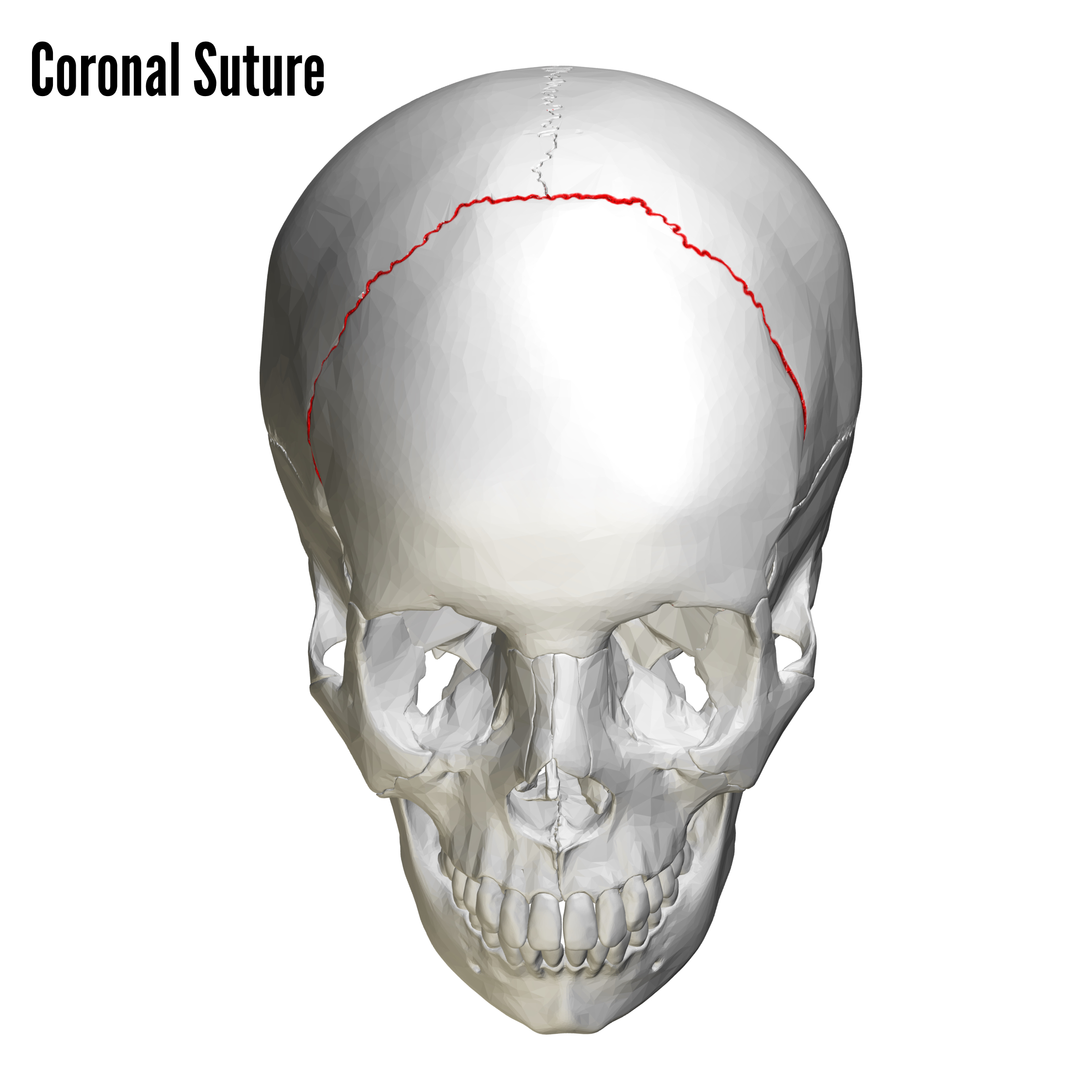
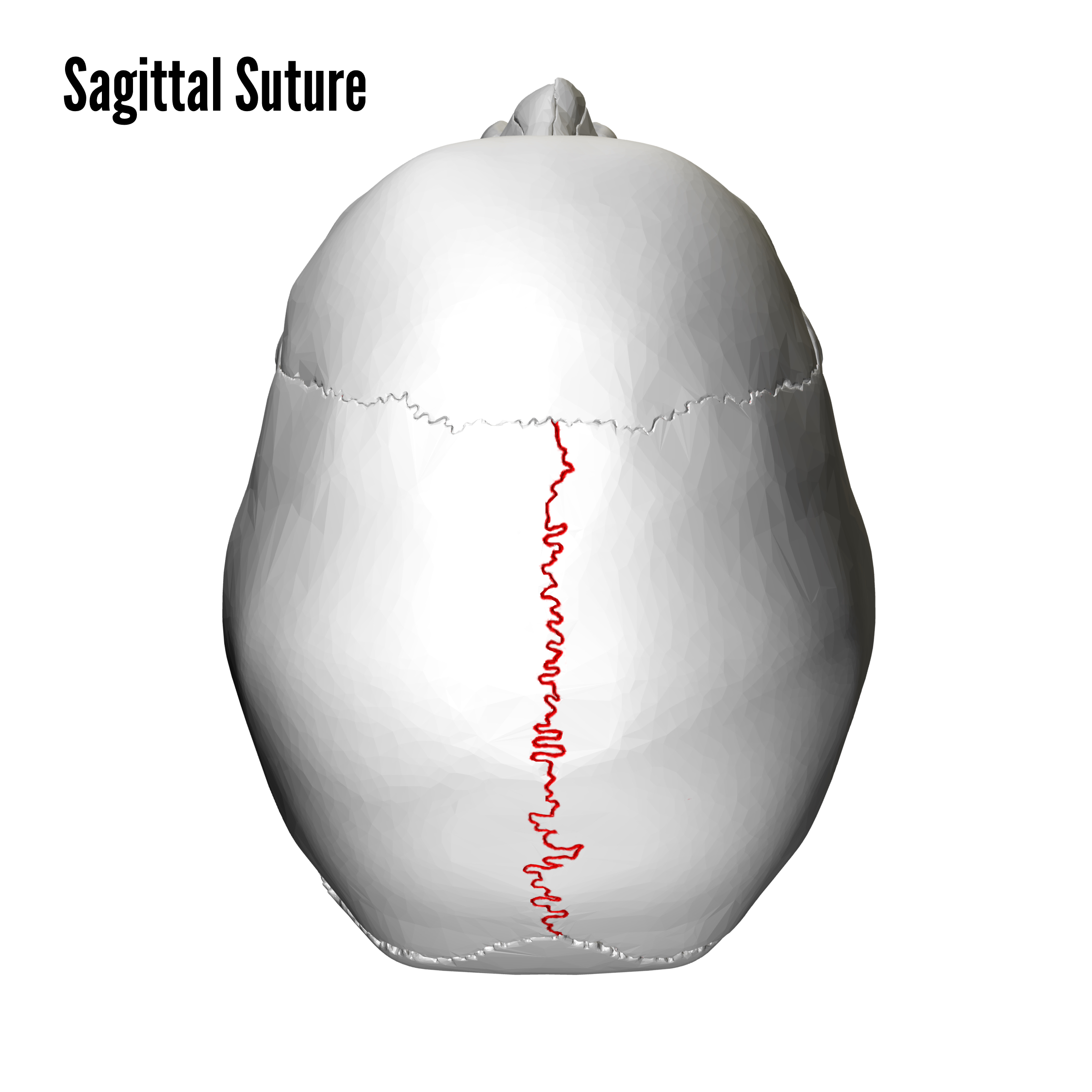
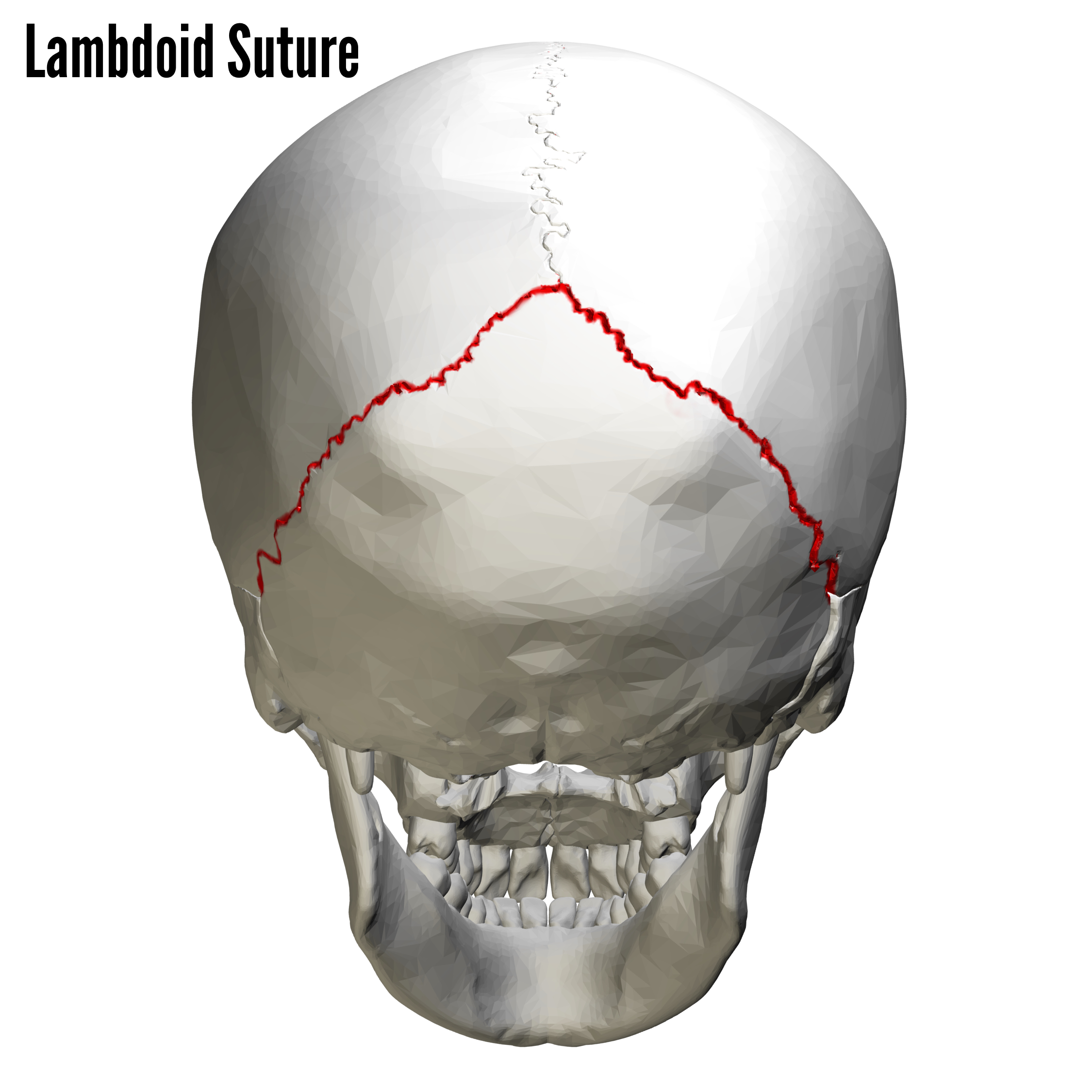
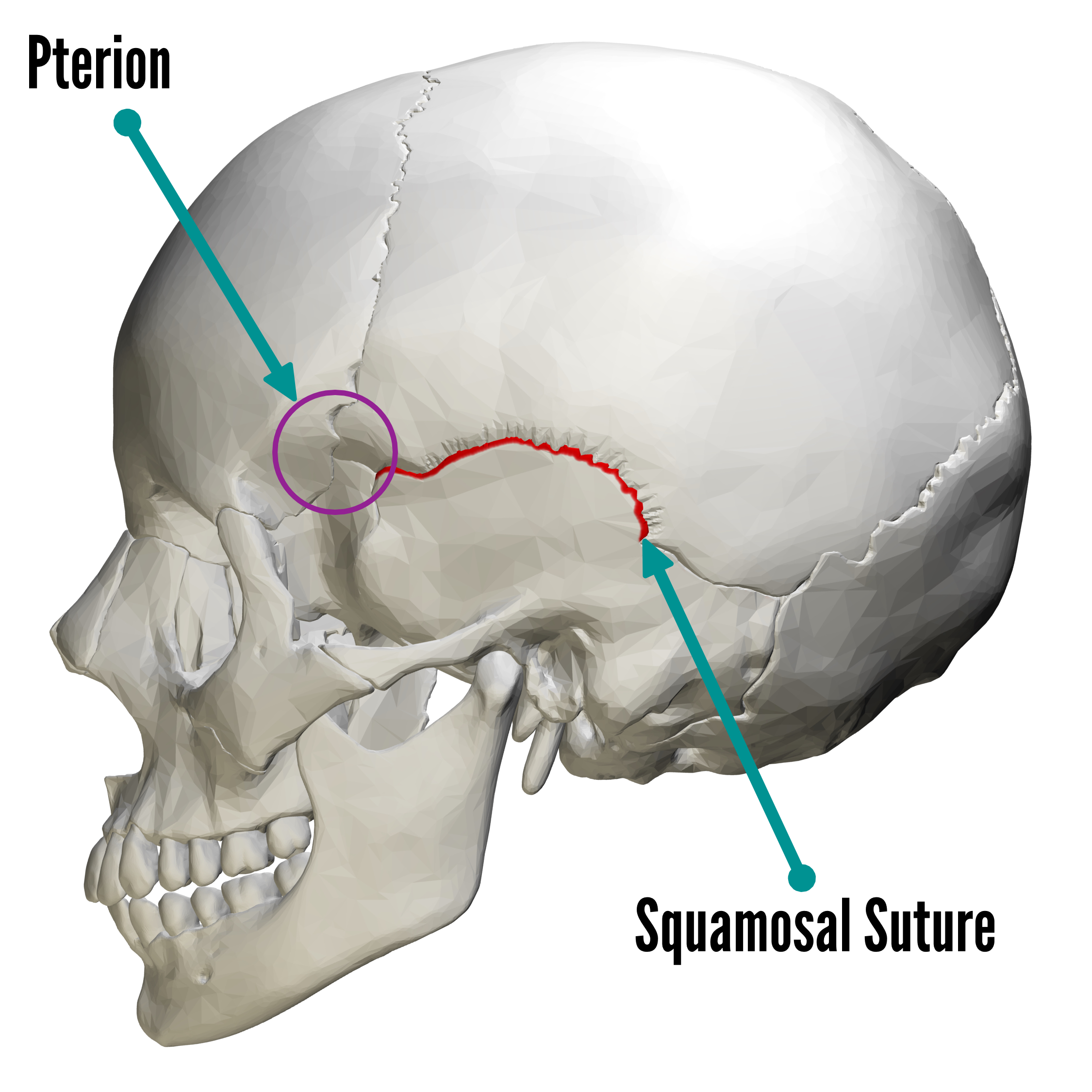
We have briefly mentioned several of the sutures of the skull. They are shown in the pictures below for ease of learning.
Frontal (metopic) suture
- Fused frontal bones that may sometimes be seen in normal adult skulls (Figure 10)

Coronal suture
- Between the frontal and parietal bones
Sagittal suture
- Between the parietal bones
Lambdoidal suture
- Between the parietal and occipital bones
Squamosal suture
- Between the temporal and parietal bones
Clinical relevance: Endocrine dysfunction
Disease such as acromegaly and osteopetrosis can lead to overgrowth of bones. In the skull, this has an important consequence of narrowing the foramen responsible for nerve and blood vessel transmission. Reduced visual acuity and deafness can be consequences of such diseases.
Clinical relevance: Skull fractures
Regions of the skull where multiple bones join together can be relatively weak and predispose that region to fracture.
One of the weakest points on the human skull is the ‘temple’ region. This is known anatomically as the pterion (see the image above) and is formed by the junction of the frontal, parietal and sphenoid bones. Not only is it weak due to the number of bones converging here, but the bones themselves are very thin.
Another fracture type to be aware of is a ‘blowout’ fracture. A blowout fracture occurs when the maxilla at the floor of the eye is fractured and the eye appears to sink into the depression (enophthalmos). The patient will also experience significant diplopia. If either enophthalmos or diplopia are present, surgical intervention is warranted.
References
Reference texts
- Sinnatamby, C. S. (2011). Last’s Anatomy, International Edition: Regional and Applied. Elsevier Health Sciences.
- Moore, K. L., Dalley, A. F., & Agur, A. M. (2013). Clinically oriented anatomy. Lippincott Williams & Wilkins.
Images
- Henry Vandyke Carter. License: [Public domain]
- LadyofHats Mariana Ruiz Villarreal. License: [Public domain]
- BodyParts3D/Anatomography. License: [CC BY-SA]
- Vassil. License: [Public domain]
- Matthew S. Cain (2008), A photograph of the lower inner surface of a human skull, public domain via Wikimedia.org [LINK] (modified by Melissa Gough, 2019)
