
- 📖 Geeky Medics OSCE Book
- ⚡ Geeky Medics Bundles
- ✨ 1300+ OSCE Stations
- ✅ OSCE Checklist PDF Booklet
- 🧠 UKMLA AKT Question Bank
- 💊 PSA Question Bank
- 💉 Clinical Skills App
- 🗂️ Flashcard Collections | OSCE, Medicine, Surgery, Anatomy
- 💬 SCA Cases for MRCGP
To be the first to know about our latest videos subscribe to our YouTube channel 🙌
Introduction
There are three major groups of back muscles:
- Superficial: attached to the shoulder girdle
- Intermediate: attached to the posterior thorax
- Deep: attached to the vertebral column
The first two groups (superficial and intermediate) are referred to as the extrinsic back muscles. The deep group are the intrinsic muscle group. This article will focus on the deep group.
The deep back muscles lie immediately adjacent to the vertebral column and ribs. They are divided into three groups, as shown below.
Superficial group:
- Splenius capitis
- Splenius cervicis
Intermediate group:
- Iliocostalis (lumborum, thoracis, cervicis)
- Longissimus (thoracis, cervicis, capitis)
- Spinalis (thoracis, cervicis, capitis)
Deep group:
- Semispinalis (thoracis, cervicis, capitis)
- Multifidus
- Rotatores
- Interspinales, interansversarii, levatores costarum
This article will discuss the muscles in each of these three intrinsic back muscle subgroups.
A small note on nomenclature that makes learning the orientation of muscles easier: capitis refers to head, and any muscle with capitis in the name will insert into the head. Cervicis refers to the cervical spine, thoracis the thoracic spine and lumborum the lumbar spine. Respectively, muscles with these names will insert into the cervical, thoracic or lumbar spines.
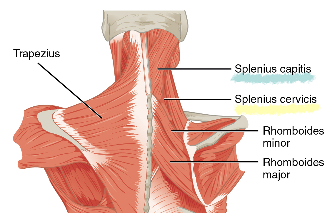
Superficial: splenius capitis
Splenius capitis is one of the deep back muscles that is associated with rotating and extending the head and neck. It is a long, broad, strap-like muscle found deep to the trapezius muscle.
Origin and insertion
Splenius capitis originates from the spinous processes of C7-T4 and the nuchal ligament. It splays laterally to insert onto the mastoid process and the lateral part of the superior nuchal line of the occipital bone.
Fibre orientation
The fibres for splenius capitis splay superolaterally from the midline of the spine.
Function
The function of splenius capitis unilaterally is to cause ipsilateral rotation of the head. Acting bilaterally, splenius capitis causes extension of the head and cervical spine.
Innervation
The sensorimotor innervation of splenius capitis is derived from the dorsal rami of C3 and C4.
Arterial supply
Muscular branches of the aorta.
Superficial: splenius cervicis
The splenius cervicis is similar to the splenius capitis. However, instead of attaching to the head, it attaches to the transverse processes of the cervical vertebra.
Origin and insertion
Originating from the spinous processes of T3-T6 vertebra, the splenius cervicis then inserts onto the transverse processes of C1-C4.
Fibre orientation
The fibres of splenius cervicis travel superolaterally from the midline of the spine.
Function
Splenius cervicis functions unilaterally to ipsilaterally rotate the cervical spine. Bilaterally, it causes extension of the cervical spine.
Innervation
The sensorimotor innervation of the splenius cervicis is derived from the dorsal rami of C5-C8 (common variants include C4-T6).
Blood supply
The splenius cervicis is supplied by the occipital or transverse cervical arteries. Occasionally, it can be supplied by both.
Clinical relevance: splenius capitis and cervicis
Splenius capitis and cervicis are strong muscles that assist with major head and neck movements. Posterior cervical laminectomies or discectomies, as well as suboccipital cranial surgeries, can lead to significant access trauma to these muscles manifesting as fibrofatty replacement.
This may manifest with both poor head and neck extension, with patients appearing to ‘look at the ground.’ In these patients, this damage can be a significant cause of pain.1,2
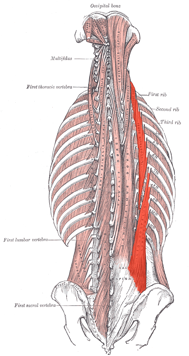
Intermediate: iliocostalis
Iliocostalis is the most lateral muscle of the intermediate group, also known as the erector spinae group. The most medial is the spinalis muscle group. A useful mnemonic for these muscles, from lateral to medial, is “I Love Sex” for Iliocostalis, Longissimus and Spinalis.
The iliocostalis muscle is large enough that it is broken into three distinct components:
- Iliocostalis lumborum
- Iliocostalis thoracis
- Iliocostalis cervicis
Origin and insertion
- Iliocostalis cervicis originates from the posterior aspect of ribs 3-6 and inserts into the transverse processes of C4-C6.
- Iliocostalis thoracis originates from the posterosuperior borders of the angle of ribs 6-12, and inserts into the posterosuperior border of ribs 1-6.
- Iliocostalis lumborum originates from the deep thoracolumbar fascia, iliac crest and sacrum, and inserts into the posteroinferior aspect of ribs 6-12.
- Take away: iliocostalis spans from the ilium to the superior ribs and cervical vertebra.
Fibre orientation
The fibres of the iliocostalis muscle group are flat and sheet-like.
Function
Iliocostalis functions unilaterally to cause ipsilateral lateral flexion of the spine. Bilaterally, it causes extension of the spine.
Innervation
The sensorimotor innervation of the iliocostalis muscles is derived segmentally from the dorsal rami of spinal nerves C4-L5.
Blood supply
The intercostal and lumbar arteries provide segmental vascular supply to the iliocostalis muscle group.
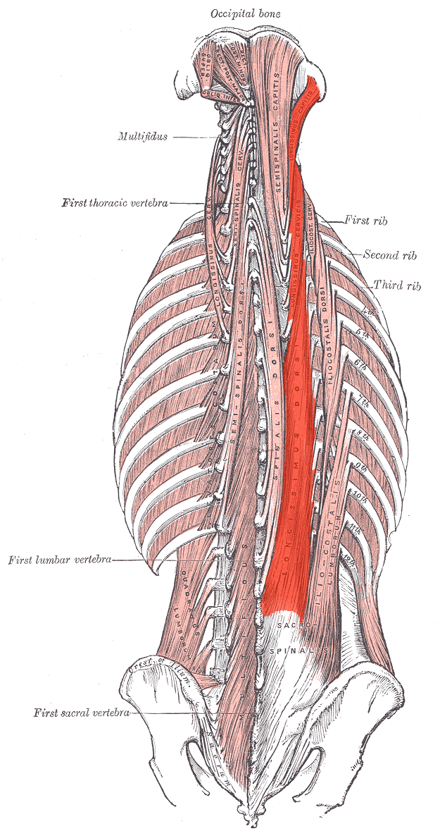
Intermediate: longissimus
Lying medial to the iliocostalis muscle group, longissimus is also formed of distinct muscular segments.
These are divided into:
- Longissimus capitis
- Longissimus cervicis
- Longissimus thoracis
Origin and insertion
- Longissimus capitis originates from the transverse processes of T1-T5 and inserts onto the posterior margin of the mastoid process.
- Longissimus cervicis originates from the transverse processes of T1-T5 and inserts onto the transverse processes of C2-C6.
- Longissimus thoracis originates from the transverse processes of L1-L5 and the thoracodorsal fascia and inserts onto the transverse processes of T1-T12.
- Take away: the longissimus muscle group spans from the thoracolumbar fascia midline of the lumbar vertebra to the transverse processes of all thoracic vertebrae. Longissimus is long.
Fibre orientation
The longissimus muscle group is a long, thick muscle that travels longitudinally along the length of the vertebral column.
Function
Unilaterally, longissimus assists the other erector spinae muscles in ipsilateral lateral flexion of the spine. Bilaterally, it assists in the extension of the spine.
Innervation
Longissimus derives its segmental innervation from the dorsal rami of spinal nerves along the length of the spine.
Blood supply
Longissimus is supplied by the lateral sacral artery and segmentally by perforating branches of the posterior intercostal arteries.
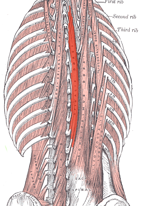
Intermediate: spinalis
The most medial of the erector spinae muscles, spinalis is also the smallest of this group. It is formed of the distinct groups:
- Spinalis capitis
- Spinalis cervicis
- Spinalis thoracis
Origin and insertion
- Spinalis capitis originates from the spinous processes of C1, C3 and C4, and inserts onto the inferior nuchal line and nuchal ligament.
- Spinalis cervicis originates from the spinous processes of C4-C7 and inserts onto the spinous processes of C3-C5.
- Spinalis thoracis originates from the spinous processes of T7-L1 and inserts onto the spinous processes of T1-T6.
- Take away: the spinalis muscle group attaches spinous processes in adjacent regions of the spine.
Fibre orientation
The fibres of the spinalis group are small-calibre strap muscles.
Function
Unilaterally, the spinalis muscles assist the erector spinae group in ipsilateral lateral flexion. Bilaterally, they assist in the extension of the spine.
Innervation
The spinalis group is supplied segmentally by the dorsal rami of spinal nerves along the length of the spine.
Blood supply
The spinalis group is supplied by the lateral sacral artery, and segmentally by the posterior intercostal arteries.
Clinical relevance: erector spinae muscles
The erector spinae muscles are the primary postural and antigravity muscles of the spine. They derive their innervation from the deeper cerebral tracts such as the reticulospinal and vestibulospinal tracts. Lesions anywhere along these pathways from their primary nuclei in the brainstem or in their spinal cord tract can manifest as poor posture and difficulty maintain an upright stance during locomotion.2
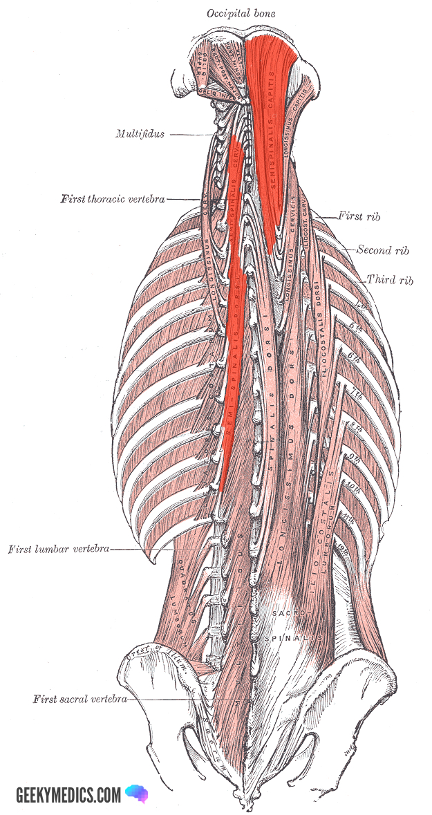
Deep: semispinalis capitis and cervicis
The semispinalis muscles originate on the transverse processes, and course medially to attach closer to the midline of the spine. This contrasts with the splenius muscles, which begin on the midline of the spine and course laterally.
Origin and insertion
- Semispinalis capitis originates on the transverse processes of C7-T6 and courses medially to insert between the superior and inferior nuchal lines of the skull.
- Semispinalis cervicis originates on the transverse processes of T6-T12 and inserts onto the spinous processes of C1-C5.
- Take away: the semispinalis muscle group passes over the thoracic and cervical spines, running deep to the erector spinae muscle group.
Fibre orientation
The fibres of semispinalis capitis and cervicis course superomedially from the transverse processes as a long, thin muscle.
Function
Acting unilaterally, the semispinalis capitis acts in ipsilateral lateral flexion and rotation. Bilaterally, it assists in the extension of the spine.
Innervation
Semispinalis capitis is supplied by the greater occipital nerve.
Blood supply
There is no specific artery that supplies the semispinalis group, but they derive supply largely from the occipital artery.
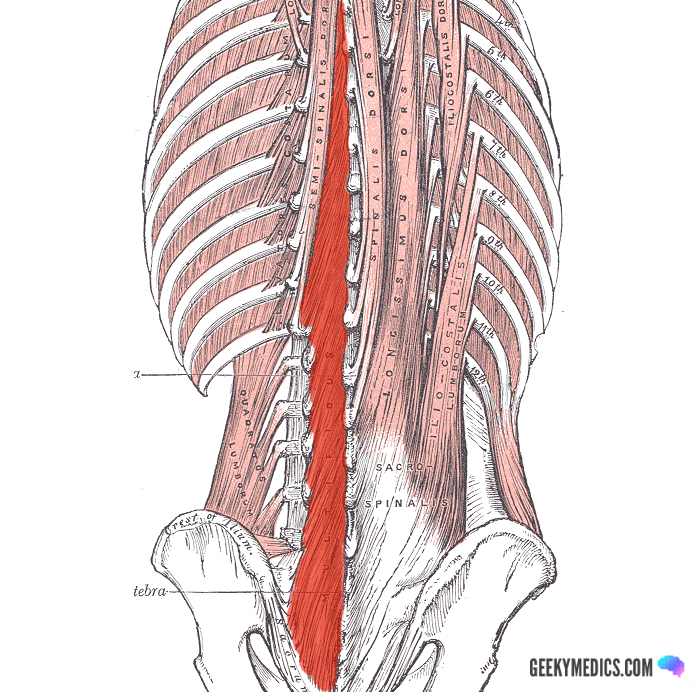
Deep: multifidus
Multifidus is a muscle that traverses the entire spine from the sacrum to the cervical region. Its primary action is proprioceptive in nature.
Origin and insertion
Multifidus originates from the sacrum, iliac crest and erector spinae aponeurosis, and inserts into the spinous processes of each vertebra.
Fibre orientation
The fibres of multifidus are short and thick, traversing each intervertebral space.
Function
Multifidus has a very weak rotation, extension and lateral flexion movement. Its primary function is proprioception for the vertebral column.
Innervation
The dorsal rami of spinal nerves along the vertebral column supply sensorimotor innervation to multifidus.
Blood supply
The vascular supply of multifidus varies regionally.
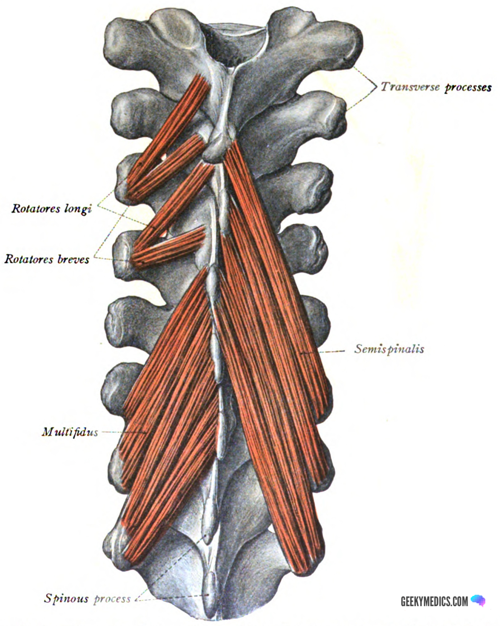
Deep: rotatores
Rotatores is a small muscle that runs from transverse processes of inferior vertebra to the spinous process of superior vertebrae. It is a rotation and proprioceptive muscle.
Origin and insertion
Most prominent in the thoracic region, rotatores originates from the transverse processes of all vertebrae and inserts into the spinous processes of the superiorly-located vertebra. It usually spans 1 (rotatores brevis) or 2 (rotatores longus) vertebral levels.
Fibre orientation
Rotatores are narrow, strap-like muscles.
Function
Rotatores functions to rotate the spine, but also acts proprioceptively.
Innervation
The dorsal rami of the spinal nerves in each region of the spine supply rotatores.
Blood supply
The vascular supply of rotatores varies regionally.
Deep: interspinales, intertransversarii and levatores costarum
These three muscle groups are small, proprioceptive muscles that have minimal agonist action. They are of vital importance in maintaining an upright posture with continuous biofeedback to the antigravity muscles. They will only briefly be mentioned.
The interspinal muscles run between adjacent spinous processes (sometimes skipping a spinous process) and provide proprioceptive information about the level of flexion, extension or rotation present.
The intertransversarii muscles run between adjacent transverse processes and provide proprioceptive information on the level of lateral flexion or rotation present in the spine.
Levatores costarum are small muscles originating on the transverse process of lower cervical and thoracic vertebrae. They have two heads, longus and brevis, that travel to the rib of the same vertebral level (brevis) or to the rib of a single vertebral level down (longus). They very weakly assist in elevation of the ribs and act mainly as proprioceptive muscles that provide information on the level of thoracic expansion.
Quiz
Put your knowledge to the test with our quiz on the deep muscles of the back.
References
Reference images
- OpenStax College. License: [CC BY 3.0]
- Erector spinae muscles: Henry Vandyke Carter. License: [Public domain]
- Semispinalis muscles: Henry Vandyke Carter. License: [Public domain]
- Multifidus: Uwe Gille. License: [Public domain]
- Rotatores: Dr Johannes Sobotta. License: [Public domain]
Reference texts
- Sinnatamby, C. S. (2011). Last’s Anatomy, International Edition: Regional and Applied. Elsevier Health Sciences.
- Moore, K. L., Dalley, A. F., & Agur, A. M. (2013). Clinically oriented anatomy. Lippincott Williams & Wilkins.
