
- 📖 Geeky Medics OSCE Book
- ⚡ Geeky Medics Bundles
- ✨ 1300+ OSCE Stations
- ✅ OSCE Checklist PDF Booklet
- 🧠 UKMLA AKT Question Bank
- 💊 PSA Question Bank
- 💉 Clinical Skills App
- 🗂️ Flashcard Collections | OSCE, Medicine, Surgery, Anatomy
- 💬 SCA Cases for MRCGP
To be the first to know about our latest videos subscribe to our YouTube channel 🙌
Introduction
Although uncommon, dermatological emergencies are important to recognise as they can be life-threatening and easily missed in clinical practice.
This article will cover three dermatological emergencies:
- Eczema herpeticum
- Erythroderma
- Stevens-Johnson syndrome (SJS) and toxic epidermal necrolysis (TEN)
Eczema herpeticum
Eczema herpeticum is a complication of atopic eczema that occurs with infection of the herpes simplex virus (HSV).
Aetiology
Eczema herpeticum (EH) is thought to be due to a reduced level of immunity to HSV in patients with atopic dermatitis.¹
Risk factors
EH can affect any age but most commonly affects infants and children. It is also more common in those with severe atopic dermatitis/eczema.¹
Other conditions related to EH include burns, pemphigus vulgaris and cutaneous T-cell lymphoma.
Clinical features
History
Typical symptoms of eczema herpeticum include:¹
- General malaise
- Fever
- New itchy, painful lesions
- Gritty or sore eyes (if eye involvement present)
Symptoms tend to develop 5-12 days following contact with HSV.¹
Clinical examination
Typical clinical findings of eczema herpeticum include:
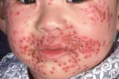
- Groups of itchy painful blisters, erosions and crusted papules (Figure 1)
- Local lymphadenopathy near the site of the lesions
- Evidence of secondary bacterial infection (e.g. cellulitis/impetigo)
Differential diagnoses
The clinical presentation of eczema herpeticum can be mistaken for impetigo. The typical golden crusting of impetigo can also mask the primary herpetic lesions.
Investigations
Diagnosis is mainly based on clinical findings. However, viral and bacterial swabs can be taken from the base of a new blister.
- Viral swabs: the presence of HSV type 1 or 2 would confirm the diagnosis.
- Bacterial swabs: may reveal secondary infection with staphylococci or streptococci.¹
Management
All patients with eczema herpeticum should be referred to a dermatologist for urgent assessment. If there is any ocular involvement an ophthalmology review should also be requested.
Medical management
The mainstay of treatment is antiviral medication (e.g. aciclovir). In serious cases, intravenous aciclovir can be considered.²
If a secondary bacterial infection is present, antibiotic treatment may be required.
Complications
Complications of eczema herpeticum can include:
- Herpes hepatitis
- Encephalitis
- Disseminated intravascular coagulation
- Death (very rare)
Erythroderma
Erythroderma is intense redness of the skin covering at least 90% of the skin surface area, usually secondary to pre-existing inflammatory skin disease.³
Aetiology
The pathology of erythroderma is still not fully understood. It is thought to be a complex process leading to rapid epidermal cell turnover.³
Risk factors
Erythroderma usually occurs in those with inflammatory skin disease. The most common precipitant is psoriasis.⁴
Other causes include eczema, drug eruptions (e.g. allopurinol, gold, sulfonamide, sulfonylureas, isoniazid), and cutaneous lymphoma (Sezary syndrome).
Clinical features
History
Typical symptoms of erythroderma include:
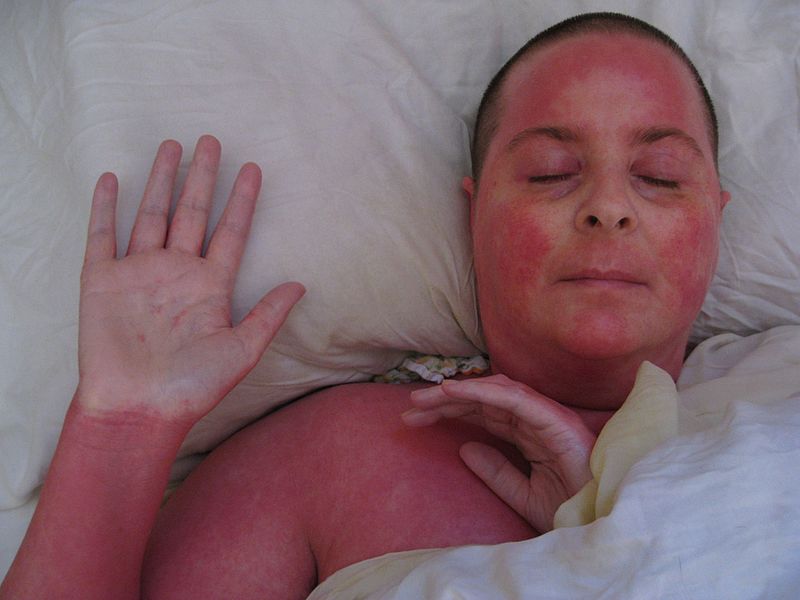
- Red, painful and itchy skin over a large area (Figure 2)
- Malaise
Other important areas to cover in the history include:
- Past medical history (e.g. history of inflammatory skin diseases such as psoriasis or eczema)
- Drug history (e.g. the recent commencement of a new drug)
Clinical examination
Typical clinical findings of erythroderma include:
- Hot, erythematous skin covering at least 90% of the skin surface
- The skin will often show signs of desquamation or peeling of the skin
- Generalised lymphadenopathy
Investigations
The diagnosis of erythroderma is usually clinical, however, baseline observations and blood tests should be completed. If the diagnosis is unclear, a skin biopsy should be considered.
Management
All patients with suspected erythroderma should be reviewed by a dermatologist.
Patients with erythroderma who are systemically unwell will require admission to a specialist burns unit or intensive care unit.
Medical management
Emollients and cool, wet dressings are the mainstays of treatment. Any underlying causes should be treated (e.g. discontinuing suspected trigger medication). Topical corticosteroids may be used in some cases.
Patients should be nursed in a warm room (~30°C) and have their fluid balance, electrolytes and body temperature monitored closely.
Complications
Complications of erythroderma can include:
- Dehydration
- Electrolyte imbalance
- Secondary bacterial infection
- Hypothermia secondary to impaired thermoregulation
- Cardiac failure
- Death
Stevens-Johnson syndrome and toxic epidermal necrolysis
Stevens-Johnson syndrome (SJS) and toxic epidermal necrolysis (TEN) are variants of the same condition. They are severe mucocutaneous reactions, almost always secondary to medications.⁵
Aetiology
The mechanisms of SJS and TEN are not fully understood. SJS and TEN most commonly occur due to medications including:
- Allopurinol
- Anti-epileptic drugs
- Sulfonamides
- Antivirals
- Nonsteroidal anti-inflammatories
- Salicylates
- Sertraline
- Imidazole
Risk factors
SJS and TEN are statistically more common in females and in those with HIV.⁶
Infections such as mycoplasma and cytomegalovirus can also trigger the condition.⁵
Clinical features
History
Typical symptoms of SJS and TEN include:
- Prodromal flu-like/non-specific upper respiratory tract illness.
- A painful rash starting on the trunk which then spreads over several hours to days onto the face and limbs.
- Mouth ulcers or soreness.
- Painful or irritated eyes.
Other important areas to cover in the history include:
- Drug history (e.g. newly commenced medication – symptoms can occur 5-28 days after starting the causative medication)
- Past medical history (e.g. HIV)
Clinical examination
Typical clinical findings of SJS/TEN include:
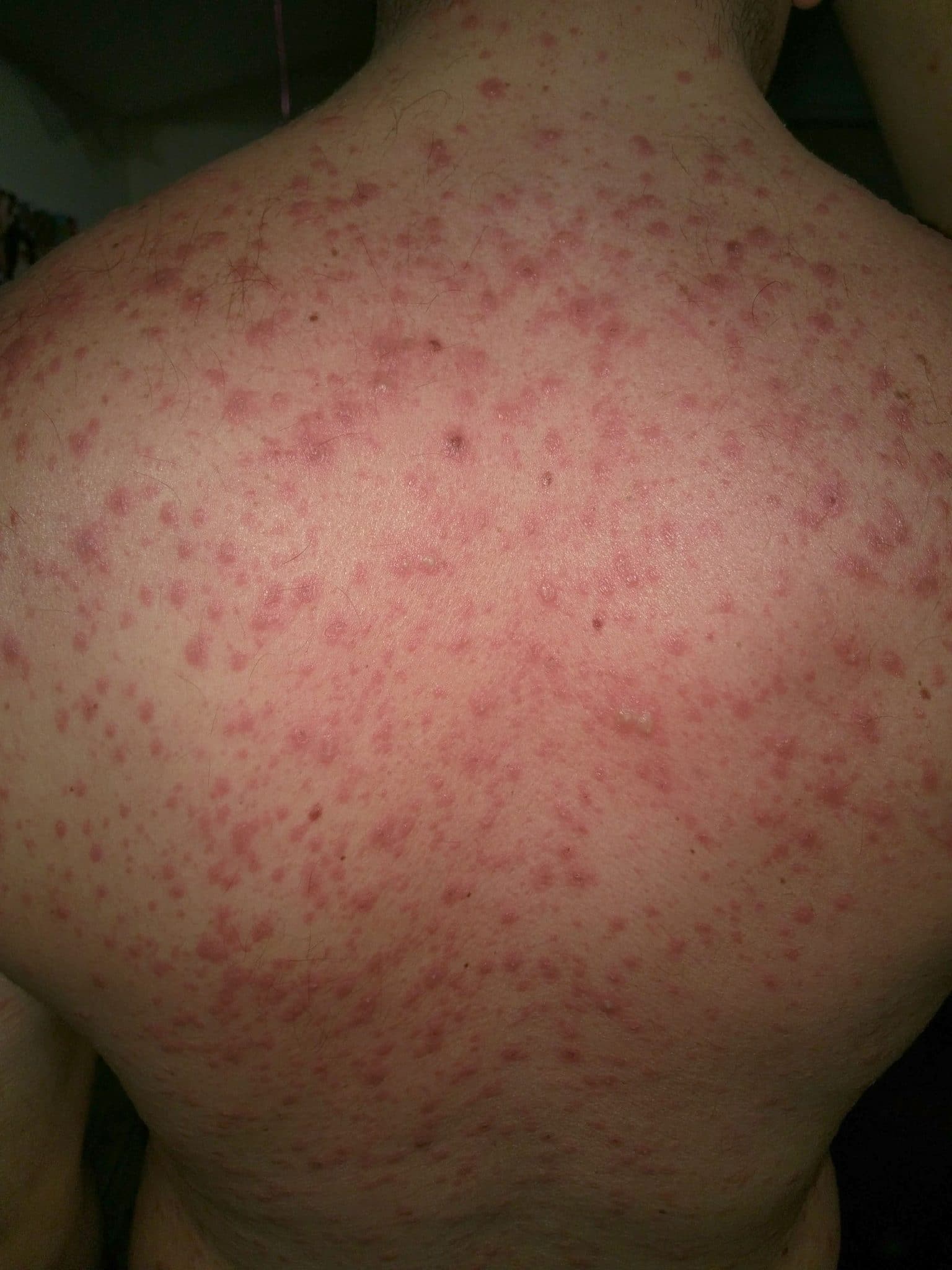
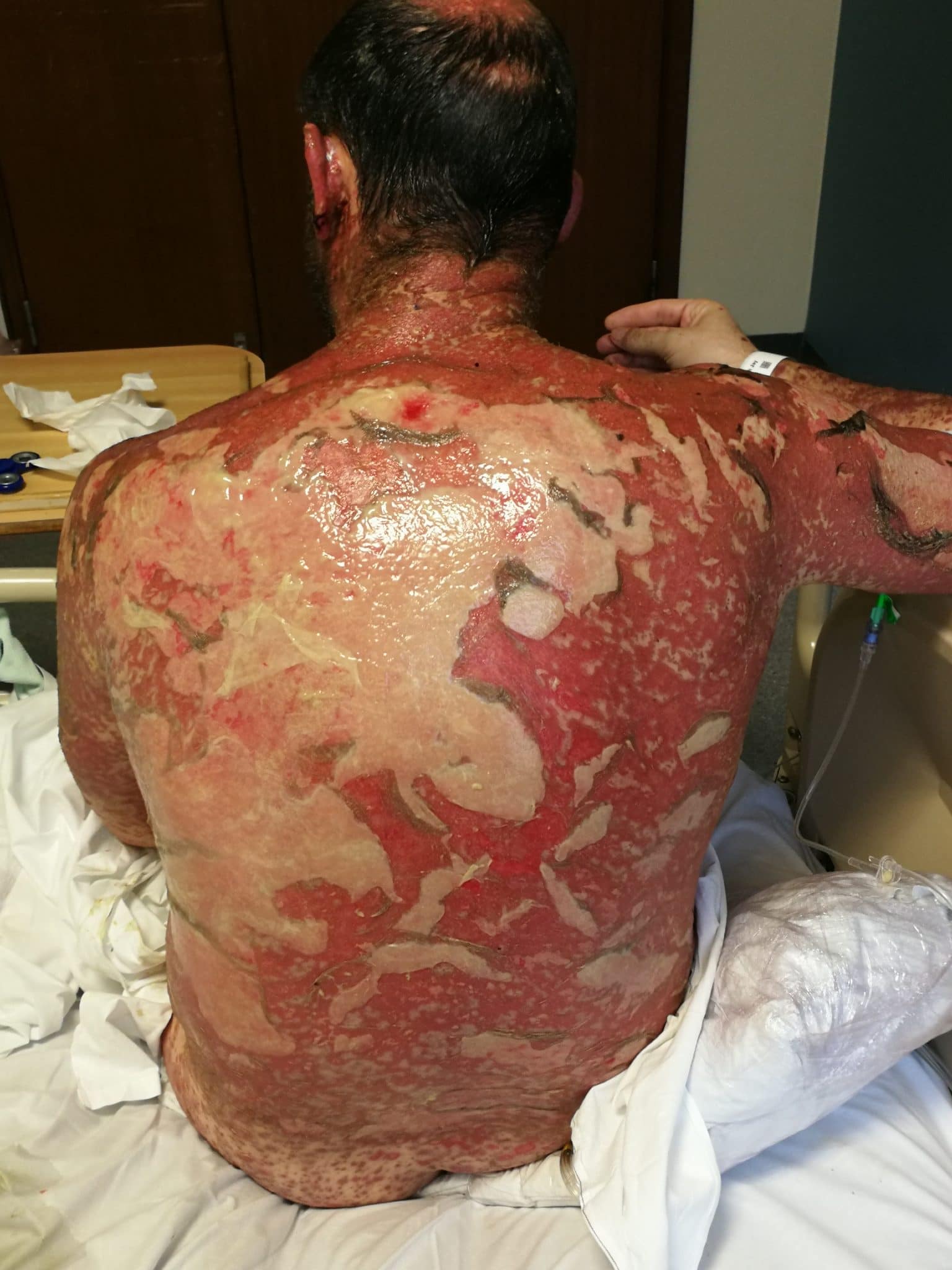
- A rash which initially starts as macules (Figure 4) which then progresses to blisters and eventually sheets of desquamation (Figure 5).
- Positive Nikolsky’s sign: gentle rubbing of the skin causing desquamation.
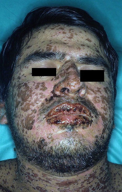
- Ulceration, erythema and blistering in the oral cavity
- Conjunctivitis or corneal ulceration
Differential diagnoses
In young children, staphylococcal scalded skin syndrome is an important differential and can present with similar clinical features. A skin biopsy with histology is required for a definitive diagnosis.
Investigations
The diagnosis is usually made clinically although a skin biopsy is required to confirm the diagnosis.
The condition is then stratified based on percentage body surface area of the detached epidermis.
Table 1. The categorisation of SJS and TEN.
| SJS | SJS/TEN | TEN |
| <10% | 10-30% | >30% |
Management
These patients require hospital admission, urgent dermatology referral and admission to a specialist burns unit or intensive care unit.
Any suspected causative medications should be stopped. Fluid balance and electrolytes should be monitored closely. Patients will require adequate analgesia as the condition is extremely painful.
Ophthalmology input will be required if there is evidence of ocular involvement (e.g. corneal ulceration).
Complications
Complications of SJS and TEN include:
- Dehydration/hypovolaemic shock
- Secondary infection of the skin or mucous membranes
- Sepsis
- Disseminated intravascular coagulation
- Thromboembolism
- Death (mortality rates are approximately 10% for SJS and ~30% for TEN)⁵
Key points
Eczema herpeticum
- A complication of atopic dermatitis most commonly occurring in infants and children.
- Clinical features include a painful, itchy, blistered and crusted rash.
- Treatment is with antivirals (intravenous or oral) depending on the spread and/or eye involvement.
Erythroderma
- Intense erythema and desquamation of skin covering at least 90% of skin surface usually secondary to inflammatory skin disease.
- Hospital admission is required for those who are systemically unwell with close monitoring of fluid balance and electrolytes.
- Treatment options include emollients, wet wraps and topical corticosteroids.
Stevens-Johnson syndrome and toxic epidermal necrolysis
- Usually secondary to medications and much more common in those with HIV.
- Presents with a prodromal illness followed by a rash.
- Target-looking macules progressing to blisters and desquamation.
- Classified according to percentage epidermal detachment of the body surface area.
- Treatment involves discontinuing causative medication, analgesia and monitoring of fluid balance and electrolytes.
Reviewer
Dr Thomas King
Dermatology SpR
Editor
Dr Chris Jefferies
References
Text references
- DermnetNZ. Eczema herpeticum. Published in 2010. Available from: [LINK]
- NICE. Quality statement 7: Treatment of eczema herpeticum. Published in 2013. Available from: [LINK]
- DermnetNZ. Erythroderma. Last updated in 2016. Available from: [LINK]
- Patient Professional. Erythrodermic Psoriasis. Published in 2015. Available from: [LINK]
- Dermnet NZ. Stevens-Johnson Syndrome/Toxic Epidermal Necrolysis. Last updated 2016. Available from: [LINK]
- Patient Professional. Stevens-Johnson Syndrome. Published in 2016. Available from: [LINK]
Images references
- Figure 1. Mohammad2018. Eczema herpeticum. License: [CC BY-SA]. Available from: [LINK]
- Figure 2. SalishSea2/Corinna Kennedy. Red (burning) Skin Syndrome from Topical Steroids. License: [CC BY-SA]. Available from: [LINK]
- Figure 3. Dr Thomas Habif. Stevens-johnson-syndrome. License: [CC BY-SA]. Available from: [LINK]
- Figure 4. Jay2Base. Early-stage blisters on the back TENS patient. License: [CC BY-SA]. Available from: [LINK]
- Figure 5. Jay2Base. TENS patient on day 10. [CC BY-SA]. Available from: [LINK]
