
- 📖 Geeky Medics OSCE Book
- ⚡ Geeky Medics Bundles
- ✨ 1300+ OSCE Stations
- ✅ OSCE Checklist PDF Booklet
- 🧠 UKMLA AKT Question Bank
- 💊 PSA Question Bank
- 💉 Clinical Skills App
- 🗂️ Flashcard Collections | OSCE, Medicine, Surgery, Anatomy
- 💬 SCA Cases for MRCGP
To be the first to know about our latest videos subscribe to our YouTube channel 🙌
Introduction
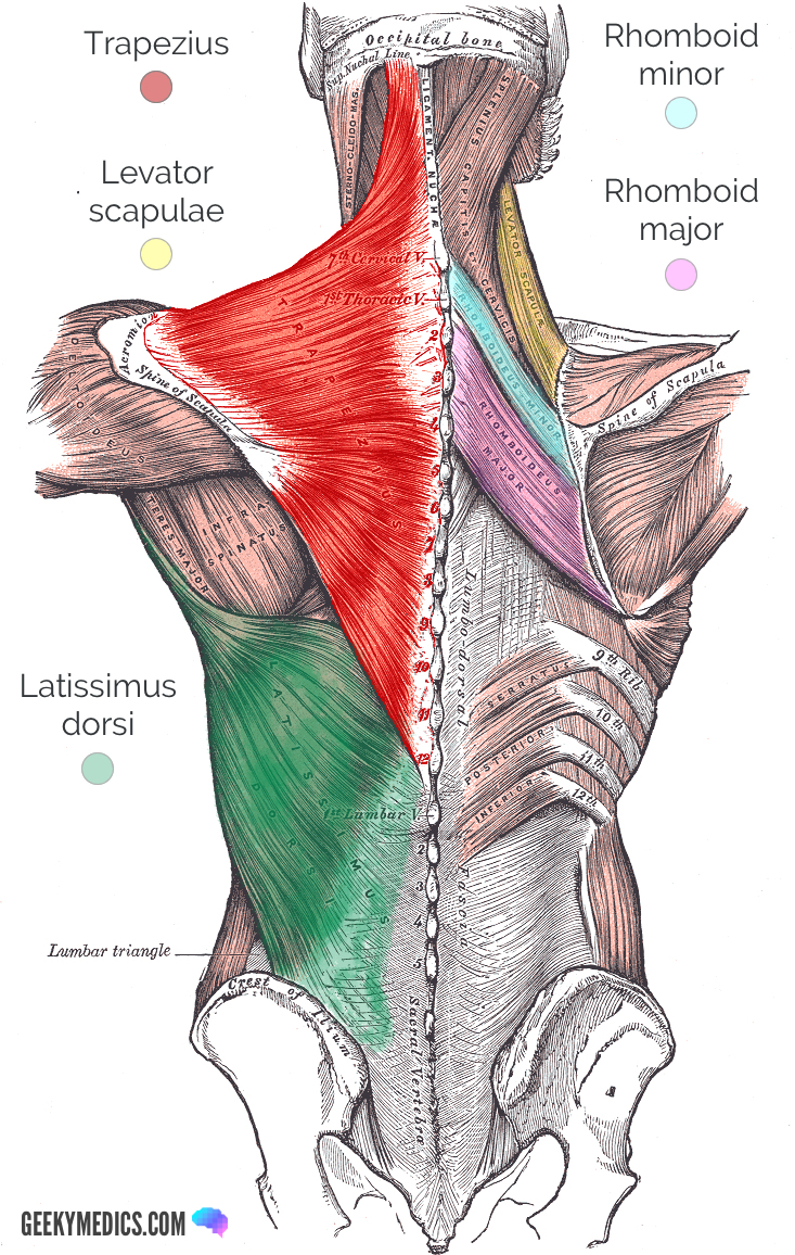
The shoulder muscles can be classified into extrinsic and intrinsic categories.
The extrinsic muscles of the shoulder include:
- Trapezius
- Latissimus dorsi
- Levator scapulae
- Rhomboid major and rhomboid minor
These muscles extend from the torso to the bones of the shoulder.
The intrinsic muscles of the shoulder include:
- Supraspinatus
- Infraspinatus
- Deltoid
- Teres minor
- Teres major
- Subscapularis
Furthermore, the extrinsic shoulder muscles can be sub-classified into superficial and deep muscles. Trapezius and latissimus dorsi comprise the superficial extrinsic muscles. The deep layer includes the levator scapulae and rhomboid muscles.
This article will primarily highlight the origin, insertion, innervation, action and blood supply of the extrinsic shoulder muscles.
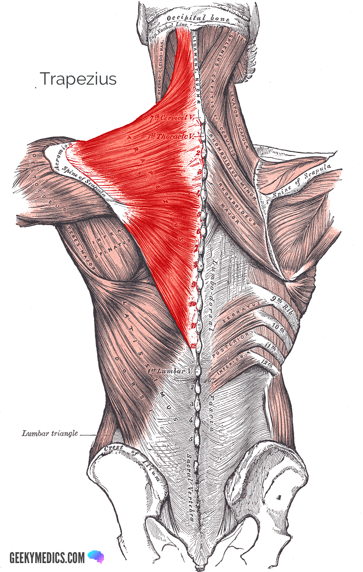
Trapezius
The trapezius muscle is a broad, diamond-shaped muscle that comprises of three functional and anatomically distinct parts: upper (or superior), middle (or transverse) and lower (or inferior).
Origin and insertion
The upper fibres originate from the external occipital protuberance, C7 spinous process, ligamentum nuchae, as well as the superior nuchal line located on the occipital bone. The fibres of the upper trapezius insert into the lateral third of the clavicle.
The spinous processes of C7, T1, T2 and T3 anchor the middle fibres of this muscle, and then insert into the medial aspect of the acromion and the posterior margin of the spine of the scapula.
The inferior fibres of the trapezius originate from T4-T12 spinous processes and insert into the spine of the scapula.
Innervation
The trapezius muscle receives motor innervation via the accessory nerve and proprioceptive fibres from C3 and C4 spinal nerves.
Function
The upper part of the trapezius elevates and rotates the scapula. These functions are necessary for abduction of the arm. The middle fibres achieve retraction of the scapula. Inferior fibres depress the scapula.
Blood supply
The trapezius receives blood from the superficial branch of the transverse cervical artery.
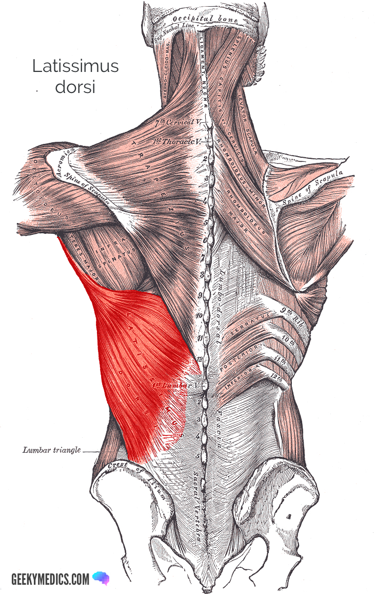
Latissimus dorsi
The Latissimus dorsi muscle spans from the lower back to the upper arm and is partially covered by the trapezius. It is the largest muscle of the upper body.
Origin and insertion
The fibres of this muscle originate from the spinous processes of T7-L5, the inferior angle of the scapula, inferior 3-4 ribs, thoracolumbar fascia, as well as the iliac crests. The latissimus dorsi inserts into the intertubercular groove of the humerus.
Innervation
Cervical nerves 6, 7 and 8 innervate the latissimus dorsi through the thoracodorsal nerve.
Function
This muscle functions to extend, abduct, and internally rotate the shoulder joint. The latissimus dorsi also transversely extends and flexes the shoulder joint from an extended position.
Blood supply
The thoracodorsal branch of the subscapular artery provides the arterial supply to this muscle.
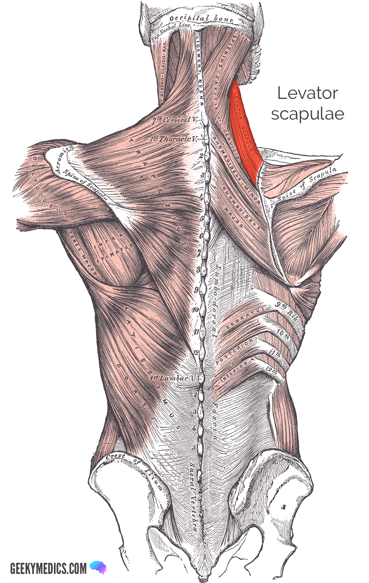
Levator scapulae
The levator scapulae is located on the postero-lateral aspect of the neck.
Origin and insertion
This muscle originates from the posterior tubercles of the transverse processes of C1-C4. The levator scapulae inserts into the medial border of the scapula.
Innervation
Branches from the dorsal scapular nerve (C5), as well as cervical nerves C3 and C4 provide innervations to this muscle.
Function
As the name would indicate, the levator scapulae acts to elevate the scapula. This muscle also assists in rotating the scapula.
Blood supply
The arterial supply to the levator scapulae is provided by the dorsal scapular artery.
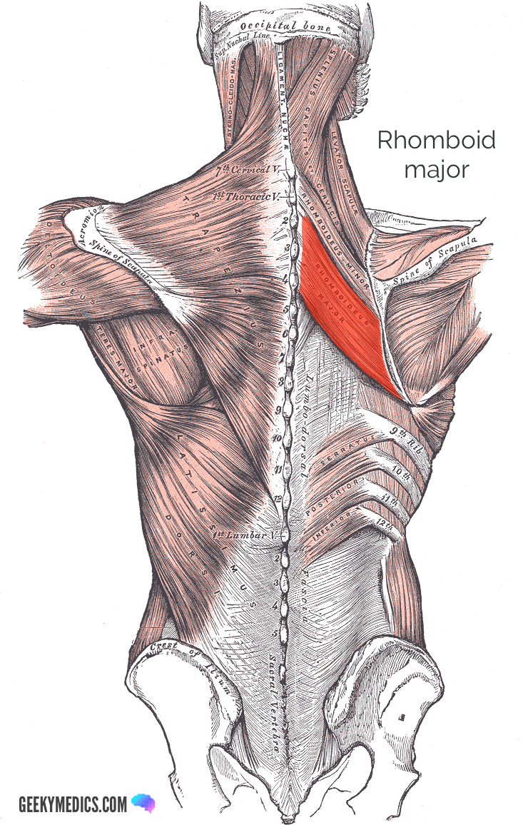
Rhomboid major
The rhomboid major muscle is located in the back, inferior to rhomboid minor, and extends from the vertebrae to the scapula.
Origin and insertion
This muscle originates from T2-T5 spinous processes, as well as from the supraspinatus ligament. Rhomboid major then inserts into the medial border of the scapula (inferior to the insertion of rhomboid minor).
Innervation
Innervation is provided to this muscle by the dorsal scapular nerve (C5).
Function
Rhomboid major works in conjunction with other muscles to ensure that the scapula is attached to the ribcage. This muscle also retracts and rotates the scapula.
Blood supply
The dorsal scapular artery provides the arterial supply to rhomboid major.
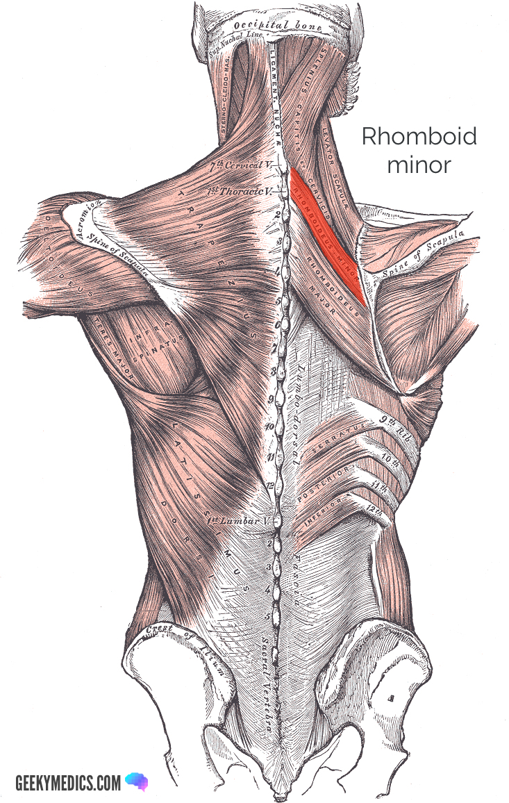
Rhomboid minor
The rhomboid minor muscle lies superior to the rhomboid major muscle and inferior to levator scapulae in the back. It spans from the vertebra to the scapula.
Origin and insertion
The rhomboid minor muscle originates from the inferior nuchal ligament and C7-T1 spinous processes. This muscle then inserts into the medial border of the scapula superior to rhomboid major.
Innervation
This muscle receives innervation via the dorsal scapular nerve.
Function
Rhomboid major works in conjunction with other muscles to fix the scapula to the thoracic wall. It also functions to rotate and retract the scapula.
Blood supply
A deep branch of the transverse cervical artery provides the arterial supply to rhomboid minor.
References
Reference images
- Trapezius: By Mikael Häggström, used with permission. License: [Public domain]
- Latissimus dorsi: By Mikael Häggström, used with permission. License: [Public domain]
- Levator scapulae: modified by Uwe Gille. License: [Public domain]
- Rhomboid major: modified by Uwe Gille. License: [Public domain]
- Rhomboid minor: modified by Uwe Gille. License: [Public domain]
Reference texts
- Arthur, F et al. (2010). Clinically oriented anatomy. 6th edition. Philadelphia, PA: Lippincott Williams & Wilkins, Wolters Kluwer
- Drake, R et al. (2010). Gray’s Anatomy for students. 2nd edition. Philadelphia, PA: Churchill Livingstone Elsevier
- Moore, K et al. (2011). Essential clinical anatomy. 4th ed. Baltimore, MD: Lippincott Williams and Wilkins
- Wikipedia (2019). Latissimus Dorsi muscle. Available from: [LINK]
- Wikipedia (2019). Rhomboid minor muscle. Available from: [LINK]
