

- 📖 Geeky Medics OSCE Book
- ⚡ Geeky Medics Bundles
- ✨ 1300+ OSCE Stations
- ✅ OSCE Checklist PDF Booklet
- 🧠 UKMLA AKT Question Bank
- 💊 PSA Question Bank
- 💉 Clinical Skills App
- 🗂️ Flashcard Collections | OSCE, Medicine, Surgery, Anatomy
- 💬 SCA Cases for MRCGP
To be the first to know about our latest videos subscribe to our YouTube channel 🙌
Introduction
Henoch-Schönlein purpura (HSP) is an IgA vasculitis which commonly presents with glomerulonephritis, abdominal pain, arthralgia and purpura.
It is the most common vasculitis in children, with an annual incidence of 20 per 100,000 in children under 17.1
Aetiology
HSP is an immune-mediated small vessel vasculitis. IgA and complement components (C3) are deposited in vessel walls leading to inflammation.
The mechanism underlying this is not entirely clear, but HSP can follow an upper respiratory tract infection, so it is thought to be triggered by an abnormal immune system response.2
Risk factors
The most notable risk factor for HSP is a preceding upper respiratory tract infection.
Other risk factors include:1
- Age: the incidence of HSP peaks in those aged 4-6 years old
- Male sex: HSP is more common in boys, with a male-to-female ratio of approximately 1.5:1
Clinical features
History
Typical symptoms of HSP include:
- Purpuric and petechial rash: most commonly on the lower limbs and buttock
- Abdominal pain: commonly associated with nausea and vomiting and less commonly bloody diarrhoea may be seen.
- Joint pain: arthralgia occurs in the knees and ankles and may be associated with peri-articular oedema.
- Frothy urine: suggestive of proteinuria due to renal involvement
- Haematuria: due to renal involvement
- Fever: low-grade fever may be present
Other important areas to cover in the history include:
- Preceding upper respiratory tract infection: URTI symptoms may occur 7-14 days before the presentation of HSP symptoms
Clinical examination
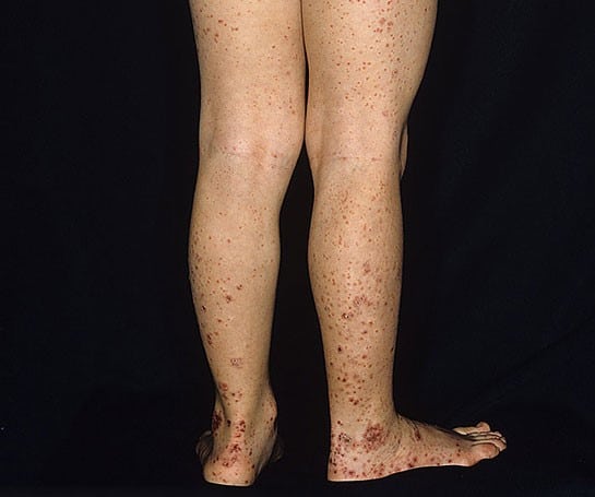
Rash1,2,5
Petechia and purpura most commonly affect the lower limbs and buttocks, particularly the extensor surfaces. However, this is not always the case, and it can occur on the upper limbs in up to one-third of cases.
Petechiae vs purpura4
Petechiae are non-blanching pinpoint spots <3mm in diameter
Purpura are non-blanching spots >3mm in diameter and are commonly palpable
Joints
Arthralgia occurs in about 80% of HSP cases and can occur in any joint, but the hips, knees, and ankles are most commonly affected.
Along with arthralgia, swelling and tenderness can occur, but erythema is less likely.
Joint involvement in HSP is transient and doesn’t persist after the acute illness with no long-term damage to the joint. 1,2,5
Abdomen
Gastrointestinal symptoms occur in 50% of cases and tend to present around seven days after the onset of the rash.
The presentation ranges from mild symptoms such as abdominal pain, nausea and vomiting to severe presentations including gastrointestinal haemorrhage and intussusception.1,3,5
Renal
Approximately 50% of HSP patients develop renal involvement, which is on a wide clinical spectrum:2
- Microscopic or macroscopic haematuria
- Proteinuria
- Nephrotic syndrome: proteinuria, oedema and hypoalbuminemia
- Nephritic syndrome: oedema, haematuria and hypertension
- Renal failure
Differential diagnoses
It is important to consider a wide range of differential diagnoses in the context of a child with a non-blanching rash. All children with a non-blanching rash require a same-day paediatric assessment.3.4
For more information, see the Geeky Medics guide to non-blanching rashes.
Meningococcal septicaemia
Meningococcal septicaemia is an important differential diagnosis to exclude when considering a non-blanching rash.
The child will be systemically unwell with a high-grade fever and signs of shock (hypotension and tachycardia). In contrast, the child is likely to be clinically stable in HSP.
On examination, reduced consciousness, photophobia and neck stiffness suggest a diagnosis of meningitis.
Idiopathic thrombocytopenic purpura (ITP)
ITP is a fall in the patient’s platelet count (thrombocytopenia), causing a purpuric rash. It is commonly triggered by a viral infection. Children with ITP are generally well but can present with bleeding (e.g. epistaxis).
Haemolytic uraemic syndrome (HUS)
HUS is a triad of thrombocytopenia, acute kidney injury (AKI) and microangiopathic haemolytic anaemia caused by an E. coli infection.
Like HSP, these children present with abdominal pain and a petechial rash. However, the key distinguishing feature of HUS is the presence of bloody diarrhoea.
Mechanical causes
Forceful coughing and vomiting can cause a petechial rash in the distribution of the superior vena cava. A rash is seen on the head, neck and shoulder rather than the lower limbs like HSP.
It is always important to consider non-accidental injury as a cause of a petechial rash in a paediatric setting.
Investigations
Bedside investigations
Relevant bedside investigations include:5
- Blood pressure: if elevated, this suggests renal involvement.
- Urinalysis: proteinuria or haematuria indicate renal involvement. It is important to note that an early morning urine sample is the most reliable and should be used for a protein: creatinine ratio.
Laboratory investigations
Relevant laboratory investigations include:1,5
- U&E: to assess renal function
- Full blood count and clotting studies: likely to be normal in the context of HSP but required to exclude other causes of a non-blanching rash (e.g. ITP)
- Liver function tests: including albumin to assess for hypoalbuminemia, which is suggestive of nephrotic syndrome
- Urine microscopy: assesses for red cell casts
- Anti-streptolysin O titre: can be performed to look for group A streptococcal antibodies but is rarely performed in clinical practice.
Imaging
Relevant imaging investigations include:5
- Abdominal ultrasound: may be performed to exclude intussusception as a complication in the context of severe abdominal pain.
Other investigations
Other relevant investigations include:5
- Renal biopsy: only performed if there is significant ongoing proteinuria or impaired renal function under specialist guidance.
Diagnosis
The Paediatric Rheumatology European Society has produced a set of diagnostic criteria which state that palpable purpura and one of the following should be present for diagnosis:7
- Diffuse abdominal pain
- Arthralgia
- Renal involvement
- Typical histopathology (glomerulonephritis or vasculitis)
Management
Most children with HSP have a complete spontaneous recovery within weeks to months but may require supportive care:6
- Paracetamol can be used for joint and abdominal pain
- Prednisolone may be considered for severe pain, but it is important to note that there is no evidence that steroids improve overall outcomes
- NSAIDs (e.g. such as ibuprofen) should be used cautiously in those with renal involvement
Follow up6
Children with HSP must be followed up closely as renal involvement may not manifest until weeks later.
Specific protocols vary between hospitals, but it is generally recommended that children with HSP have a urine dip and blood pressure reading regularly for six months after diagnosis.
Children with concerning features for renal involvement (e.g. proteinuria >2+, frank haematuria or hypertension) must be re-reviewed in secondary care.
Children can then be fully discharged if they have two consecutive normal urine dips and a normal blood pressure at six months.
Complications
Complications of Henoch-Schönlein purpura include:1,2,6
- Relapse: relapse rates are 30 – 40%, but relapses tend to be shorter and less severe than the original presentation.
- Renal failure: approximately 1% of HSP cases result in end-stage renal failure, this is more likely in children who initially present with nephrotic and nephritic syndrome
Key points
- HSP is an IgA vasculitis which can be preceded by an upper respiratory tract infection.
- HSP commonly presents with a purpuric petechial rash, renal involvement, arthralgia and abdominal pain.
- Management of HSP is conservative and supportive, with no evidence steroids improve outcomes.
- It is important to monitor blood pressure and urine for signs of renal involvement for at least six months following diagnosis.
- Complications include relapse and renal failure
Reviewer
Dr Alex Brightwell
Consultant Paediatrician
Editor
Dr Chris Jefferies
References
- Up to Date: IgA vasculitis (Henoch-Schönlein purpura): Clinical manifestations and diagnosis. Published in 2023. Available from: [LINK]
- National Library of Medicine: Henoch Schönlein Purpura. Published in 2022. Available from: [LINK]
- Dr Bethany Mitchell. Non-blanching Rashes. Published 2021. Available from: [LINK]
- Kingston Hospital NHS Foundation Trust: Non-blanching rashes in children. Published 2023. Available from: [LINK]
- BMJ Best Practice. IgA vasculitis (Henoch-Schonlein purpura). Published 2021. Available from: [LINK]
- Cymru NHS. Guidelines for the management of Henoch Schonlein purpura. Published 2011. Available from: [LINK]
- Patient.info. Henoch-Schönlein purpura. Published 2019. Available from: [LINK]
Image references
