

- 📖 Geeky Medics OSCE Book
- ⚡ Geeky Medics Bundles
- ✨ 1300+ OSCE Stations
- ✅ OSCE Checklist PDF Booklet
- 🧠 UKMLA AKT Question Bank
- 💊 PSA Question Bank
- 💉 Clinical Skills App
- 🗂️ Flashcard Collections | OSCE, Medicine, Surgery, Anatomy
- 💬 SCA Cases for MRCGP
To be the first to know about our latest videos subscribe to our YouTube channel 🙌
Introduction
Herpes zoster ophthalmicus (HZO) refers to shingles affecting the ophthalmic branch (V1) of the trigeminal nerve.
HZO accounts for 10-20% of all shingles, which itself has a lifetime incidence of 30%, contributing significantly to the global health burden.1,2
HZO can involve any part of the eye or orbit, ranging from potentially benign, to sight- or life-threatening complications.
Aetiology
Varicella zoster virus (VZV), like herpes simplex virus (HSV), is also a dsDNA virus of the herpes group.
Primary infection with VZV manifests as chickenpox (varicella). Following resolution, viruses establish latent infection within sensory nerve ganglions. Reactivation of such dormant viruses results in shingles (herpes zoster). Involvement of the ophthalmic branch of the trigeminal nerve (V1) results in herpes zoster ophthalmicus (HZO).
Individuals with no previous VZV infection may contract chickenpox from those with shingles. Transmission is by direct contact or droplet spread.
Risk factors
The incidence and severity of HZO increase with advancing age, with a sharp increase in incidence in individuals aged 60 and above1. Other groups at risk of severe infection include:
- Immunosuppressed (e.g. HIV, chemotherapy)
- Pregnant patients
- Neonates
Ocular involvement occurs in approximately 50% of patients with HZO. There is no correlation between the severity of skin rash and the extent of ocular complications.
Clinical features
History3
Typical symptoms of HZO include:
- Viral prodromal phase: fever, malaise, headache
- Pre-herpetic neuralgia: paraesthesia and pain along the affected dermatome of V1. If this pain persists for longer than one month after the rash has healed, it is termed post-herpetic neuralgia.
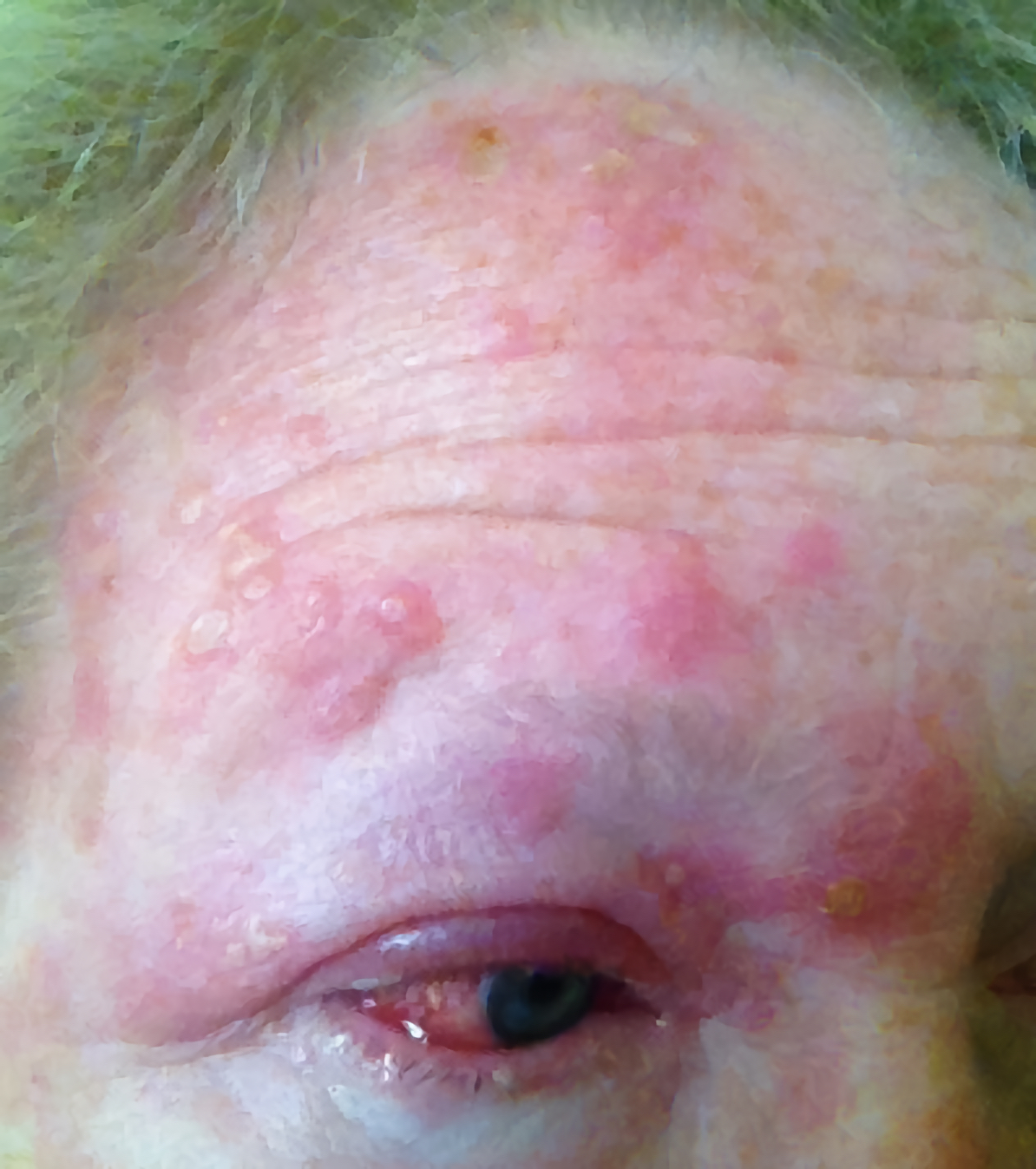
- Rash: a characteristic, unilateral rash following the dermatomal distribution of V1
- Zoster sine herpete: neuropathic pain not accompanied by a characteristic rash
HZO may involve any part of the globe or none. Symptoms may vary depending on the structures affected, and this can guide clinical examination:
- Isolated conjunctivitis and corneal involvement present with mildly reduced visual acuity, red eye, sharp pain, foreign body sensation and epiphora
- The presence of photophobia, dull eye pain and new floaters may indicate uveitis warranting urgent ophthalmological opinion. This may be 1-3 weeks following the onset of the rash.
- Symptoms of diplopia may indicate associated extraocular muscle palsy.
Clinical examination3
Patients with suspected HZO should undergo a comprehensive examination including visual acuity testing, colour vision assessment, anterior segment examination (using an Arclight or a direct ophthalmoscope on +10 lens setting), slit lamp examination, fundoscopy, and testing of extra-ocular eye muscles.
Patients with suspected cerebral involvement should undergo a full neurological examination.
Typical clinical findings of HZO on examination may include:
- Eyelid erythema and oedema (may be severe)
- Skin lesions in the following chronological order: macules-> papules -> vesicles -> pustules -> crusted lesions in the upper lid and along the rest of the V1 dermatomal distribution
- Conjunctiva: diffuse or circumlimbal injection
- Cornea: fluorescein staining reveals punctate keratitis (small dots when viewed under high magnification) and pseudodendrites (lacks terminal bulbs as in HSV)
- Signs of anterior uveitis and posterior segment involvement (retinitis, optic neuritis etc)
- Cranial nerve palsy
- Reduced corneal sensation
Hutchinson’s sign
Hutchinson’s sign refers to cutaneous lesions on the tip, side or root of the nose which indicates involvement of the V1 nasal branch. This is a strong predictor of ocular involvement.
Investigations
The diagnosis of HZO is clinical in the vast majority of cases with the identification of characteristic skin lesions.
Relevant investigations may include:
- Conjunctival or skin swabs of lesions can be sent for confirmatory viral PCR
- Suspected cerebral involvement (rare) will require neuroimaging and/or lumbar puncture
Management
All cases of HZO with intraocular involvement should be promptly referred to an ophthalmologist.
Antiviral therapy
Antiviral therapy is the mainstay of treatment of HZO. When given within 72 hours of the onset of the rash, it decreases the severity and duration of both the acute illness and post-herpetic neuralgia.4
Concurrent use of oral steroids may confer additional benefits in decreasing acute pain and accelerating the resolution of the rash. However oral steroids do not decrease the incidence or severity of post-herpetic neuralgia.5
A typical antiviral regimen would be oral aciclovir 800mg five times a day for 7 – 10 days. Alternatively, oral valaciclovir 1g three times a day may be used. Severe cases may require intravenous therapy.
Supportive treatment
Supportive treatments such as cold compresses, oral analgesics and topical preservative-free lubricants should be prescribed.
Severe pain and post-herpetic neuralgia may benefit from opioid analgesics, tricyclic antidepressants, carbamazepine or gabapentin.4 A referral to a specialist pain clinic may be required for some patients.
Other therapy
Topical steroids, cycloplegics and intraocular pressure lowering drops are used in cases with deeper ocular involvement (e.g. in stromal keratitis and uveitis). Oral steroids are indicated in the treatment of certain intraocular manifestations such as retinitis.
Follow-up of HZO should be tailored to the individual patient, depending on the extent, severity and chronicity of their symptoms.
Complications
Complications of HZO include:
- Postherpetic neuralgia: the most common complication, affecting up to 75% of individuals over 70 years of age
- Repeat corneal ulcers following resolution of the acute episode, resulting in corneal thinning and perforation
- Vision-threatening involvement of the posterior segment of the eye (e.g. acute retinal necrosis, progressive outer retinal necrosis and optic neuritis)
- Cranial nerve palsy of the nerves supplying the extraocular muscles: third, fourth and sixth nerve.
- Systemic complications include pneumonitis, encephalitis, vasculitis, stroke and Guillain-Barré syndrome
Key points
- Herpes zoster ophthalmicus is the term used to describe shingles affecting the dermatomal distribution of the V1 nerve
- HZO is a common presentation of shingles (10-20%) and mostly affects individuals aged 60 or over
- HZO is a clinical diagnosis made on the identification of a rash in the characteristic distribution.
- Lesions along the tip, root or side of the nose are a strong predictor of ocular involvement (Hutchinson’s sign).
- HZO has a broad range of ocular and systemic manifestations from benign to sight- and life-threatening, therefore thorough history and neuro-ophthalmic examination are required.
- Antiviral therapy and supportive measures are the mainstays of treatment. Significant ocular involvement requires prompt referral for specialist advice and treatment.
- Post-herpetic neuralgia is the most common long-term complication of HZO and may be challenging to treat in a subset of patients.
Reviewer
Dr Vishal Vohra
Editor
Dr Chris Jefferies
References
- Liesegang TJ. Herpes Zoster Ophthalmicus. Natural History, Risk Factors, Clinical Presentation, and Morbidity. Ophthalmology 2008;115(2 SUPPL.).
- Curran D, Callegaro A, Fahrbach K, et al. Meta-Regression of Herpes Zoster Incidence Worldwide. Infect Dis Ther 2022;11(1):389–403.
- Kanski JJ, Bowling B. Clinical Ophthalmology: A Systematic Approach. Edinburgh, Elsevier/Saunders; 2015.
- Dworkin RH, Schmader KE, Goldstein EJC. Treatment and Prevention of Postherpetic Neuralgia. Clinical Infectious Diseases [Internet] 2003;36(7):877–82.
- Wood MJ, Johnson RW, McKendrick MW, Taylor J, Mandal BK, Crooks J. A Randomized Trial of Acyclovir for 7 Days or 21 Days with and without Prednisolone for Treatment of Acute Herpes Zoster. New England Journal of Medicine [Internet] 1994;330(13):896–900.
Image references
- Figure 1. Burntfingers, CC BY-SA 4.0 via Wikimedia Commons
