

- 📖 Geeky Medics OSCE Book
- ⚡ Geeky Medics Bundles
- ✨ 1300+ OSCE Stations
- ✅ OSCE Checklist PDF Booklet
- 🧠 UKMLA AKT Question Bank
- 💊 PSA Question Bank
- 💉 Clinical Skills App
- 🗂️ Flashcard Collections | OSCE, Medicine, Surgery, Anatomy
- 💬 SCA Cases for MRCGP
To be the first to know about our latest videos subscribe to our YouTube channel 🙌
Introduction
The hip joint connects the pelvis to the lower limb. It is a synovial ball and socket joint made up of the head of the femur and the acetabulum of the pelvis. The hip is designed to be highly stable and allows sufficient weight-bearing, with limited mobility.
There are many movements at the hip including flexion, extension, adduction and abduction, internal and external rotation, and circumduction. The joint is stabilised by a series of muscles and ligaments.
Articular surfaces
The hip is a multiaxial ball and socket joint. The articulating surfaces are the acetabulum of the pelvis and the head of the femur. Both of these surfaces are covered by a layer of hyaline cartilage, which helps to reduce friction and aid movement of the joint.
The acetabulum lies on the inferolateral aspect of the bony pelvis and forms a ‘cup’ for articulation with the head of the femur. The non-articular portion of the acetabulum contains loose connective tissue.
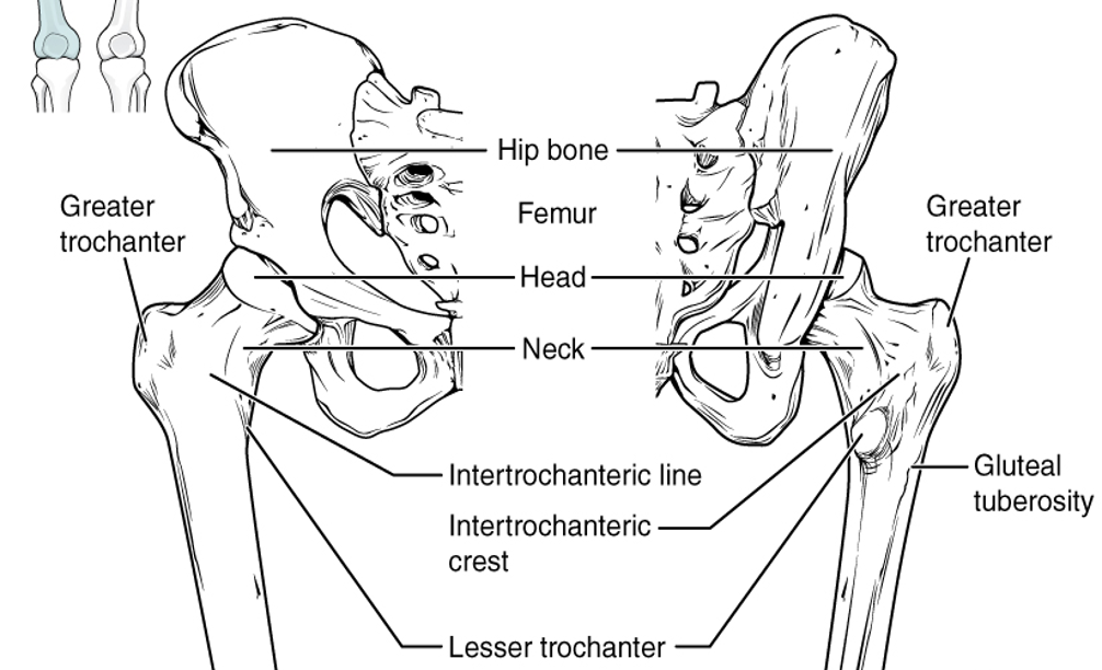
Clinical relevance: fractured neck of femur
A fractured neck of femur is usually caused by a fall, especially in older people. However, it can be caused by other conditions such as cancer. Fractures can be intracapsular or extracapsular depending on the location of the break, and management is guided by this.
The leg is typically held in a shortened and externally rotated position and patients are unable to weight bear.
Intracapsular fractures may be associated with avascular necrosis. This is because the blood supply to the head of the femur runs retrospectively up the femoral neck into the joint capsule, and can be disrupted following a fracture.
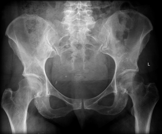
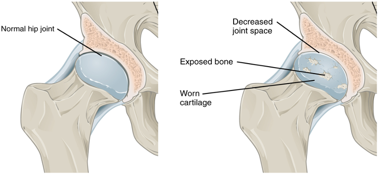
Clinical relevance: osteoarthritis of the hip
Osteoarthritis is a degenerative disease that primarily occurs in synovial joints. It involves degeneration of the articular surfaces. Symptoms of severe osteoarthritis include joint stiffness, reduced range of joint movement and joint pain.
Typical X-ray findings of osteoarthritis include:
- Joint space narrowing
- Subchondral sclerosis: hardening of the bone beneath the joint surface
- Osteophyte formation: small bony growths around the edges of the joint
- Bony cyst formation
For more information, see the Geeky Medics guides to osteoarthritis and interpreting a hip X-ray.
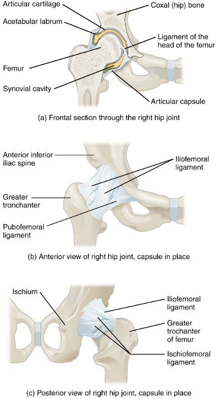
Ligaments
There are several ligaments which act to stabilise the hip joint.
The inferior aspect of the acetabulum is formed by the transverse acetabular ligament and helps to stabilise the inferior part of the hip joint.
The fovea of the femoral head is connected to the acetabulum via the ligament of the head of the femur (ligamentum teres). This ligament contains a branch of the obturator artery, which supplies the head of the femur.
The iliofemoral, pubofemoral and ischiofemoral ligaments spiral around the outer surface of the joint and act together to stabilise the hip.
Table 1. Ligaments of the hip
| Location on the hip joint | Origin | Insertion | |
|
Iliofemoral ligament |
Anterior |
Between anterior superior iliac spine (ASIS) and acetabulum |
Intertrochanteric line of the femur
|
|
Pubofemoral ligament |
Anteroinferior |
Iliopubic eminence
|
Combines with the deep iliofemoral ligament
|
|
Ischiofemoral ligament |
Posterior |
Ischium |
Greater trochanter of the femur
|
Bursae
Bursae are sacs filled with synovial fluid that act to reduce friction within the joint. The two main bursae in the hip are the trochanteric and iliopsoas bursae.
Clinical relevance: bursitis
Bursitis involves inflammation of a bursa and commonly occurs in the hip as trochanteric bursitis.
Patients commonly report a dull aching pain on the lateral aspect of their hip, which is worse when they lie on the affected side. Treatment of simple bursitis is rest, ice, and over-the-counter painkillers such as paracetamol or ibuprofen.
Blood supply and innervation
Blood supply to the hip joint
Arterial supply to the hip is mainly from the obturator artery, as well as the medial and lateral circumflex arteries, branches of the femoral artery, and the superior and inferior gluteal arteries. Branches of these arteries form an anastomotic network around the joint.
Innervation of the hip joint
Nerves supplying the hip joint include branches of the femoral nerve, obturator nerve and the superior gluteal nerve.
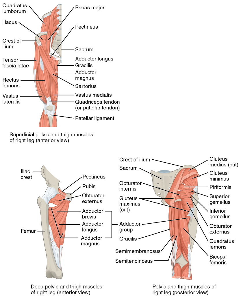
Muscles
There are several groups of muscles that act to move the hip joint. Movements at the hip joint include flexion, extension, internal and external rotation, adduction and abduction.
Table 2. Muscles acting on the hip joint.
|
Muscle group |
Muscle |
Action on the hip |
|
Deep gluteal
|
Piriformis |
External rotation, abduction |
|
Obturator internus |
||
|
Gemellus superior + inferior |
||
|
Quadratus femoris |
External rotation |
|
|
Superficial gluteal
|
Gluteus maximus |
Extension, internal rotation, abduction |
|
Gluteus medius |
Internal rotation, abduction |
|
|
Gluteus minimus |
||
|
Anterior compartment of the thigh
|
Psoas major |
Flexion |
|
Iliacus |
||
|
Sartorius |
||
|
Rectus femoris |
||
|
Medial compartment of the thigh
|
Gracilis |
Adduction |
|
Pectineus |
Adduction, flexion |
|
|
Adductor longus |
Adduction, internal rotation |
|
|
Adductor brevis |
||
|
Adductor magnus |
||
|
Obturator externus |
External rotation |
|
|
Posterior compartment of the thigh
|
Biceps femoris |
Extension, external rotation |
|
Semitendinosus |
Extension, internal rotation |
|
|
Semimembranosus |
Editor
Dr Chris Jefferies
References
Reference texts
- Richard L. Drake, A. Wayne Vogl, Adam W. M. Mitchell. Gray’s Anatomy for Students (Third Edition). Elsevier 2015.
- OpenStax. Anatomy and Physiology. Available from: [LINK]
- NHS.uk. 2022. Bursitis. Available from: [LINK]
Image references
- Openstax. Anatomy and Physiology. 8.4 Bones of the Lower Limb. Licence: [CC-BY-SA]
- Gaillard, F., 2022. Neck of femur fracture | Radiology Reference Article | Radiopaedia.org. Licence: [CC BY-NC-SA 3.0].
- Openstax. Anatomy and Physiology. 9.4 Synovial Joints. Licence: [CC-BY-SA]
- Openstax. Anatomy and Physiology. 9.6 Anatomy of Selected Synovial Joints. Licence: [CC-BY-SA].
- Openstax. Anatomy and Physiology. 11.6 Appendicular Muscles of the Pelvic Girdle and Lower Limbs. Licence: [CC-BY-SA].
