

- 📖 Geeky Medics OSCE Book
- ⚡ Geeky Medics Bundles
- ✨ 1300+ OSCE Stations
- ✅ OSCE Checklist PDF Booklet
- 🧠 UKMLA AKT Question Bank
- 💊 PSA Question Bank
- 💉 Clinical Skills App
- 🗂️ Flashcard Collections | OSCE, Medicine, Surgery, Anatomy
- 💬 SCA Cases for MRCGP
To be the first to know about our latest videos subscribe to our YouTube channel 🙌
The muscles of facial expression (also known as the mimetic muscles) can generally be divided into three main functional categories: orbital, nasal and oral.
These muscles are all innervated by the facial nerve (CN VII).¹
These striated muscles broadly originate from the surface of the skull and insert onto facial skin. Their contraction uniquely pulls on facial skin in order to exhibit various facial expressions.
Orbital Facial Muscles
The orbital facial muscles comprise of three main muscles:²
- Occipitofrontalis (frontalis contributes to this functional group)
- Orbicularis oculi
- Corrugator supercilii
As well as controlling the movement of the eyelids, these muscles also play a role in protecting the cornea from injury.
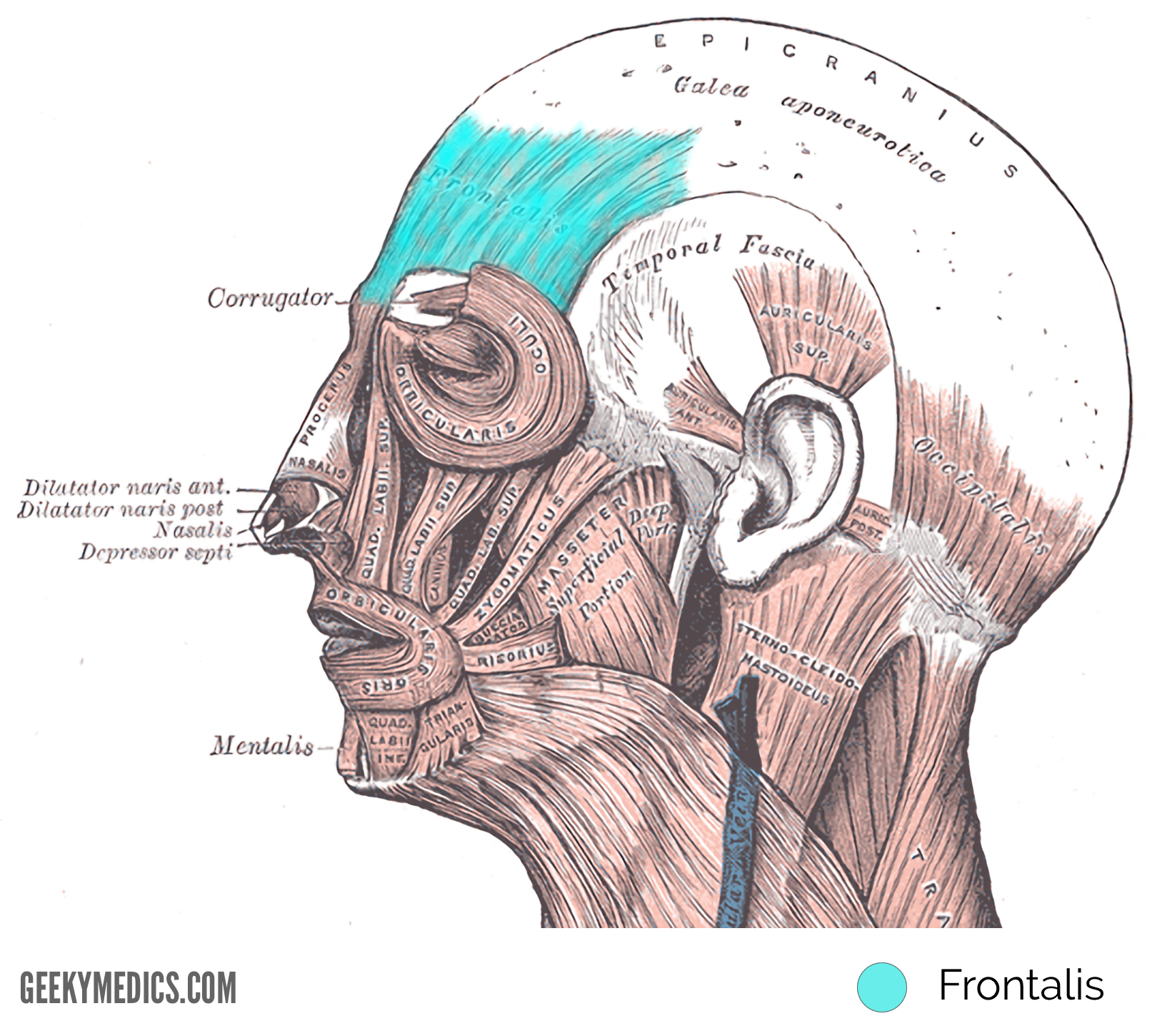
Occipitofrontalis
The occipitofrontalis muscle comprises of two main sections. These sections include the occipital (located posteriorly) and frontal (located anteriorly) bellies. The frontal belly is the major contributor to facial expression.¹
Origin
- The occipital belly originates from the occipital bone, as well as the mastoid process of the temporal bone.
- The frontal belly originates from the epicranial aponeurosis.¹
Insertion
- The occipital part inserts into the epicranial aponeurosis and the frontal belly inserts into the fascia of the facial muscles surrounding the eyes and the skin above the eyes.²
Action
- Contraction of this muscle raises the eyebrows and wrinkles the forehead.
Innervation
- The temporal branch of the facial nerve innervates the frontalis and the posterior auricular branch of the facial nerve innervates the occipitalis.¹
Blood supply
- The occipital belly is supplied by the occipital artery and the frontal belly is supplied by the supraorbital and supratrochlear arteries.¹
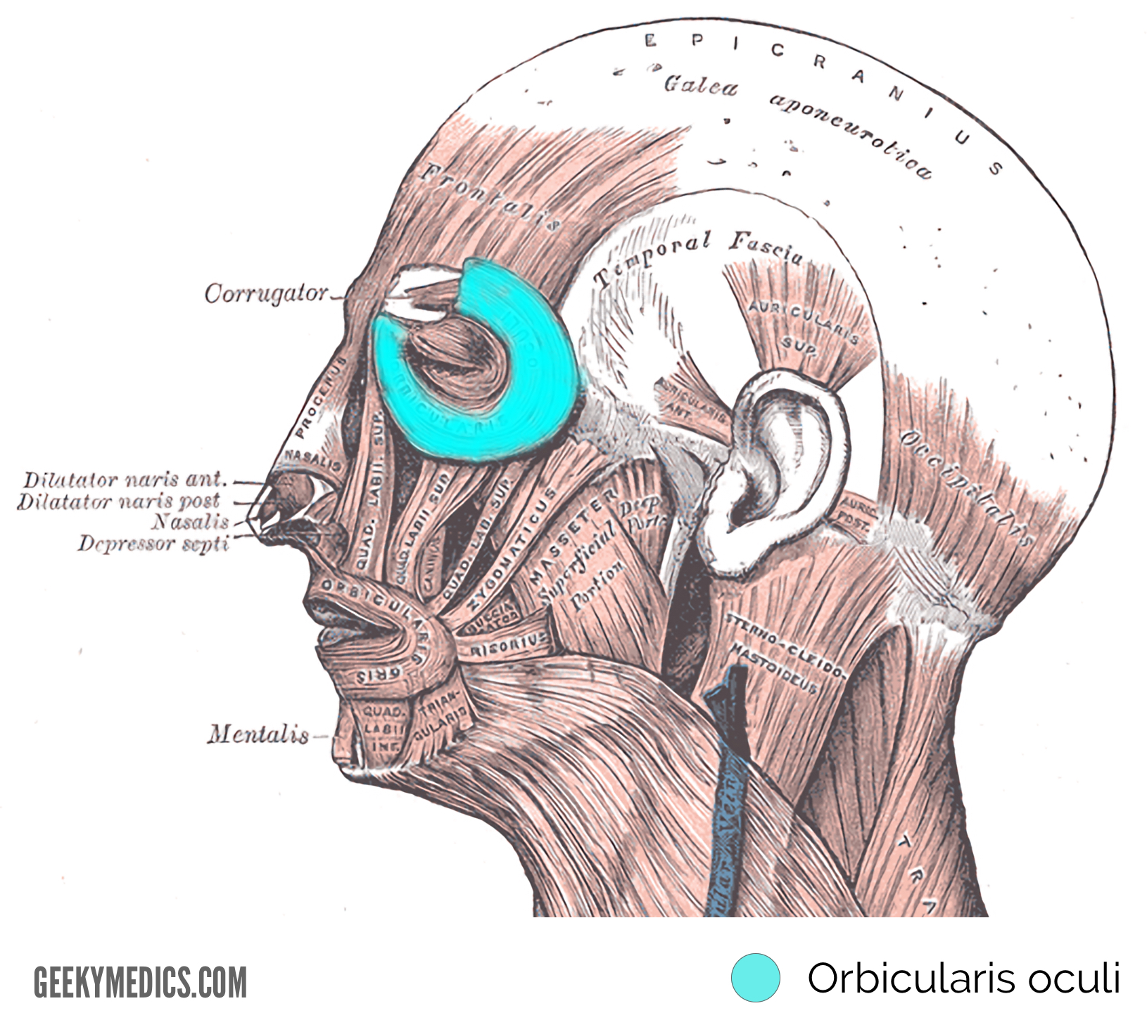
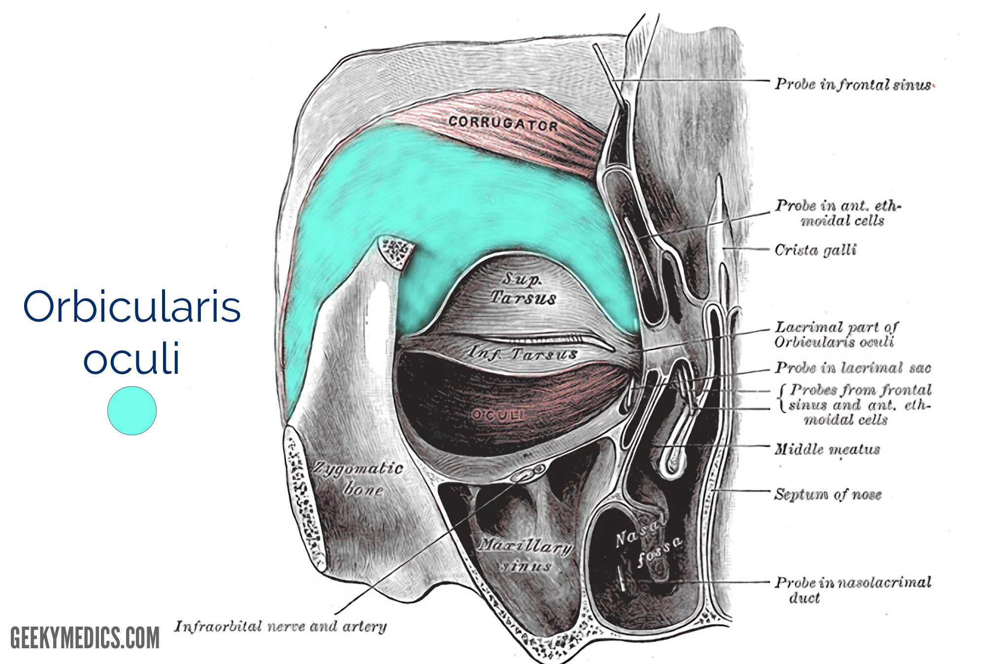
Orbicularis oculi
This muscle comprises of three main sections. These sections include the orbital orbicularis, palpebral orbicularis and lacrimal orbicularis.¹
Origin
- This muscle originates from the nasal portion of the frontal bone, frontal process of the maxilla, medial palpebral ligament, as well as the lacrimal crest and lacrimal bone.²
Insertion
- The insertion site of this muscle includes the skin overlying the circumference of the orbit, the orbital septum, the temporal aspect of the orbit and inferiorly towards the cheek.
Action
- This muscle mainly functions to close the eye.²
- The palpebral section functions voluntarily, as well as involuntarily during such actions as in blinking.
- The orbital section requires conscious effort.
- The lacrimal section controls the tear pump mechanism that filters into the lacrimal sac.
Innervation
- The temporal and zygomatic branches of the facial nerve.¹
Blood supply
- Branches of the facial, superficial temporal, maxillary and ophthalmic arteries.¹
Corrugator supercilii
Origin
- This muscle originates at the medial end of the supraorbital ridge.
Insertion
- The skin of the forehead near the eyebrow acts as the insertion site for this muscle.
Action
- Contraction of this muscle assists in wrinkling the forehead and drawing the eyebrows downwards and medially to assist in shielding the eyes from bright light.¹
Innervation
- The temporal branch of the facial nerve.²
Blood supply
- The ophthalmic artery and branches from the superficial temporal artery.²

Nasal Muscles
The nasal facial muscles are responsible for the movements of the nose and the surrounding skin. Three main muscles comprise this group, including nasalis, procerus, and the depressor septi nasi.
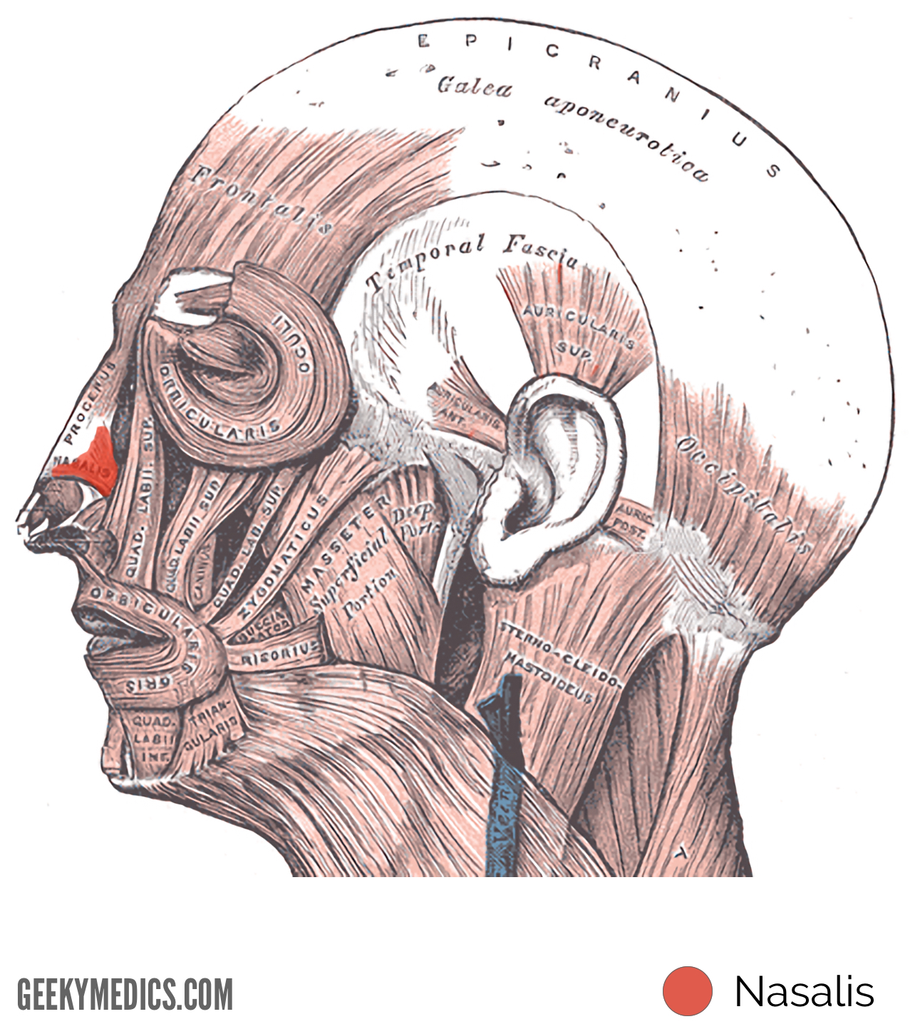
Nasalis
This is the largest of the nasal facial muscles that comprises of two main groups. These two groups include the transverse and alar sections.¹
Origin
- The nasalis muscle originates from the incisive fossa of the maxilla.²
Insertion
- Insertion occurs at an aponeurosis over the bridge of the nose (which blends with counterpart), as well as at the skin around the alar part of the nose.
Action
- Compression of the nasal bridge
- Depression of the corners of the nostrils
- Depression of the tip of the nose
Innervation
- The buccal branch of the facial nerve.¹
Blood supply
- The superior labial branch of the facial artery.
- The infraorbital branch of the maxillary artery.
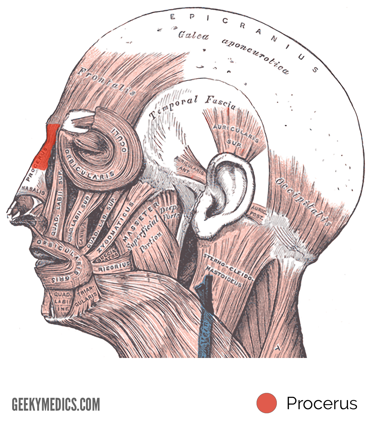
Procerus
Origin
- The fascia overlying the inferior section of the nasal bone and the superior section of the lateral nasal cartilage.²
Insertion
- The inferior skin of the forehead on either side of the midline between the eyebrows.
Action
- Contraction of this muscle assists in drawing the medial aspect of the eyebrows inferiorly. This contributes to the expression of frowning.²
Innervation
- The temporal branch and lower zygomatic branches of the facial nerve.²
Blood supply
- The angular and lateral nasal branches of the facial artery.²
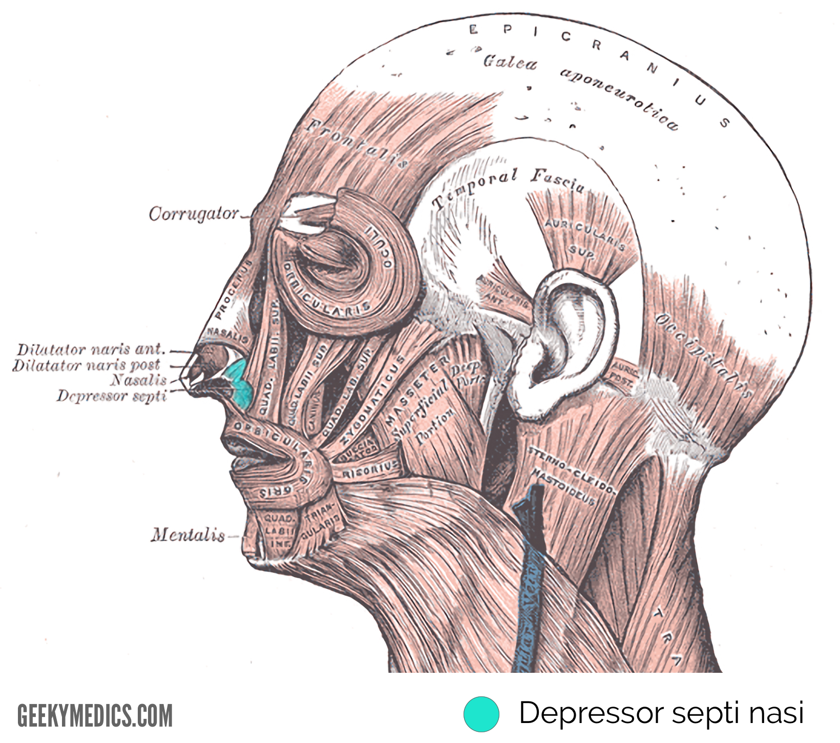
Depressor septi nasi
Origin
- The incisive fossa of the maxilla is the site of origination of this muscle.¹
Insertion
- The nasalis muscle and nasal septum.¹
Action
- Contraction of this muscle widens the nasal aperture.
Innervation
- The buccal branch of the facial nerve.¹
Blood supply
- The superior labial branch of the facial artery
Oral Muscles
The oral muscles are responsible for the movement of the lips and mouth. This group comprises of the following muscles: orbicularis oris, buccinators, depressor anguli oris, levator anguli oris, risorius, zygomaticus major and minor, levator labii superioris, levator labii superioris alaeque nasi, depressor labii inferioris, mentalis and platysma.²
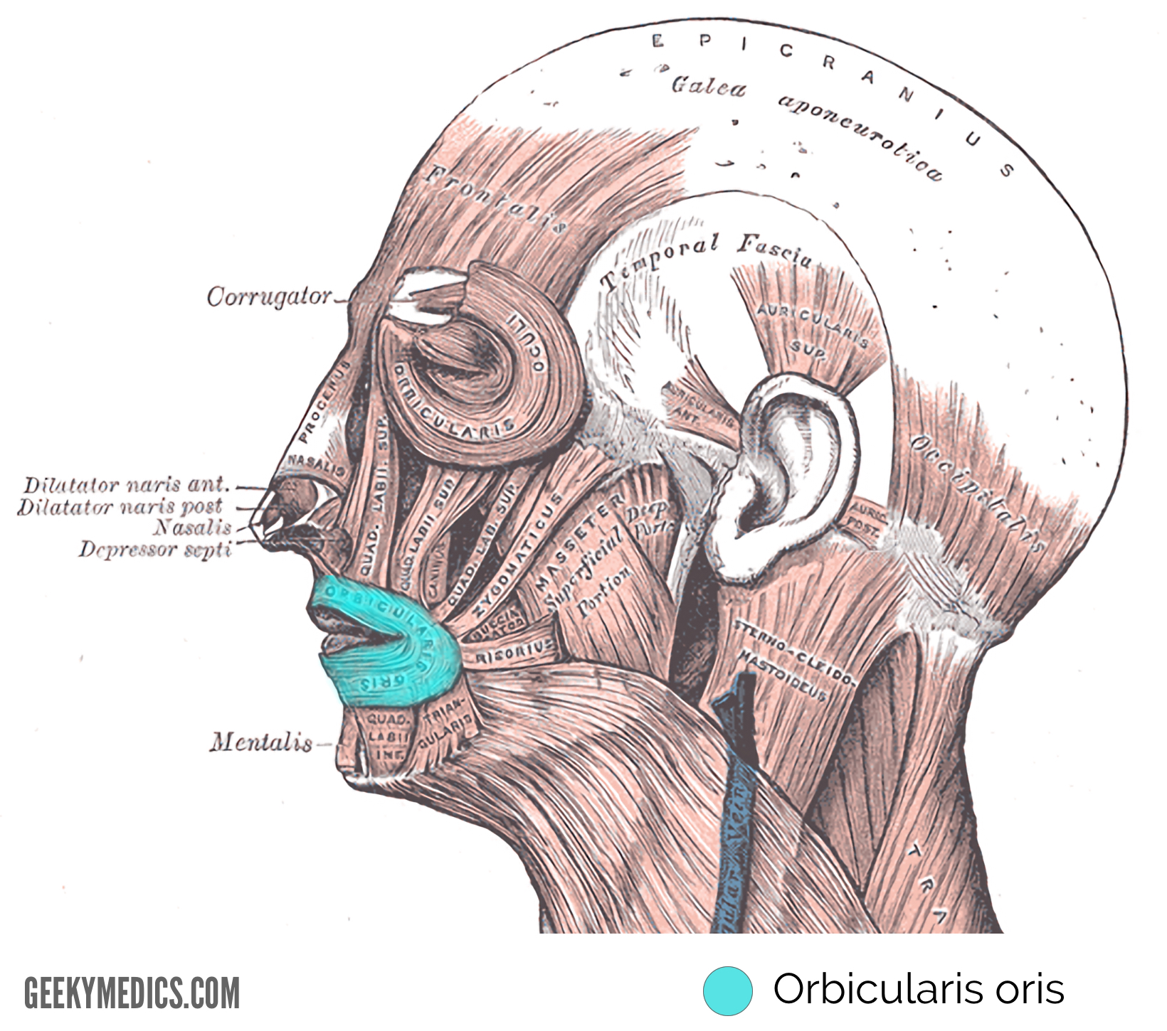
Orbicularis oris
This sphincter-like muscle is located around the circumference of the mouth.
Origin
- The medial aspect of the maxilla and mandible and the modiolus.¹
Insertion
- The skin surrounding the lips is the insertion site of the orbicularis oris muscle.¹
Action
- The contraction of this muscle puckers the lips and closes the mouth.
Innervation
- The buccal branch of the facial nerve.¹
Blood supply
- The superior and inferior labial branches of the facial artery.¹
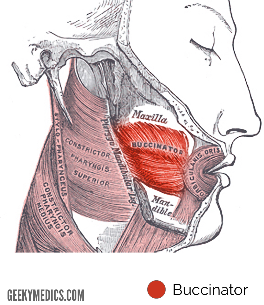
Buccinator
The buccinator is located between the maxilla and mandible. The buccinator forms the anterior aspect of the cheek and the lateral aspect of the oral cavity. Several structures penetrate the buccinators, including the parotid duct, molar glands of the cheeks, as well as the buccal branch of the mandibular nerve.
Origin
- The outer surfaces of the alveolar processes of the mandible and maxilla, and the pterygomandibular raphe.
Insertion
- The orbicularis oris muscle and modiolus act as the insertion site for this muscle.
Action
- Contraction of this muscle compresses the cheeks against the teeth (this action is especially useful in mastication and whistling).
Innervation
- The buccal branch of the facial nerve.¹
Blood supply
- The buccal branch of the maxillary artery.¹
Depressor anguli oris
Origin
- The mental tubercle of the mandible.²
Insertion
- The modiolus and angle of the mouth.
Action
- Contraction of this muscle causes depression of the angle of the mouth which contributes to frowning.
- This muscle is in direct opposition to the actions of the levator angulioris muscle.
Innervation
- The buccal and mandibular branches of the facial nerve.
Blood supply
- The inferior labial branch of the facial artery and mental branch of the maxillary artery.

Levator anguli oris
Origin
- The canine fossa of the maxilla.¹
Insertion
- The modiolus and angle of the mouth.
Action
- Contraction of this muscle elevates the angle of the mouth contributing to smiling.²
Innervation
- The zygomatic and buccal branches of the facial nerve.
Blood supply
- The superior labial branch of the facial artery and the infraorbital branch of the maxillary artery.²

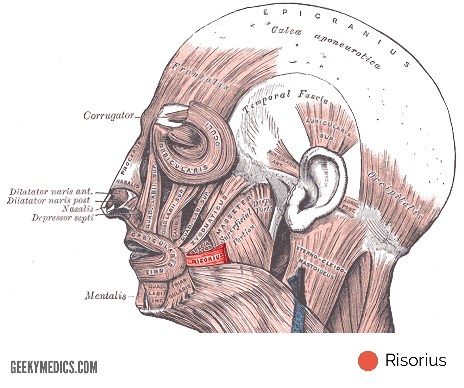
Risorius
Origin
- The parotid fascia.
Insertion
- The modiolus of the mouth.
Action
- This muscle draws the angle of the mouth backwards.²
Innervation
- The buccal branch of the facial nerve.²
Blood supply
- The superior labial branch of the facial artery.²
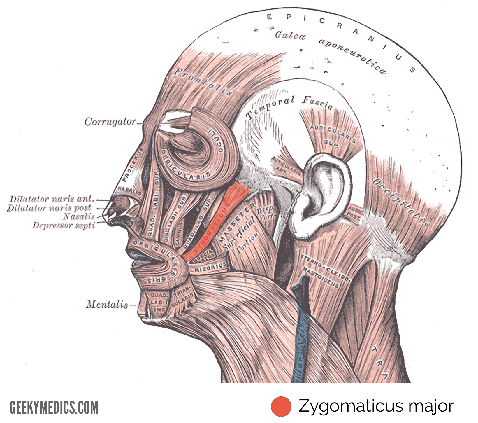
Zygomaticus major
Origin
- The zygomatic bone.
Insertion
- The modiolus of the mouth.
Action
- This muscle helps to facilitate smiling by pulling the angles of the mouth superiorly and laterally.
Innervation
- The zygomatic and buccal branches of the facial nerve.
Blood supply
- The superior labial branch of the facial artery.¹
Zygomaticus minor
Origin
- The zygomatic bone.¹
Insertion
- This muscle inserts into the skin of the lateral upper lip.
Action
- Contraction of this muscle aids in the elevation of the upper lip.
Innervation
- The zygomatic and buccal branches of the facial nerve.
Blood supply
- The superior labial branch of the facial artery.²

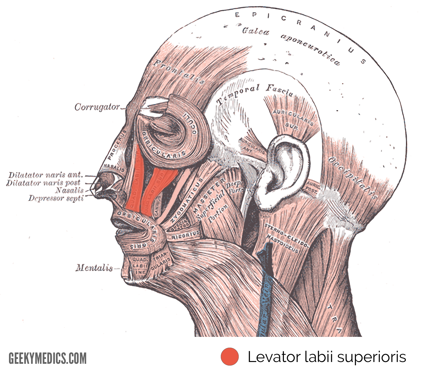
Levator labii superioris (also known as the quadratus labii superioris)
Origin
- The maxilla and zygomatic bone superior to infraorbital foramen.
Insertion
- This muscle inserts into the skin and muscle of the upper lip.¹
Action
- Contraction causes elevation of the upper lip.
Innervation
- The zygomatic and buccal branches of the facial nerve.¹
Blood supply
- The facial artery and the infraorbital branch of the maxillary artery.
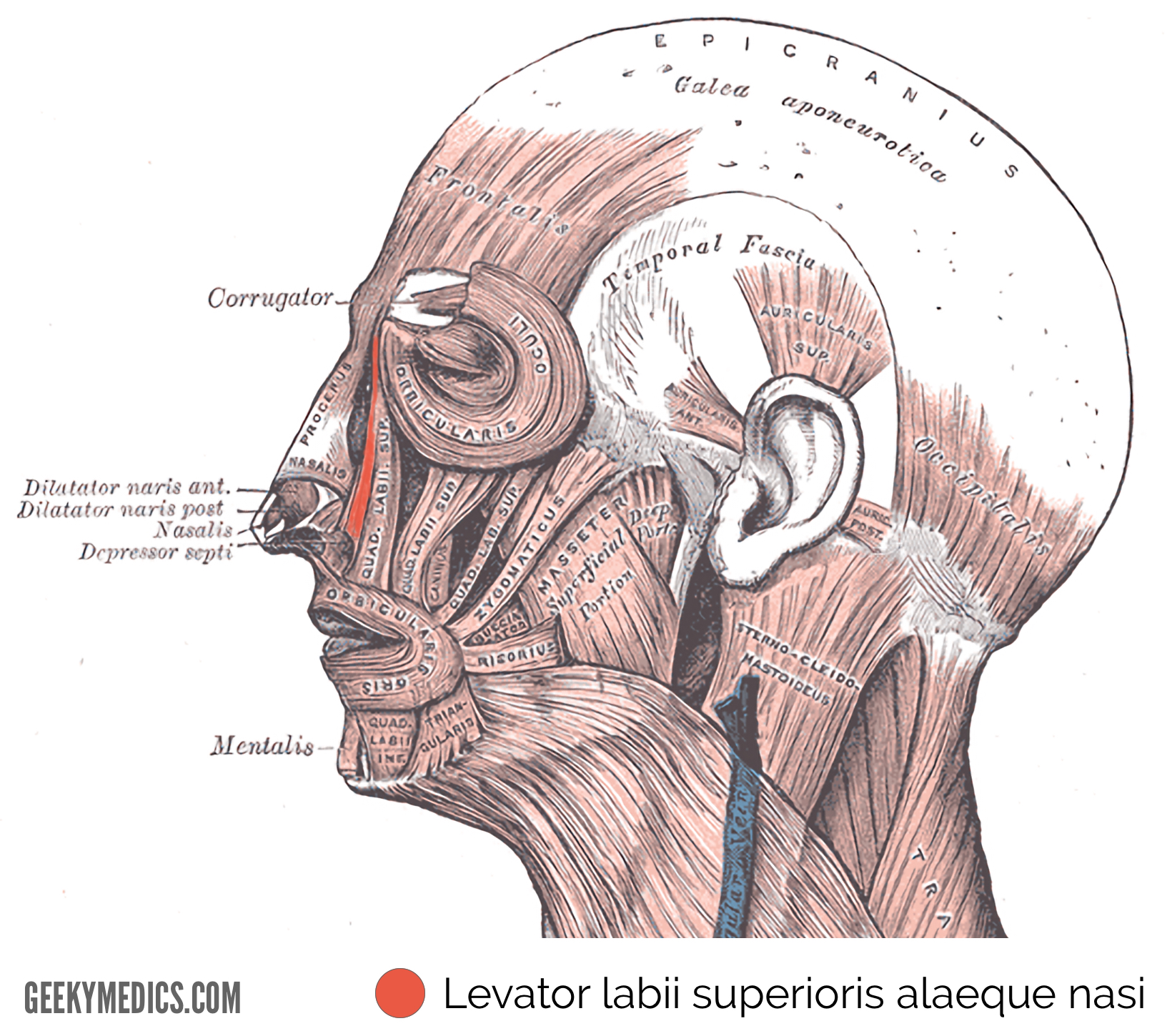
Levator labii superioris alaeque nasi
Origin
- The frontal process of the maxilla.²
Insertion
- Skin of the alar cartilage of nose and skin of the upper lip.¹
Action
- This muscle facilitates the expression of ‘snarling’ by causing dilation of the nostrils, as well as elevation of the wings of the nose and upper lip.
Innervation
- The zygomatic and buccal branches of the facial nerve.
Blood supply
- The facial artery and the infraorbital branch of the maxillary artery.²
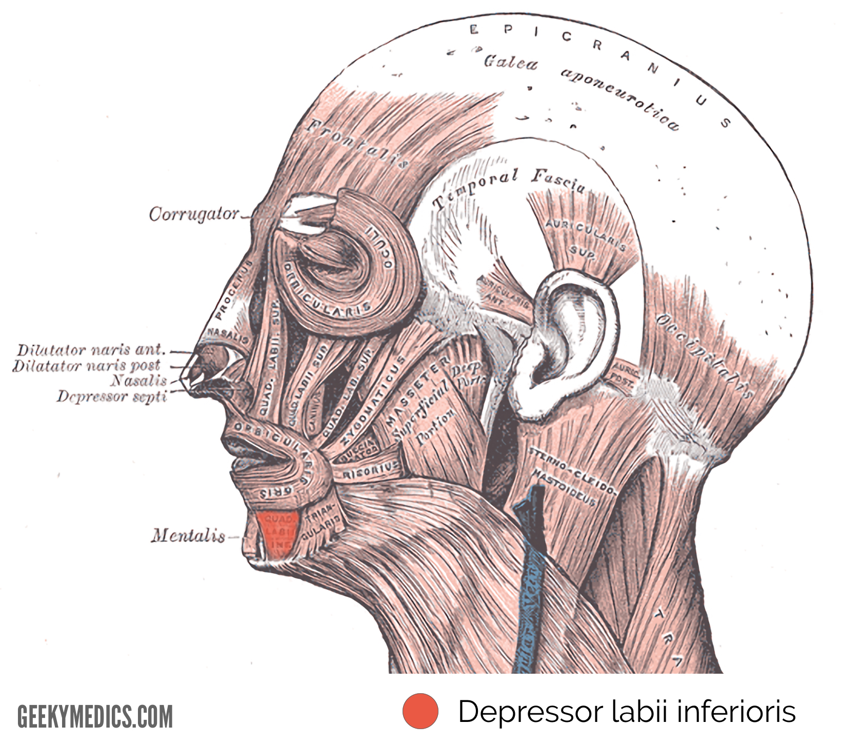
Depressor labii inferioris
Origin
- The mandible (specifically between the mental foramen and the symphysis).²
Insertion
- The skin of the lower lip. Its fibres blend with the fibres of the orbicularis oris muscle at the insertion point.
Action
- Contraction of this muscle depresses the lower lip.
Innervation
- The mandibular branch of the facial nerve.¹
Blood supply
- The inferior labial branch of the facial artery and the mental branch of the maxillary artery.
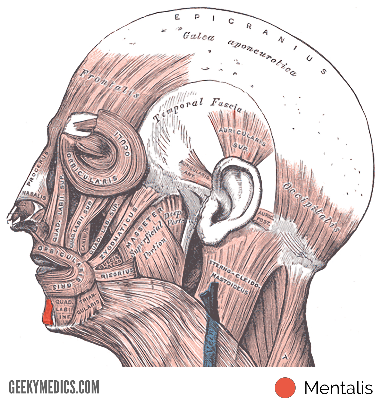
Mentalis
Origin
- The anterior aspect of the mandible.²
Insertion
- The skin of the chin.¹
Action
- Contraction of this muscle causes protrusion of the lower lip, as well as elevation and wrinkling of the skin of the chin.
Innervation
- The mandibular branch of the facial nerve.
Blood supply
- The inferior labial branch of the facial artery and the mental branch of the maxillary artery.²
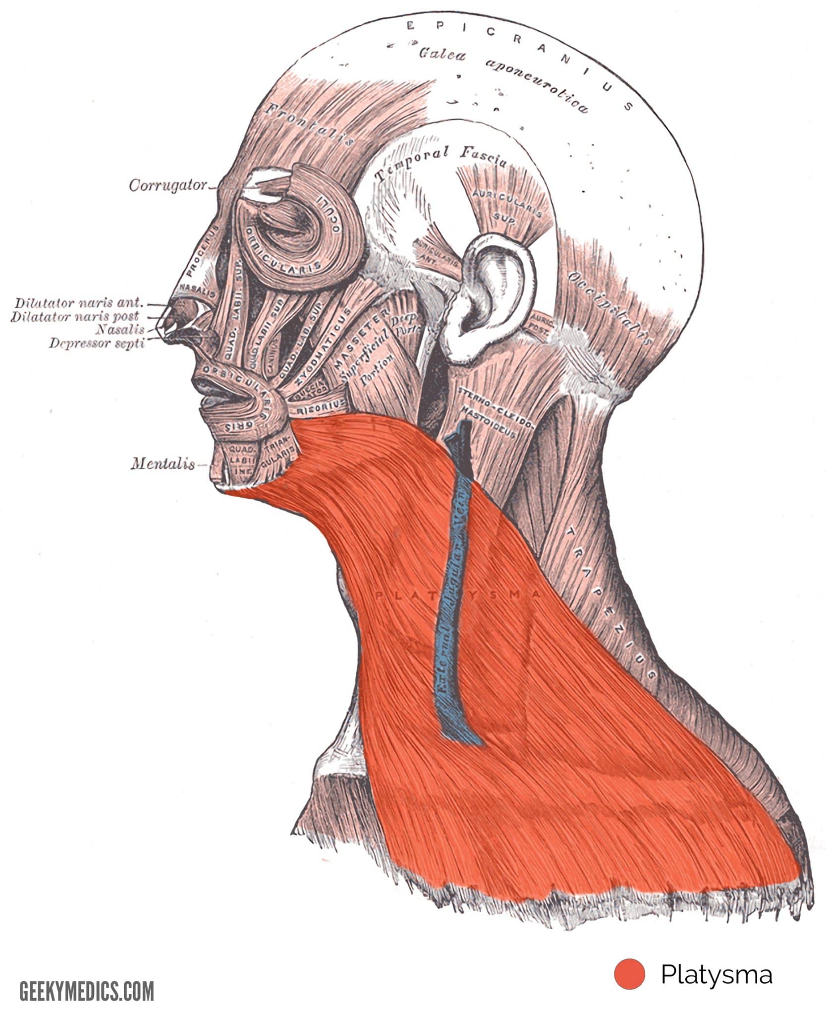
Platysma
Origin
- The skin and fascia of the infraclavicular and supraclavicular regions.²
Insertion
- The base of the mandible, skin of the cheek and lower lip, angle of the mouth, as well as the orbicularis oris muscle.²
Action
- Draws the corners of the mouth downwards which assists in creating the expression of melancholy.¹
- Tenses the skin of the neck when the teeth are clenched.¹
- Depresses the lower jaw.¹
Innervation
- The cervical branch of the facial nerve.²
Blood supply
- Branches of the submental and suprascapular arteries.²
Reviewer
Dr James Nott
Anatomy Lecturer & Anatomy Lead at Geeky Medics
References
- Drake, R. L., Wayne Vogl, A., Mitchell, A. (2009). Gray’s anatomy for students second edition. Elsevier health sciences.
- Gilroy, A. M. (2017). Anatomy – an essential textbook (2nd edition). Thieme medical publishers.
