
- 📖 Geeky Medics OSCE Book
- ⚡ Geeky Medics Bundles
- ✨ 1300+ OSCE Stations
- ✅ OSCE Checklist PDF Booklet
- 🧠 UKMLA AKT Question Bank
- 💊 PSA Question Bank
- 💉 Clinical Skills App
- 🗂️ Flashcard Collections | OSCE, Medicine, Surgery, Anatomy
- 💬 SCA Cases for MRCGP
To be the first to know about our latest videos subscribe to our YouTube channel 🙌
Introduction
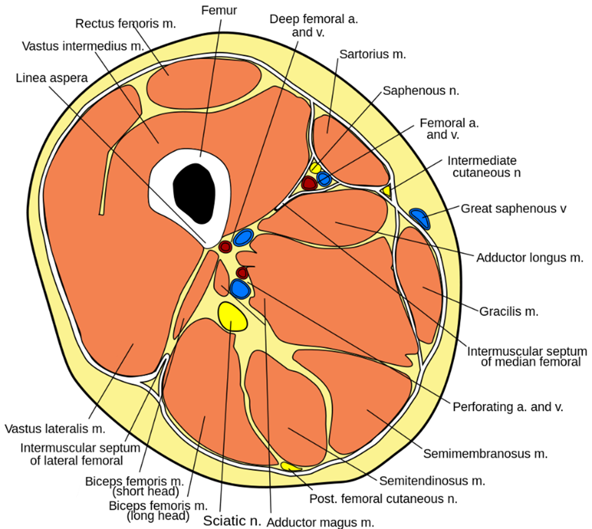
The thigh is the part of the leg that runs between the hip and the knee. It consists of three muscular compartments: anterior, posterior, and medial (Figure 1). This article will focus on the anterior compartment of the thigh.
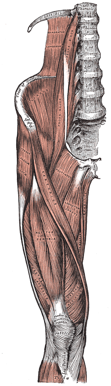
The muscles that make up the anterior compartment of the thigh are sartorius and the quadriceps femoris muscles (rectus femoris, vastus lateralis, vastus medialis, vastus intermedius). Psoas major and iliacus muscles also pass into the upper aspect of the anterior compartment.
The main action of this muscle group is to flex the thigh at the hip and extend the thigh at the knee. Innervation to this muscle group is the femoral nerve (L2, L3, L4). The blood supply is derived from the femoral artery.
Iliopsoas
The iliopsoas is made up of the psoas major and iliacus muscles.
These both originate as separate muscles on the posterior abdominal wall. From their point of origin, they descend and pass laterally to the inguinal ligament into the upper aspect of the anterior compartment of the thigh, where they converge into a common tendon. This common tendon inserts onto the lesser trochanter of the femur.
Psoas major
Origin
- Posterior abdominal wall: T12 to L5
Insertion
- Lesser trochanter of the femur
Innervation
- Anterior rami of L1, L2 and L3
Action
- Flexion of the hip
Iliacus
Origin
- Posterior abdominal wall: in the iliac fossa
Insertion
- Lesser trochanter of the femur
Innervation
- Femoral nerve (L2, L3)
Action
- Flexion of the hip
Quadriceps femoris
Quadriceps femoris is a muscle group comprised of four individual muscles which make up the bulk of the anterior compartment of the thigh.
These muscles are rectus femoris, vastus medialis, vastus intermedius and vastus lateralis. This muscle group works to extend the leg at the knee, as well as stabilising the patella during knee movement.
Rectus femoris
Origin
- Rectus femoris has two heads of origin, one from the anterior inferior iliac spine (straight head) and the other from the ilium immediately superior to the acetabulum (reflected head)
- The two heads converge to form a common muscle
Insertion
- The rectus femoris tendon converges with the quadriceps femoris tendon to form the patellar tendon
- The patellar tendon inserts onto the base of the patella
Innervation
- Femoral nerve (L2, L3, L4)
Action
- Flexion of the hip
- Extension of the knee
Vastus medialis
Vastus medialis is the most medial muscle in the quadriceps femoris group, hence its name. It sits lateral to sartorius and medial to rectus femoris.
Origin
- The vastus medialis has a long origin along the femur.
- It starts on the intertrochanteric line, descends posteroinferiorly along the pectinate line and along the linea aspera, down to the medial supracondylar line.
Insertion
- It inserts onto the medial aspect of the quadriceps tendon and the medial border of the patella.
Innervation
- Femoral nerve (L2, L3, L4)
Action
- Extension of the knee
Vastus intermedius
Vastus intermedius gets its name as it is the middle of the three vastus muscles. It lies behind rectus femoris and medial to the vastus lateralis muscle.
Origin
- The muscle originates from the upper two-thirds of the anterolateral surface of the femur.
Insertion
- Vastus intermedius inserts onto the quadriceps femoris tendon, as well as the lateral border of the patella and the lateral condyle of the tibia.
Innervation
- Femoral nerve (L2, L3, L4)
Action
- Extension of the knee
Vastus lateralis
Vastus lateralis is the largest of the quadriceps femoris muscle group. As its name suggests, it is the most lateral muscle in the vastus group and lies on the outside of the thigh.
Origin
- The muscle originates along the femur, starting from the superior intertrochanteric line, before moving laterally to the lateral gluteal tuberosity and down the lateral aspect of the linea aspera.
Insertion
- Vastus lateralis inserts onto the quadriceps femoris tendon and the lateral aspect of the patella.
Innervation
- Femoral nerve (L2, L3, L4)
Action
- Extension of the knee
Sartorius
Sartorius is the most superficial muscle of the anterior compartment and runs obliquely from the anterior superior iliac spine on the pelvis (lateral) to the tibia (medial). It is not part of the quadriceps femoris group as it does not have an insertion onto the common quadriceps tendon.
In the upper third of the thigh, the medial border of sartorius forms the lateral wall of the femoral triangle.
Origin
- Originates on the anterior superior iliac spine (ASIS) on the pelvis
Insertion
- Inserts onto the medial aspect of the tibia, just inferior to the tibial tuberosity
Innervation
- Femoral nerve (L2, L3)
Action
- Flexion of the hip
- Flexion of the knee
- Abduction of the thigh
- Lateral rotation of the thigh
Clinical relevance: compartment syndrome1
Compartment syndrome occurs when there is a build-up of pressure inside a fascial compartment in a limb.
Pressure in the compartment builds and eventually compresses veins and nerves, reducing blood flow away from the muscles and sensation. When pressure is above diastolic blood pressure, arteries will also become compressed. Causes of compartment syndrome include limb trauma, haemorrhage and compression (e.g. a plaster cast).
The main symptom of compartment syndrome is pain out-of-keeping with physical examination findings. Due to nerve compression, numbness or paraesthesia may also occur. Passive stretching of the muscles in the affected compartment will exacerbate the pain.
Acute compartment syndrome is a surgical emergency and must be treated quickly to avoid tissue necrosis. The only treatment is to relieve the pressure within the fascial compartment by performing a fasciotomy. This involves making an incision into the skin to open the compartment and relieve the pressure. Sometimes these incisions are left open for several days to allow the pressure to subside completely.
Summary table
Table 1. The muscles of the anterior thigh.
| Muscle | Origin | Insertion | Innervation | Function |
|
Psoas major |
Posterior abdominal wall |
Lesser trochanter of the femur |
Anterior rami of L2-L4 |
Flexion of the hip |
|
Iliacus |
Posterior abdominal wall |
Lesser trochanter of the femur |
Femoral nerve (L2, L3) |
Flexion of the hip |
|
Rectus femoris |
Straight head: ASIS Reflected head: ilium, just superior to acetabulum |
Quadriceps femoris tendon |
Femoral nerve (L2-L4) |
Flexion of the hip Extension of the knee |
|
Vastus medialis |
From intertrochanteric line, descends posteroinferiorly along the pectinate line and along the linea aspera, down to the medial supracondylar line. |
Quadriceps femoris tendon Medial border of patella |
Femoral nerve (L2-L4) |
Extension of the knee |
|
Vastus intermedius |
Upper two-thirds of the anterolateral surface of the femur |
Quadriceps femoris tendon Lateral border of patella Lateral tibial condyle |
Femoral nerve (L2-L4) |
Extension of the knee |
|
Vastus lateralis |
The superior intertrochanteric line, before moving laterally to the lateral gluteal tuberosity and down the lateral aspect of the linea aspera. |
Quadriceps femoris tendon Lateral border of the patella |
Femoral nerve (L2-L4) |
Extension of the knee |
|
Sartorius |
Anterior superior iliac spine |
Medial surface of tibia, just below tibial tuberosity |
Femoral nerve (L2, L3) |
Flexion of the hip Flexion of the knee |
Editor
Dr Chris Jefferies
References
- OrthoInfo. Compartment Syndrome. Published October 2009. Available from: [LINK]
- Richard L. Drake, A. Wayne Vogyl, Adam W.M. Mitchell. Gray’s Anatomy for Students 3rd Anterior compartment of the thigh, Lower Limb. Pages 589-953. Elsevier. Published in 2015.
Image references
- Figure 1. Marshall Strother. Thigh cross-section. License: [CC BY 3.0].
- Figure 2. Henry Vandyke Carter. Gray430. License: [Public domain]
