
- 📖 Geeky Medics OSCE Book
- ⚡ Geeky Medics Bundles
- ✨ 1300+ OSCE Stations
- ✅ OSCE Checklist PDF Booklet
- 🧠 UKMLA AKT Question Bank
- 💊 PSA Question Bank
- 💉 Clinical Skills App
- 🗂️ Flashcard Collections | OSCE, Medicine, Surgery, Anatomy
- 💬 SCA Cases for MRCGP
To be the first to know about our latest videos subscribe to our YouTube channel 🙌
Introduction
In this article, we will provide a basic overview of the muscles of the gluteal region while integrating clinical anatomical pathology to describe certain features.
The gluteal region refers to the general region of the posterior buttocks, lying external to the pelvic cavity.
Its borders are:
- Anterior: pelvic girdle
- Superior: iliac crest
- Inferior: gluteal folds
Contained within the region are muscles, lymphatic vessels and neurovascular structures.
The gluteal region communicates with the pelvic cavity via the greater and lesser sciatic foramina, which several neurovascular structures traverse.
Most of these muscles are supplied by two branches of the internal iliac artery, and they mimic the two major nerves that supply these muscles – the superior and inferior gluteal nerves.
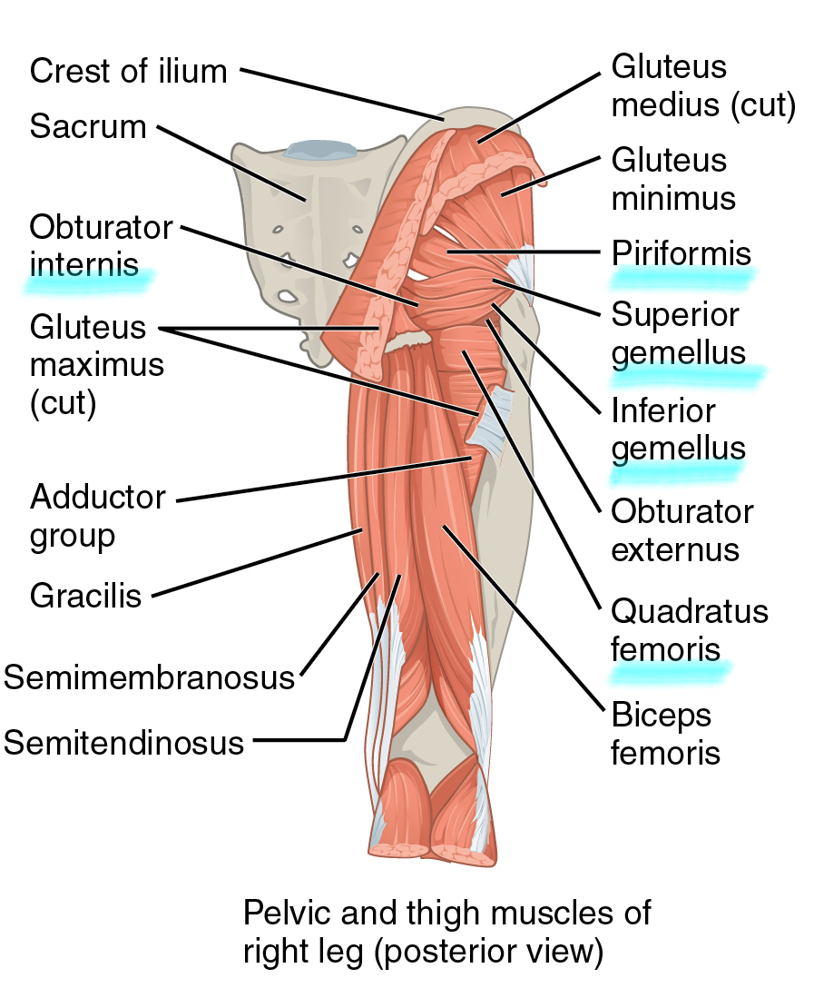
Superficial gluteal muscle group
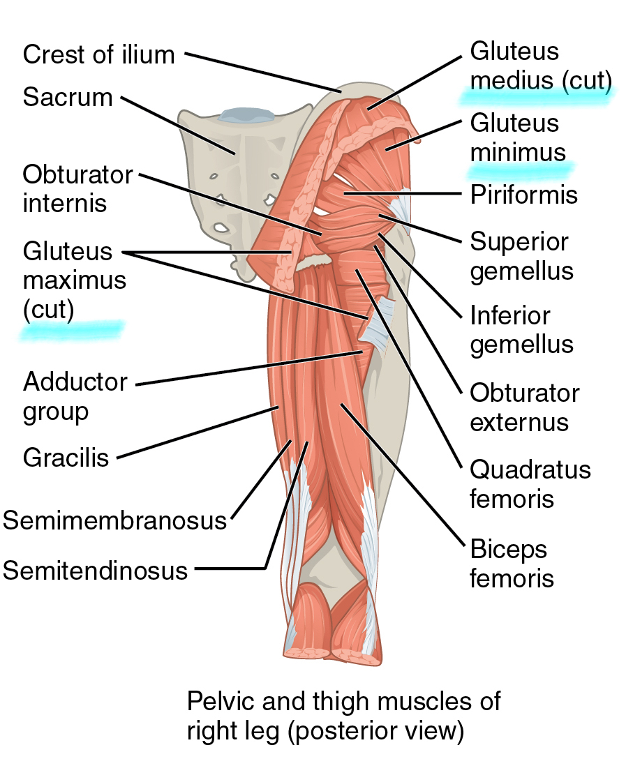
Gluteus maximus
As the largest muscle of the gluteal region, gluteus maximus is a powerful muscle involved in both primary hip movements and stabilisation of the hip.
Gluteus maximus lies superficial to the other gluteal muscles.
- Function: extension of the hip, assists in external rotation, abduction (superior fibres) and adduction (inferior fibres)
- Origin: dorsal aspects of the ilium, sacrum and coccyx
- Insertion: iliotibial band and gluteal tuberosity of the dorsal femur
- Innervation: inferior gluteal nerve (L5, S1, S2)
As the gluteus maximus also inserts into the iliotibial band, which crosses the knee joint, it is involved in stabilisation of this joint.
Gluteus medius and minimus
Gluteus medius and minimus are extremely important abductors of the hip, and their dysfunction can lead to significant difficulty in walking (ataxia).
- Function: abduction of the hip joint
- Origin: gluteal fossa of the ilium
- Insertion: lateral aspect of the greater trochanter of the femur
- Innervation: superior gluteal nerve (L4, L5, S1)
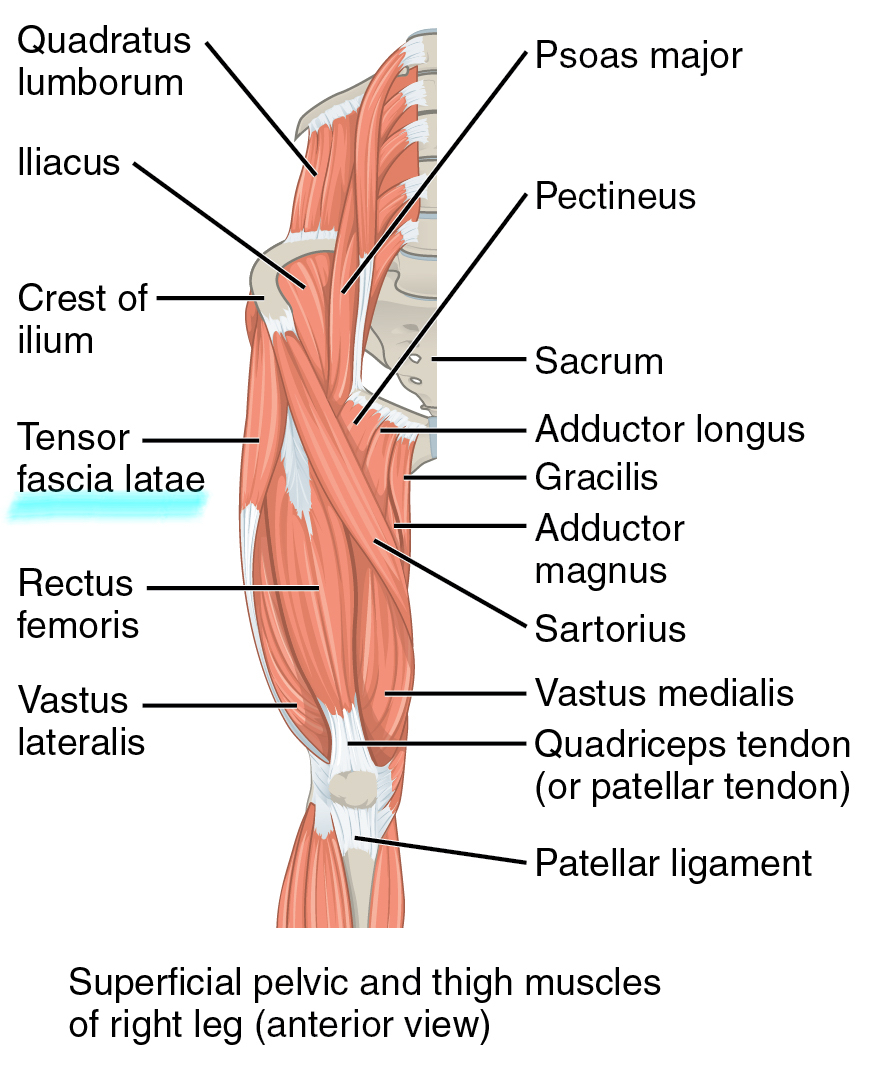
Tensor fasciae latae
Tensor fascia latae is the most anterior of the superficial gluteal muscles.
- Function: flexion, medial rotation and abduction of the hip, stabilisation of the knee via the iliotibial band
- Origin: iliac crest between the anterior superior iliac spine and iliac crest tuberculum
- Insertion: anterior aspect of the iliotibial tract
- Innervation: superior gluteal nerve (L4, L5, S1)
Deep gluteal muscle group
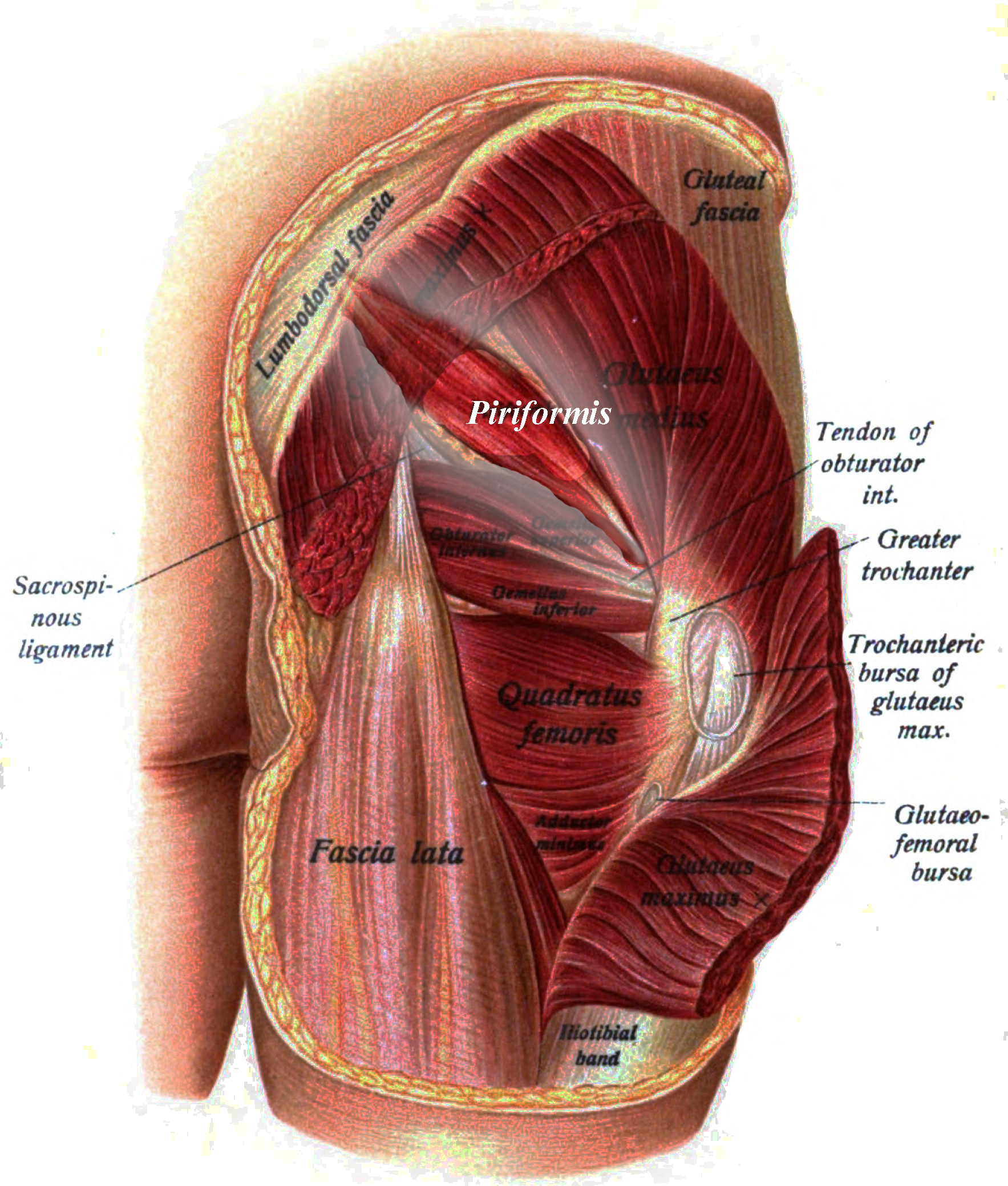
The deep gluteal muscles all contribute to external rotation of the hip. They are covered dorsally by the gluteus maximus and are only seen in cadaveric specimens where this muscle has been dissected away.
Piriformis
Piriformis is the most superior muscle of the deep gluteal group. It is named after its pear-like shape (yes, the fruit!).
- Function: external rotation and abduction of the hip
- Origin: anterolateral aspect of the sacrum
- Insertion: superior aspect of the greater trochanter of the femur
- Innervation: nerve to piriformis (S1, S2)
Piriformis is also an important anatomical landmark for it divides the greater sciatic foramen into supra- and infra-piriform regions. Nerves of the pelvic region, gluteal region and posterior thigh can be grouped as supra-piriform or infra-piriform.
Supra-piriform:
- Superior gluteal nerve (and vessels)
Infra-piriform:
- Inferior gluteal nerve (and vessels)
- Posterior cutaneous nerve of the thigh
- Pudendal nerve (and internal pudendal vessels)
- Sciatic nerve
The pudendal vessel follows an odd path, where it exits through the supra-piriform foramen and re-enters the pelvis in the infra-piriform foramen.
Hypertrophy of the piriformis muscle can lead to compression of the neurovascular structures in the infrapiriform foramen, a condition known as piriformis syndrome.
Gemellus superior
There are two gemelli (singular: gemellus) muscles. The superior muscle lies superior to the obturator internus, and the inferior muscle lies inferior to the obturator internus. They are fan-shaped muscles converging towards their insertion on the femur.
- Function: external rotation and abduction of the hip
- Origin: gluteal surface of the ischial spine
- Insertion: greater trochanter of the femur
- Innervation: nerve to obturator internus (L5, S1, S2)
Obturator internus
Obturator internus originates on the internal surface of the pelvis. Obturator externus originates on the external surface of the pelvis. Both attach to the femur to contribute to femoral rotation.
- Function: external rotation and slight abduction of the hip
- Origin: internal surface of the obturator membrane and adjacent pubic, ischial and iliac surfaces
- Insertion: greater trochanter of femur
- Innervation: nerve to obturator internus (L5, S1, S2)
Gemellus inferior
The second of the gemelli muscles lies below the obturator internus tendon.
- Function: external rotation and abduction of the hip
- Origin: gluteal surface of the ischial tuberosity
- Insertion: greater trochanter of the femur
- Innervation: nerve to quadratus femoris (L4, L5, S1)
Quadratus femoris
The last of the deep gluteal muscles, quadratus femoris is named after its flat, quadrilateral shape.
- Function: external rotation of the hip
- Origin: lateral aspect of the ischium
- Insertion: intertrochanteric crest of the femur
- Innervation: nerve to quadratus femoris (L4, L5, S1)
Tips for the gluteal muscles
The deep gluteal region has several muscles that may appear difficult to differentiate. One method to identify these muscles is to locate the piriformis – the cone-shaped muscle lying on top of the sciatic nerve. The order of the deep gluteal muscles inferior to piriformis follows the mnemonics “GOGO-Q” or “GO In, GO Out-Q” for superior-to-inferior:
- Gemellus superior
- Obturator internus
- Gemellus inferior
- Obturator externus (outside; not one of the deep gluteal muscles)
- Quadratus femoris
Movements of the hip muscles
- Muscles passing the hip anteriorly cause flexion at the hip
- Muscles passing the hip posteriorly cause extension at the hip (look at gluteus maximus)
- Muscles passing laterally cause abduction at the hip (look at gluteus medius and minimus)
- Small muscles that are lower on the femur are often more responsible for rotation (the deep gluteal muscles)
Clinical relevance: Trendelenburg’s sign
Patients with impaired hip abduction may present with an abnormal gait (ataxia). Impaired hip abduction is commonly due to damage to the superior gluteal nerve. This may occur secondary to pelvic fractures, space-occupying lesions and as a complication of hip surgery.
In these patients, Trendelenburg’s sign is said to be positive and may be demonstrated by asking the patient to stand on the affected leg only, when this is done the pelvis drops to the unaffected sign.
See our hip examination guide for more information.
Editor
William Maish
References
Reference images
- OpenStax College. License: [CC BY 3.0]
- Johannes Sobotta. License: [Public domain]
Reference texts
- Gray’s anatomy for students: third edition
