
- 📖 Geeky Medics OSCE Book
- ⚡ Geeky Medics Bundles
- ✨ 1300+ OSCE Stations
- ✅ OSCE Checklist PDF Booklet
- 🧠 UKMLA AKT Question Bank
- 💊 PSA Question Bank
- 💉 Clinical Skills App
- 🗂️ Flashcard Collections | OSCE, Medicine, Surgery, Anatomy
- 💬 SCA Cases for MRCGP
To be the first to know about our latest videos subscribe to our YouTube channel 🙌
This guide provides an overview of the recognition and immediate management of post-operative bleeding using an ABCDE approach.
The ABCDE approach can be used to perform a systematic assessment of a critically unwell patient. It involves working through the following steps:
- Airway
- Breathing
- Circulation
- Disability
- Exposure
Each stage of the ABCDE approach involves clinical assessment, investigations and interventions. Problems are addressed as they are identified and the patient is re-assessed regularly to monitor their response to treatment.
This guide has been created to assist students in preparing for emergency simulation sessions as part of their training, it is not intended to be relied upon for patient care.
Background
Types of bleeding
Post-operative bleeding can be divided into primary, reactive and secondary bleeding.
Primary bleeding
Primary bleeding refers to bleeding that occurs during the surgical procedure. The surgical team manages this bleeding intraoperatively. The estimated intraoperative blood loss and any transfusions that were administered should be documented on the operation note.
Reactive bleeding
Reactive bleeding refers to bleeding within 24 hours of the operation.
During surgery patients often become relatively hypotensive and vasoconstricted. In the post-operative period, as blood pressure rises and vasodilatation occurs, a damaged blood vessel may subsequently begin to bleed.
Secondary bleeding
Secondary bleeding refers to bleeding occurring within 7-10 days after the operation. Secondary bleeding is often associated with wound infection.
Clinical signs of post-operative bleeding
Typical clinical signs associated with post-operative bleeding include:
- Tachycardia
- Hypotension (typically develops late, only after a significant volume of blood has been lost)
- Tachypnoea
- Cool peripheries
- Pre-syncope/syncope
- Confusion/agitation
- Swelling and/or bruising at the wound site (secondary to haematoma formation)
- Bleeding from the wound site
- Increasing tenderness at the wound site
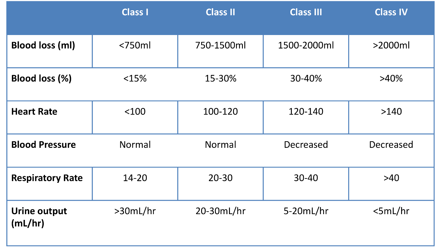
Classification of haemorrhagic shock
It is useful to have an understanding of how haemorrhagic shock is classified when interpreting clinical findings. It is important to note that a patient’s blood pressure can appear relatively normal despite the presence of significant blood loss. As a result, a normal blood pressure reading in isolation should not provide reassurance that bleeding is unlikely to be significant.
Tips before you begin
General tips for applying an ABCDE approach in an emergency setting include:
- Treat all problems as you discover them.
- Re-assess regularly and after every intervention to monitor a patient’s response to treatment.
- Make use of the team around you by delegating tasks where appropriate.
- All critically unwell patients should have continuous monitoring equipment attached for accurate observations.
- Clearly communicate how often would you like the patient’s observations relayed to you by other staff members.
- If you require senior input, call for help early using an appropriate SBARR handover structure.
- Review results as they become available (e.g. laboratory investigations).
- Make use of your local guidelines and algorithms in managing specific scenarios (e.g. acute asthma).
- Any medications or fluids will need to be prescribed at the time (in some cases you may be able to delegate this to another member of staff).
- Your assessment and management should be documented clearly in the notes, however, this should not delay initial clinical assessment, investigations and interventions.
Initial steps
Acute scenarios typically begin with a brief handover from a member of the nursing staff including the patient’s name, age, background and the reason the review has been requested.
You may be asked to review a patient with post-operative bleeding due to tachycardia, hypotension, bleeding from the wound site and/or increasing pain.
Introduction
Introduce yourself to whoever has requested a review of the patient and listen carefully to their handover.
Interaction
Introduce yourself to the patient including your name and role.
Ask how the patient is feeling as this may provide some useful information about their current symptoms.
Preparation
Make sure the patient’s notes, observation chart and prescription chart are easily accessible.
Review the operation note including:
- Operative site
- Estimated intraoperative blood loss
- Intraoperative complications
- Surgeon’s name and contact details
Ask for another clinical member of staff to assist you if possible.
If the patient is unconscious or unresponsive, start the basic life support (BLS) algorithm as per resuscitation guidelines.
Catastrophic bleeding
If catastrophic bleeding is identified when approaching the patient, apply direct pressure to the bleeding site and ask another member of staff to take over this role whilst you complete a full ABCDE assessment.
Airway
Clinical assessment
Can the patient talk?
Yes: if the patient can talk, their airway is patent and you can move on to the assessment of breathing.
No:
- Look for signs of airway compromise: these include cyanosis, see-saw breathing, use of accessory muscles, diminished breath sounds and added sounds.
- Open the mouth and inspect: look for anything obstructing the airway such as secretions or a foreign object.
Interventions
Regardless of the underlying cause of airway obstruction, seek immediate expert support from an anaesthetist and the emergency medical team (often referred to as the ‘crash team’). In the meantime, you can perform some basic airway manoeuvres to help maintain the airway whilst awaiting senior input.
Head-tilt chin-lift manoeuvre
Open the patient’s airway using a head-tilt chin-lift manoeuvre:
1. Place one hand on the patient’s forehead and the other under the chin.
2. Tilt the forehead back whilst lifting the chin forwards to extend the neck.
3. Inspect the airway for obvious obstruction. If an obstruction is visible within the airway, use a finger sweep or suction to remove it.
Jaw thrust
If the patient is suspected to have suffered significant trauma with potential spinal involvement, perform a jaw-thrust rather than a head-tilt chin-lift manoeuvre:
1. Identify the angle of the mandible.
2. With your index and other fingers placed behind the angle of the mandible, apply steady upwards and forward pressure to lift the mandible.
3. Using your thumbs, slightly open the mouth by downward displacement of the chin.
Oropharyngeal airway (Guedel)
Airway adjuncts are often helpful and in some cases essential to maintain a patient’s airway. They should be used in conjunction with the maneuvres mentioned above as the position of the head and neck need to be maintained to keep the airway aligned.
An oropharyngeal airway is a curved plastic tube with a flange on one end that sits between the tongue and hard palate to relieve soft palate obstruction. It should only be inserted in unconscious patients as it is otherwise poorly tolerated and may induce gagging and aspiration.
To insert an oropharyngeal airway:
1. Open the patient’s mouth to ensure there is no foreign material that may be pushed into the larynx. If foreign material is present, attempt removal using suction.
2. Insert the oropharyngeal airway in the upside-down position until you reach the junction of the hard and soft palate, at which point you should rotate it 180°. The reason for inserting the airway upside down initially is to reduce the risk of pushing the tongue backwards and worsening airway obstruction.
3. Advance the airway until it lies within the pharynx.
4. Maintain head-tilt chin-lift or jaw thrust and assess the patency of the patient’s airway by looking, listening and feeling for signs of breathing.
Nasopharyngeal airway (NPA)
A nasopharyngeal airway is a soft plastic tube with a bevel at one end and a flange at the other. NPAs are typically better tolerated in patients who are partly or fully conscious compared to oropharyngeal airways. NPAs should not be used in patients who may have sustained a skull base fracture, due to the small but life-threatening risk of entering the cranial vault with the NPA.
To insert a nasopharyngeal airway:
1. Check the patency of the patient’s right nostril and if required (depending on the model of NPA) insert a safety pin through the flange of the NPA.
2. Lubricate the NPA.
3. Insert the airway bevel-end first, vertically along the floor of the nose with a slight twisting action.
4. If any obstruction is encountered, remove the tube and try the left nostril.
CPR
If the patient loses consciousness and there are no signs of life on assessment, put out a crash call and commence CPR.
Re-assessment
Make sure to re-assess the patient after any intervention.
Breathing
Clinical assessment
Observations
Review the patient’s respiratory rate:
- A normal respiratory rate is between 12-20 breaths per minute.
- Tachypnoea may indicate significant blood loss (>1500ml), atelectasis or pneumonia.
Review the patient’s oxygen saturation (SpO2):
- A normal SpO2 range is 94-98% in healthy individuals and 88-92% in patients with COPD who are at high-risk of CO2 retention.
- Hypoxaemia may occur secondary to significant anaemia, atelectasis, pneumonia or bleeding within the thorax (e.g. haemothorax).
Auscultation
Auscultate the chest to screen for evidence of other respiratory pathology (e.g. unilaterally reduced air entry might represent a haemothorax). Each side of the thorax can hold up to 1.5L of fluid and as a result, a significant haemothorax can accumulate before the patient deteriorates significantly.
Investigations and procedures
Arterial blood gas
Take an ABG if indicated (e.g. low SpO2) to quantify the degree of hypoxia.
Chest X-ray
A chest X-ray may be indicated if abnormalities are noted on auscultation (e.g. to identify atelectasis or haemothorax). A chest X-ray should not delay the emergency management of post-operative bleeding.
See our CXR interpretation guide for more details.
Interventions
Oxygen
Administer oxygen if the patient has a low SpO2. This typically involves the use of a non-rebreathe mask with an oxygen flow rate of 15L. If the patient has COPD and a history of CO2 retention you should switch to a venturi mask as soon as possible and titrate oxygen appropriately.
If the patient is conscious, sit them upright as this can also help with oxygenation.
CPR
If the patient loses consciousness and there are no signs of life on assessment, put out a crash call and commence CPR.
Re-assessment
Make sure to re-assess the patient after any intervention.
Circulation
Clinical assessment
Pulse
Assess the patient’s pulse:
- Tachycardia is an early sign of volume depletion in the context of post-operative bleeding.
- The patient’s pulse may feel thready secondary to hypovolaemia.
Blood pressure
Assess the patient’s blood pressure:
- Patients with post-operative bleeding don’t typically develop hypotension until there has been significant blood loss (i.e. 1500-2000 mls).
Capillary refill time
Assess the patient’s capillary refill time (CRT):
- In the context of post-operative bleeding, the CRT may be prolonged (>2 seconds) both peripherally and centrally.
- The patient’s peripheries may also feel cool secondary to hypovolaemia and peripheral vasoconstriction.
Fluid balance
Calculate the patient’s fluid balance:
- Calculate the patient’s current fluid balance (e.g. oral fluids, intravenous fluids, urine output, drain output, stool output, vomiting) to inform resuscitation efforts.
- Urine output is maintained until there has been significant blood loss (e.g. 1500-2000 mls).
Inspection
Inspect the patient from the end of the bed and note evidence of pallor indicative of anaemia.
Investigations and procedures
Intravenous cannulation
Insert two large-bore cannulae (14-16G) and take blood tests as discussed below.
Adequate intravenous access is essential in the context of post-operative bleeding as patients can rapidly deteriorate and require large volumes of fluid and blood to be transfused.
Blood tests
Collect blood tests after cannulating the patient including:
- FBC: to assess the degree of anaemia to guide transfusion.
- U&Es: to assess renal function (e.g. pre-renal acute kidney injury).
- Group and crossmatch: to confirm the patient’s blood group and request blood products.
- LFTs: to screen for evidence of liver disease (e.g. cirrhosis).
- Coagulation screen: to screen for coagulopathy and inform resuscitation efforts.
Record an ECG
An ECG should be performed to screen for cardiac pathology such as myocardial infarction which may be precipitated by anaemia. Performing an ECG should not delay the emergency management of post-operative bleeding.
Imaging
Consider requesting imaging such as a CT scan to identify the source of bleeding to inform the need for operative intervention.
Interventions
Positional changes
In the context of hypotension, re-positioning your patient (where possible) so that they are supine with their legs elevated can improve blood pressure and major organ perfusion by re-distributing their circulating volume. This can be a useful temporary measure whilst other resuscitation efforts are commenced.
Fluid resuscitation
Hypovolaemic patients require fluid resuscitation:
- Administer a 500ml bolus of Hartmann’s solution or 0.9% sodium chloride (warmed if available) over less than 15 mins.
- Administer 250ml boluses in patients at increased risk of fluid overload (e.g. heart failure).
After each fluid bolus, reassess for clinical evidence of fluid overload (e.g. auscultation of the lungs, assessment of JVP).
Repeat administration of fluid boluses up to four times (e.g. 2000ml or 1000ml in patients at increased risk of fluid overload), reassessing the patient each time.
Seek senior input if the patient has a negative response (e.g. increased chest crackles) or if the patient isn’t responding adequately to repeated boluses (i.e. persistent hypotension).
See our fluid prescribing guide for more details on resuscitation fluids.
Blood transfusion
Blood transfusion should be guided by haemoglobin levels and the estimated volume of blood lost.
Base decisions on blood transfusion on the full clinical picture, recognising that over-transfusion may be as damaging as under-transfusion.
In the context of acute haemorrhage, O-negative blood may need to be administered if there is not adequate time for matching. This would, of course, be a senior-led decision.
Other blood products
Patients may require other blood products, depending on the scenario such as platelets (e.g. if thrombocytopenic) or fresh frozen plasma (e.g. if coagulation is abnormal).
If you feel your patient may need other blood products, discuss with the on-call haematologist.
CPR
If the patient loses consciousness and there are no signs of life on assessment, put out a crash call and commence CPR.
Re-assessment
Make sure to re-assess the patient after any intervention.
Disability
Clinical assessment
Consciousness
In the context of post-operative bleeding, a patient’s consciousness level may be reduced secondary to hypotension
Assess the patient’s level of consciousness using the AVPU scale:
- Alert: the patient is fully alert, although not necessarily orientated.
- Verbal: the patient makes some kind of response when you talk to them (e.g. words, grunt).
- Pain: the patient responds to a painful stimulus (e.g. supraorbital pressure).
- Unresponsive: the patient does not show evidence of any eye, voice or motor responses to pain.
If a more detailed assessment of the patient’s level of consciousness is required, use the Glasgow Coma Scale (GCS).
Pupils
Assess the patient’s pupils:
- Inspect the size and symmetry of the patient’s pupils
- Assess direct and consensual pupillary responses
Drug chart review
Review the patient’s drug chart for medications which may cause a reduced level of consciousness (e.g. opioids, sedatives, anxiolytics, insulin, oral hypoglycaemic medications).
Investigations and procedures
Blood glucose and ketones
Measure the patient’s capillary blood glucose level to screen for causes of a reduced level of consciousness (e.g. hypoglycaemia or hyperglycaemia).
A blood glucose level may already be available from earlier investigations (e.g. ABG, venepuncture).
The normal reference range for fasting plasma glucose is 4.0 – 5.8 mmol/l.
Hypoglycaemia is defined as a plasma glucose of less than 3.0 mmol/l. In hospitalised patients, a blood glucose ≤4.0 mmol/L should be treated if the patient is symptomatic.
See our blood glucose measurement guide for more details.
Imaging
Request a CT head if intracranial pathology is suspected after discussion with a senior.
See our guide on interpreting a CT head for more details.
Interventions
Maintain the airway
Alert a senior immediately if you have any concerns about the consciousness level of a patient. A GCS of 8 or below warrants urgent expert help from an anaesthetist. In the meantime, you should re-assess and maintain the patient’s airway as explained in the airway section of this guide.
CPR
If the patient loses consciousness and there are no signs of life on assessment, put out a crash call and commence CPR.
Re-assessment
Make sure to re-assess the patient after any intervention.
Exposure
It may be necessary to expose the patient during your assessment: remember to prioritise patient dignity and conservation of body heat.
Clinical assessment
Inspection
Inspect the patient for stigmata of coagulopathy:
- Bruising
- Petechiae (e.g. thrombocytopenia)
Inspect the patient’s wound for evidence of active bleeding:
- Blood oozing out of the wound
- Swelling of the wound (e.g haematoma)
Inspect any surgical drains for evidence of bleeding and quantify the amount of blood within them.
Temperature
Measure the patient’s temperature:
- If fever is present, make sure to consider co-existing infection.
- Patients with large bleeds are at risk of becoming hypothermic.
Rectal examination
Perform a rectal examination to assess for evidence of gastrointestinal bleeding if relevant (e.g. malaena).
Interventions
Catheterisation
Catheterise the patient to closely monitor urine output to guide fluid resuscitation and need for escalation.
Reverse hypothermia
Use blankets to re-warm patients who are mild to moderately hypothermic.
Consider active re-warming techniques in patients with severe hypothermia.
Wound swabs
If you are considering a post-op wound infection, ask for swabs to be taken from the wound site for culture and sensitivity.
CPR
If the patient loses consciousness and there are no signs of life on assessment, put out a crash call and commence CPR.
Re-assessment
Make sure to re-assess the patient after any intervention.
Reassess ABCDE
Re-assess the patient using the ABCDE approach to identify any changes in their clinical condition and assess the effectiveness of your previous interventions.
Deterioration should be recognised quickly and acted upon immediately.
Seek senior help if the patient shows no signs of improvement or if you have any concerns.
Support
You should have another member of the clinical team aiding you in your ABCDE assessment, such a nurse, who can perform observations, take samples to the lab and catheterise if appropriate.
You may need further help or advice from a senior staff member and you should not delay seeking help if you have concerns about your patient.
Use an effective SBARR handover to communicate the key information effectively to other medical staff.
Next steps
Well done, you’ve now stabilised the patient and they’re doing much better. There are just a few more things to do…
Take a history
Revisit history taking to explore relevant medical history. If the patient is confused you might be able to get a collateral history from staff or family members as appropriate.
See our history taking guides for more details.
Review
Review the patient’s notes, charts and recent investigation results.
Review the patient’s current medications and check any regular medications are prescribed appropriately.
Document
Clearly document your ABCDE assessment, including history, examination, observations, investigations, interventions, and the patient’s response.
See our documentation guides for more details.
Discuss
Discuss the patient’s current clinical condition with a senior clinician using an SBARR style handover.
Questions which may need to be considered include:
- Are any further assessments or interventions required?
- Does the patient need a referral to HDU/ICU?
- Does the patient need reviewing by a specialist?
- Should any changes be made to the current management of their underlying condition(s)?
Handover
The next team of doctors on shift should be made aware of any patient in their department who has recently deteriorated.
References
- Baskett, PJF. ABC of major trauma. Management of Hypovolaemic Shock. BMJ 1990; 300 1453-1457.
