

- 📖 Geeky Medics OSCE Book
- ⚡ Geeky Medics Bundles
- ✨ 1300+ OSCE Stations
- ✅ OSCE Checklist PDF Booklet
- 🧠 UKMLA AKT Question Bank
- 💊 PSA Question Bank
- 💉 Clinical Skills App
- 🗂️ Flashcard Collections | OSCE, Medicine, Surgery, Anatomy
- 💬 SCA Cases for MRCGP
To be the first to know about our latest videos subscribe to our YouTube channel 🙌
Introduction
Systemic lupus erythematosus (SLE) is a complex multisystem autoimmune disease. It results from a type III hypersensitivity reaction involving the formation of autoantibodies against cell nuclear autoantigens, which leads to the deposition of immune complexes throughout the body.
The prevalence of SLE is approximately 73 per 100,000 population in the United States.1
Aetiology
The exact aetiology of SLE is still unknown.
Twin studies show a higher-than-normal concordance rate in twins, which suggests that genetic factors may be responsible for the development of SLE. Carriers of the HLA-DR2 and DR3 haplotypes are more likely to develop SLE.1 A hereditary complement deficiency, as well as genes associated with the interferon pathway, are also thought to be implicated.
Environmental triggers for SLE include drugs (procainamide, hydralazine, minocycline, terbinafine, isoniazid, phenytoin, sulfasalazine, carbamazepine) and the Epstein-Barr virus.2
It is hypothesised that the combination of genetic susceptibility and environmental triggers cause a failure of cellular waste removal and a resulting breakdown of tolerance to self-antigens.
Risk factors
Risk factors for SLE include:1
- African American ethnicity
- Female sex (9:1)
- Childbearing age
- HLA-DR2/3 carriers
- Sunlight exposure
Clinical features
SLE is a common differential diagnosis for many other diseases due to its systemic manifestations and varied presentation.
The most common presentation of SLE includes constitutional symptoms (e.g. fatigue, fever, weight loss). Over 90% of patients with SLE experience these symptoms, and these are often the first symptoms patients will notice.1
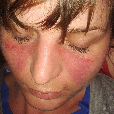
The most characteristic cutaneous feature of SLE is a malar (butterfly) rash, which is an erythematous rash of the nasal bridge and cheeks (Figure 1). The rash is often pruritic and spares the nasolabial folds.
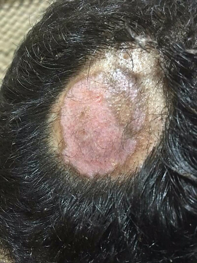
Other dermatological features of lupus are discoid erythematous plaques (discoid rash) that tend to occur in sun-exposed areas (Figure 2), oral ulcers, scarring alopecia, and photosensitivity.1
A common feature of SLE is musculoskeletal symptoms. Many patients with SLE will experience non-erosive arthritis of the small joints of the hands and feet bilaterally.2 Patients may also experience early morning stiffness characteristic of inflammatory arthritis.
Lupus has effects on most other major organ systems in the body. All three layers of the heart (pericardium, myocardium, and endocardium) can be inflamed in SLE. The most common cardiac manifestations are pericarditis with pericardial effusion, coronary artery disease, and rarely Libman-Sacks endocarditis.1
Pleuritis and pulmonary hypertension are common pulmonary manifestations of SLE, and central nervous system involvement can result in seizures and other neural abnormalities.2
Gastrointestinal involvement can lead to abdominal discomfort and vomiting that can result from enteritis, mesenteric vasculitis, or pancreatitis.
Lupus nephritis is one of the most serious consequences of SLE. The kidneys are involved in 50-70% of SLE cases. Lupus nephritis may present with haematuria, red blood cell casts, and proteinuria.2
In many patients with SLE, haematological phenomena are observed. Patients often present with anaemia, leucopenia, and thrombocytopenia.2 Antiphospholipid antibody syndrome is an associated condition that can present with lupus.
Antiphospholipid syndrome
Antiphospholipid syndrome is a clotting disorder characterised by the formation of antibodies to antiphospholipid (anticardiolipin, lupus anticoagulant, and anti-b2-microglobulin antibodies). It can be isolated or associated with SLE.
Treatment with hydroxychloroquine may be of help for patients with antiphospholipid syndrome. For those with thrombosis, additional anticoagulation is required.5
History
Typical symptoms of SLE include:
- Constitutional symptoms: fatigue, weight loss
- Musculoskeletal: arthralgia and joint stiffness
- Cardiac and pulmonary: chest pain, shortness of breath
- Other: gross haematuria, abdominal pain, dry eyes and mouth, skin and mucosal ulceration
Clinical examination
Typical findings on clinical examination may include:2,6
- Vitals: fever, hypertension
- Mucocutaneous: malar rash, discoid rash, oral ulcers, alopecia
- Other: leg oedema, point tenderness (associated fibromyalgia), Raynaud’s phenomenon
Differential diagnoses
Differentials for SLE include:1,2
- Rheumatoid arthritis
- Behcet’s disease
- Antiphospholipid antibody syndrome
- Sarcoidosis
- Rosacea
- Adult Still’s disease
- Systemic sclerosis
- Mixed connective tissue disease
- Parvovirus B19
- HIV
- Epstein-Barr virus
- Non-Hodgkin lymphoma
Investigations
Bedside investigations
Relevant bedside investigations include:
- Urinalysis: for haematuria and proteinuria
Laboratory investigations
Relevant laboratory investigations include:
- Haematology: FBC (may show anaemia, leucopenia, and thrombocytopenia)
- Biochemistry: will likely have an elevated ESR with a potentially normal CRP
- Immunology: anti-nuclear, anti-dsDNA, anti-smith, and antiphospholipid antibodies, and
complement levels (often low in SLE)
Serology in SLE
Anti-nuclear antibodies are the hallmark of SLE and have a sensitivity of over 95%.1
Following a positive anti-nuclear antibody result, screening for more specific SLE markers should be performed. These include anti-smith antibodies (>99% specificity), anti-dsDNA antibodies (95% specificity) and other antibodies known to cause rheumatological diseases.
Imaging
Use of imaging will depend on clinical presentation and may include:
- MRI brain
- Echocardiography
Diagnosis
The diagnosis of SLE is made using the Systemic Lupus Erythematosus International Collaborating Clinics Group (SLICC) Criteria.7
The diagnosis requires fulfilment of four criteria with at least one clinical and one immunological criterion fulfilled. Alternatively, lupus nephritis with the presence of anti-nuclear, anti-dsDNA antibodies will also meet the diagnostic criteria.
Management
Patients diagnosed with SLE will require education about their condition and should receive counselling on lifestyle changes that can reduce disease burden. This includes avoiding sun exposure and wearing high SPF sunscreen, regular exercise, and smoking cessation.6
Medical management
Medical management of SLE varies based on the severity of the disease.8
For mild disease, hydroxychloroquine and methotrexate are used to limit inflammation. NSAIDs may be added to further reduce inflammation and reduce pain where necessary. Low doses of prednisolone may also be used orally or as intra-articular injections. Methotrexate can be used for patients with skin and joint disease.
For moderate disease presentations, other immunosuppressants and higher doses of prednisolone may be used. These may include azathioprine, mycophenolate mofetil, and cyclosporine.
For severe cases and for instances of lupus nephritis or central nervous system involvement, high-dose oral prednisolone or intravenous methylprednisolone should be used in combination with immunosuppressants including cyclophosphamide. Biologics (e.g. rituximab, belimumab) may also be considered.
Complications
Complications that can arise from the systemic effects of SLE include:
- End-stage renal disease
- Atherosclerosis
- Recurrent miscarriage
- Permanent skin damage
- Blindness
Moreover, complications resulting from medication use are common in SLE (for example, infection because of heavy steroid use).1
Prognosis
The 10-year survival rate for SLE is around 90%.1 Lupus can contribute to early mortality from cardiovascular, renal, and infectious complications. Early diagnosis and proper management can successfully improve outcomes.
Key points
- SLE is an autoimmune disease caused by the deposition of immune complexes in tissues throughout the body.
- The cause of SLE is unknown. However, it is hypothesised that a failure to clear cellular waste leads to the loss of immune tolerance and an immune reaction against nuclear antigens.
- Risk factors include African-American ethnicity, female sex, and being of childbearing age.
- The most characteristic clinical feature of SLE is the malar rash involving the nose and cheeks.
- SLE has systemic effects throughout the body and can cause fatigue, arthritis, pericarditis, pleuritis, haematological disturbances, glomerulonephritis and central nervous system defects.
- The hallmark investigation in SLE is serology for anti-nuclear antibodies, which are required for classification as SLE under the American College of Rheumatology Classification Criteria.
- Management of SLE is with hydroxychloroquine, methotrexate and NSAIDs.
- SLE can lead to early mortality due to systemic complications including cardiovascular disease, end-stage renal disease and neurological sequelae.
Reviewer
Dr Grainne Murphy
Consultant Rheumatologist
Cork University Hospital
Editor
Dr Chris Jefferies
References
- Vaillant AAj, Goyal A, Bansal P, Varacallo M. Systemic Lupus Erythematosus. StatPearls. Updated in 2021. Available from: [LINK]
- Chee MM, Madhok R. Systemic Lupus Erythematosus. BMJ Best Practice. Reviewed in 2021. Available from: [LINK]
- Doktorinternet. Malar rash in a patient with systemic lupus erythematosus. Licence: [CC BY-SA]
- Mohammad2018. Discoid rash on head of a patient with systemic lupus erythematosus. Licence: [CC BY-SA]
- Bustamante JG, Goyal A, Bansal P, Singhal M. Antiphospholipid Syndrome. StatPearls. Updated in 2021. Available from: [LINK]
- Tidy C. Systemic Lupus Erythematosus. Edited in 2020. Available from: [LINK]
- Petri M. SLE Criteria. Systemic Lupus Erythematosus International Collaborating Clinics Group. Published in 2017. Available from: [LINK]
- Gordon C, Amissah-Arthur MB, Gayed M, Brown S, Bruce IN, D’Cruz D, et al. The British Society for Rheumatology guideline for the management of systemic lupus erythematosus in adults. Rheumatology (Oxford). 2018;57(1):e1-e45. Available from: [LINK]
