
- 📖 Geeky Medics OSCE Book
- ⚡ Geeky Medics Bundles
- ✨ 1300+ OSCE Stations
- ✅ OSCE Checklist PDF Booklet
- 🧠 UKMLA AKT Question Bank
- 💊 PSA Question Bank
- 💉 Clinical Skills App
- 🗂️ Flashcard Collections | OSCE, Medicine, Surgery, Anatomy
- 💬 SCA Cases for MRCGP
To be the first to know about our latest videos subscribe to our YouTube channel 🙌
Cranial nerve VI is the abducens nerve. To ‘abduce’ is to move away from the midline, and CN VI innervates the muscle of the eye involved in this movement. It is a somatic motor cranial nerve with a nucleus deep within the pontomedullary junction that emerges anteriorly from the brainstem. In this article, we discuss the course of the abducens nerve and the role it plays in eye movements.
Check out our summary of the cranial nerves here.
You can also check out our cranial nerve anatomy quiz here.
The abducens nucleus
The abducens nucleus is an ovoid aggregation of motor neuron cell bodies found in the dorsal pontomedullary junction. The nuclei are paired on the left and right side, and lie just lateral of the sagittal midline of the brainstem. The motor fibres of the facial nerve loop around the abducens nucleus, and these facial nerve fibres are the structure lying between the abducens nucleus and the fourth ventricle.
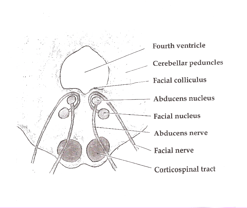
The efferent projections of the abducens nucleus form the abducens nerve, and these are described as exiting the abducens nucleus anteriorly and superiorly before coursing straight through the brainstem tissue and emerging in the cerebellopontine angle on the medial aspect of the ventral pontomedullary junction.
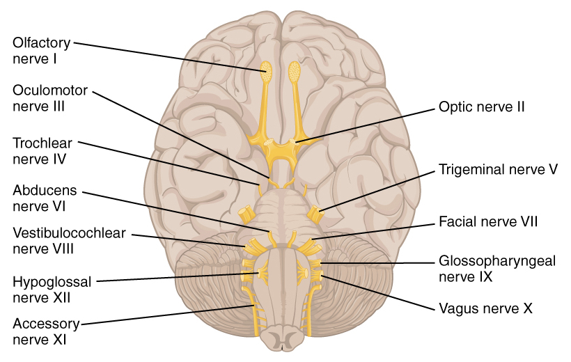
The intracranial abducens nerve
The abducens nerve fibres exit the brainstem next to the sagittal midline as a small-calibre nerve.
This nerve travels ventrally through the middle cranial fossa until it reaches the posterior clinoid process of the sphenoid bone. At the posterior clinoid process, the abducens nerve pierces a layer of dura mater. The abducens nerve then travels with the inferior petrosal sinus in Dorello’s canal, a small bony enclosure formed in the temporal bone.
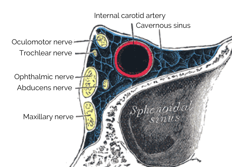
On leaving Dorello’s canal, the abducens nerve enters cavernous sinus, where it is found in close proximity to the internal carotid artery.
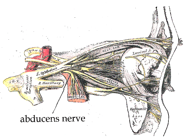
Upon exiting the cavernous sinus anteriorly, the abducens nerve travels towards a bundle of fibres formed by cranial nerves III, IV and V1, and exits the skulls through the superior orbital fissure.
The extracranial abducens nerve
As the abducens nerve passes through the superior orbital fissure, it moves along the medial surface of the lateral rectus muscle and segmentally sends motor fibres to it.
The lateral rectus muscle
The lateral rectus muscle is an extraocular muscle of the eye that originates from the annulus of Zinn, a round tendinous structure in the posterior orbital cavity that serves as a common attachment point for most of the extraocular muscles. The primary movement of the lateral rectus muscle is abduction, or lateral deviation, of the eye.
Clinical relevance – abducens nerve palsy
When examining the movements of the eye, cranial nerves III, IV and VI are all assessed together by drawing an ‘H’ shape in the air and asking patients to follow this with their eyes while keeping their head stationary.
Damage to either cranial nerve VI or the lateral rectus muscle will cause horizontal diplopia – double vision with images side-by-side. This is because the partial or complete palsy or weakness of the lateral rectus allows an unopposed pull of the medial rectus muscle in the same eye, pulling the visual axis medially.
Quite often, this presents as a significant strabismus (squint) that may be alleviated by head-turning to achieve binocular vision or patching of the eye to occlude the ‘second’ image.
Key points
- CN VI is the abducens nerve
- It originates in the pontomedullary region
- It provides general somatic efferent fibres for eye abduction
- It innervates the lateral rectus muscle
- It passes through the superior orbital fissure of the skull
References
Reference texts
- Sinnatamby, C. S. (2011). Last’s Anatomy, International Edition: Regional and Applied. Elsevier Health Sciences.
- Moore, K. L., Dalley, A. F., & Agur, A. M. (2013). Clinically oriented anatomy. Lippincott Williams & Wilkins.
- Nolte, J. (2002). The human brain: an introduction to its functional anatomy.
- Snell, R. S. (2010). Clinical neuroanatomy. Lippincott Williams & Wilkins.
Reference images
- Patrick J. Lynch. License: [CC BY]. Modified by Dr Lewis Potter.
- Btarski. License: [CC BY-SA]
- OpenStax. License: [CC BY]
- Henry Vandyke Carter. License: [Public domain]
- Btarski. License: [CC BY-SA]
