
- 📖 Geeky Medics OSCE Book
- ⚡ Geeky Medics Bundles
- ✨ 1300+ OSCE Stations
- ✅ OSCE Checklist PDF Booklet
- 🧠 UKMLA AKT Question Bank
- 💊 PSA Question Bank
- 💉 Clinical Skills App
- 🗂️ Flashcard Collections | OSCE, Medicine, Surgery, Anatomy
- 💬 SCA Cases for MRCGP
To be the first to know about our latest videos subscribe to our YouTube channel 🙌
This article will discuss the components of the heart’s conduction system including their anatomy and clinical significance.
The heart has two main types of cells:1
- Conducting cells: generate and propagate electrical impulses.
- Contractile (muscle) cells: contract following receipt of electrical impulses. These cells can also propagate and, on occasion, generate electrical impulses.
Both conducting and specialised muscle cells form the hearts conduction system and orchestrate cardiac contraction. The electrical system is intrinsic to the heart meaning that contraction can persist in the absence of neuronal input.
Components of the cardiac conduction system
The cardiac conduction system involves the spread of electrical activity from the sinoatrial node, to the atrioventricular node, down the bundle of His and along the Purkinje fibres. As the electrical activity spreads along the heart’s conduction system it initiates myocardial contraction in the surrounding myocardial tissue.
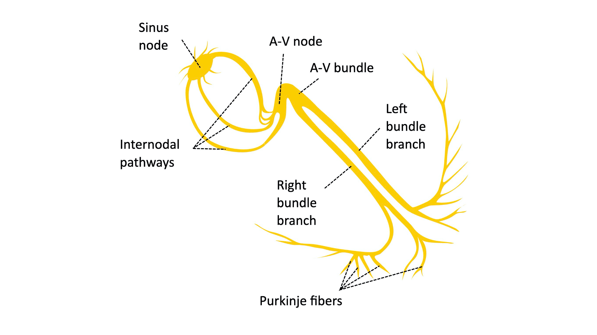
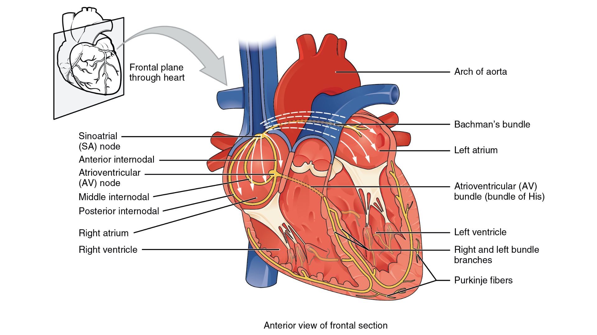
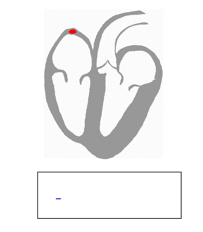
The sinoatrial node (SAN)
- The sinoatrial node (SAN) is of a group of cells found high up in the right atrium close to its junction with the superior vena cava.1,4,5
- The SAN functions as the heart’s intrinsic pacemaker, regulating heart rate.
- The SAN spontaneously generates electrical impulses which are transmitted to the right and left atrium.
- These electrical impulses stimulate the atrial myocardium to contract.
- Atrial muscle conducts relatively fast (0.5m/sec).8
Atrioventricular node (AVN)
- The atrioventricular node (AVN) is a group of specialised cells situated in the atrioventricular septum just above the coronary sinus ostium.1,4,5
- The AVN receives electrical impulses from the atria and then transmits the electrical impulse from the atria to the ventricles.
- The AVN has a slower conduction velocity (0.05m/sec) than the atria, allowing maximal ventricular filling prior to contraction.8
The bundle of His
- The bundle of His is a collection of heart muscles cells specialised for electrical conduction. These cells receive input from the AVN.1,4,5
- The bundle of His branches into the left and right bundle branches which travel down the interventricular septum.
- Branches of the bundle of His propagate impulses to the left and right ventricles respectively.
- Each branch terminates as several Purkinje fibres.
Purkinje fibres
- Purkinje fibres are situated in the subendocardium.1,4,5
- They transmit the wave of electricity to the ventricular myocardium.
- This wave of electricity results in ventricular contraction.
Summary
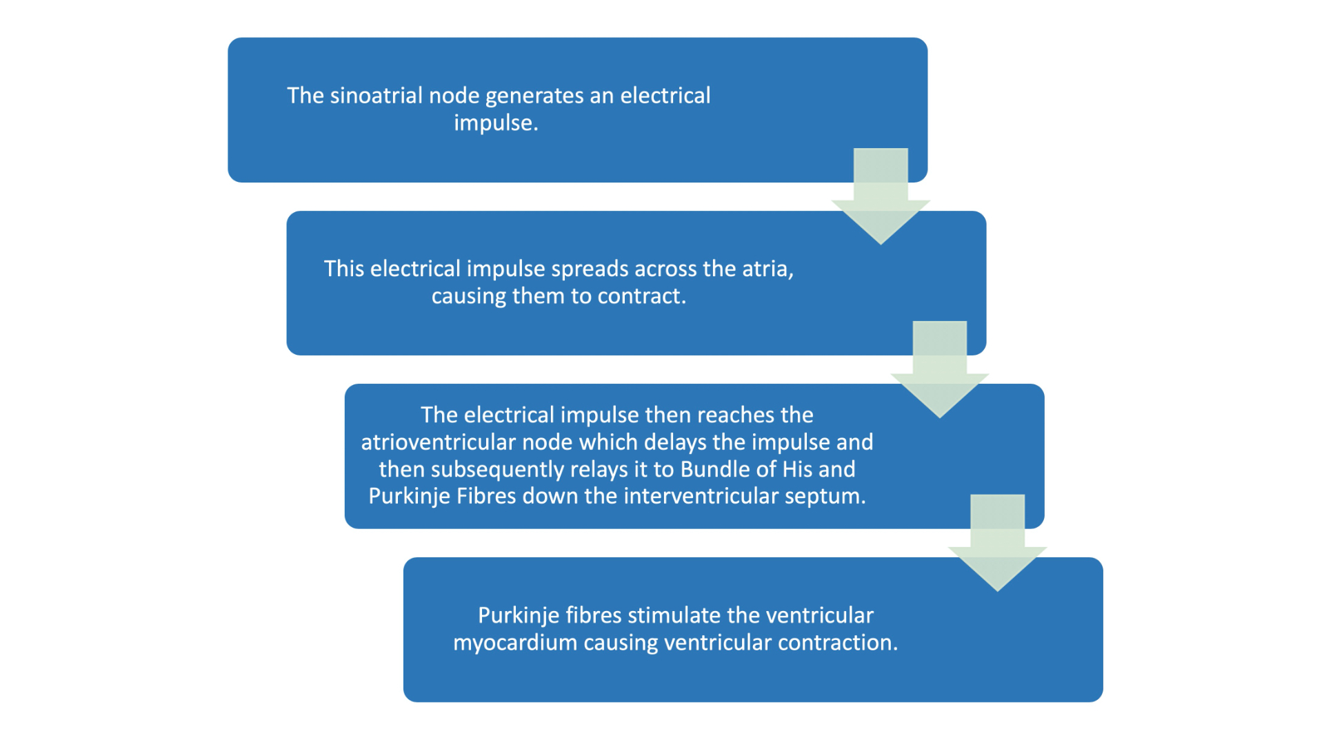
Below is a summary of the key steps involved in the cardiac conduction cycle.
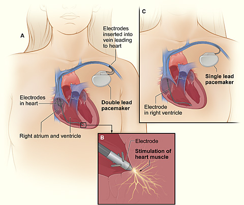
Clinical significance: Artificial pacemakers
An artificial pacemaker is a medical device that is able to monitor and regulate heart rate and rhythm. The artificial pacemaker is able to generate electrical impulses that can trigger myocardial contraction, mimicking the function of the heart’s intrinsic conduction system.
Artificial pacemakers are often used to regulate heart rate and rhythm in patients with atrioventricular block and sinus node dysfunction.7
References
- The University of Minnesota. Overview of Cardiac Conduction. Published in 2011. [LINK].
- Madhero88. Cardiac conduction system. Licence: CC BY-SA. Available from: [LINK]
- OpenStax CNX. Cardiac Muscle and Electrical Activity. Published in 2014. [LINK].
- Park D, Fishman G. The Cardiac Conduction System. Published in 2011. [LINK].
- National Heart, Lung, and Blood Institute. How the heart works. Published in 2011. [LINK].
- Kalumet. Heart’s conduction system. Licence: CC BY-SA. Available from: [LINK]
- Dalia T, Amr S. Pacemaker Indications. Published in 2020. [LINK].
- Clinical ECG Interpretation e-Book. Clinical electrocardiography and ECG interpretation. [LINK].
- Klabunde R. Normal Impulse Conduction. Published in 2012. [LINK].
