
- 📖 Geeky Medics OSCE Book
- ⚡ Geeky Medics Bundles
- ✨ 1300+ OSCE Stations
- ✅ OSCE Checklist PDF Booklet
- 🧠 UKMLA AKT Question Bank
- 💊 PSA Question Bank
- 💉 Clinical Skills App
- 🗂️ Flashcard Collections | OSCE, Medicine, Surgery, Anatomy
- 💬 SCA Cases for MRCGP
To be the first to know about our latest videos subscribe to our YouTube channel 🙌
Introduction
The pineal gland (also called the epiphysis cerebri) is a small, pinecone-shaped neuroendocrine organ in the diencephalon region of the brain (the part of the brain just above the brainstem).
The main function of the pineal gland is the regulation of the circadian rhythm (thus directing the sleep-wake cycle) by secretion of the hormone melatonin.
This article will explore the anatomy of the pineal gland, its vascular supply and its function.
Location
The pineal gland is found in the epithalamus (part of the diencephalon) and is approximately the size of a grain of rice.1
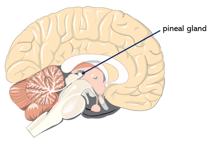
It is found between the two cerebral hemispheres, projecting from the posterior wall of the third ventricle. The pineal gland lies between the two superior colliculi (midbrain structures that play an important role in the visual pathway) and beneath the splenium of the corpus callosum.1
The location of the pineal gland within the brain is shown in Figure 1.
The pineal stalk connects the pineal gland to the rest of the brain. The pineal stalk is divided into the superior lamina and inferior lamina. Part of the third ventricle extends posteriorly into the small space between the two laminae of the pineal stalk, called the pineal recess.2
The pineal gland is a circumventricular organ, meaning that the capillaries within the pineal gland are highly permeable with no blood-brain barrier.3
With increasing age, the pineal gland can become calcified, and a calcified pineal gland may be visible on a skull X-ray.4
Histology
The main cell type in the pineal gland is the pinealocyte, a neuron-like cell type that secretes the hormone melatonin into the highly permeable capillaries within the pineal gland. Pinealocytes are arranged into lobules by connective tissue septa.
The pineal gland also contains other cells such as interstitial cells and perivascular phagocytes.
The gland is surrounded by a capsule of pia mater.1
Vascular supply
The pineal gland has a rich blood supply, with only the kidney receiving a greater blood supply relative to its mass.5
The main blood supply is from the posterior choroidal arteries (branches of the posterior cerebral arteries).6
The venous drainage of the pineal gland is to the cerebral vein of Galen, via the lateral pineal veins.6
Function
The main function of the pineal gland is the regulation of the sleep-wake cycle through the production of the hormone melatonin (a serotonin-derived hormone).
The blood concentration of melatonin rises at night and falls in the day, as seen in Figure 2.7
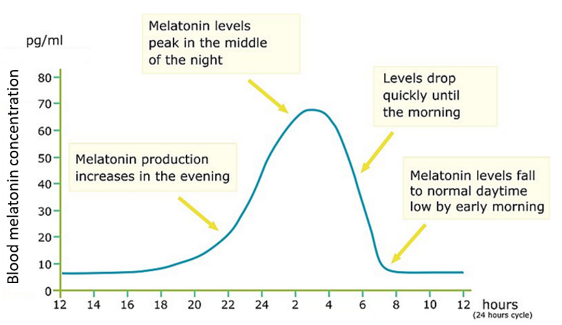
This day-night pattern is directed by light stimulating the retina. Information about the amount of light stimulating the retina is communicated to the pineal gland through the retinohypothalamic tract (via the suprachiasmatic nucleus of the hypothalamus, paraventricular nucleus of the hypothalamus and the intermediolateral nucleus of the spinal cord).7
This feature of the pineal gland led to its historical name of the ‘third eye’.
It has also been found that melatonin plays an important role in the reproductive behaviour of animals that are seasonal breeders.8 It has further been suggested that melatonin may play a role in the regulation of puberty in humans, although this idea is currently not well established and requires further investigation.9,10
Clinical relevance: Pineal gland tumours
Tumours called pinealomas can develop in the pineal gland. These tumours can increase intracerebral pressure, leading to symptoms such as headache and nausea.11
Pinealomas can compress the nearby superior colliculi, leading to Parinaud’s syndrome (a condition where the patient is unable to move their eyes up and down).12
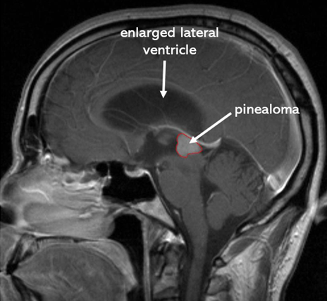
Furthermore, pinealomas can block the flow of cerebrospinal fluid through the cerebral aqueduct, which can lead to hydrocephalus (a build-up of cerebrospinal fluid within the brain’s ventricles), as seen in Figure 3.11
Key points
- The pineal gland is a small neuroendocrine organ in the diencephalon region of the brain
- The main cell type in the pineal gland is the pinealocyte
- Its main function is the production of the hormone melatonin, which regulates the sleep-wake cycle
- The pineal gland receives information about light levels in the environment from the retina via the retinohypothalamic tract
- The pineal gland may have other important endocrine functions, such as regulation of the onset of puberty, which require further investigation
Reviewer
Dr Kasra Bahadori
Editor
Dr Chris Jefferies
References
- Møller M, Baeres FM. The anatomy and innervation of the mammalian pineal gland. Cell and Tissue Research. 2002;309(1):139-50.
- Shenoy SS. Neuroanatomy, Ventricular System. StatPearls. 2021. Treasure Island (FL): StatPearls Publishing
- Gross PM, Weindl A, Knigge KM. Peering through the Windows of the Brain. Journal of Cerebral Blood Flow & Metabolism. 1987;7(6):663-72.
- Zimmerman RA, Bilaniuk LT. Age-Related Incidence of Pineal Calcification Detected by Computed Tomography. Radiology. 1982;142.
- Arendt J. Melatonin and the pineal gland: influence on mammalian seasonal and circadian physiology. Rev Reprod. 1998;3(1):13-22.
- Duvernoy HM, Parratte B, Tatu L, Vuillier F. The human pineal gland: relationships with surrounding structures and blood supply. Neurol Res. 2000;22(8):747-90.
- Claustrat B, Leston J. Melatonin: Physiological effects in humans. Neurochirurgie. 2015;61(2-3):77-84.
- Luboshitzky R, Lavie P. Melatonin and sex hormone interrelationships–a review. J Pediatr Endocrinol Metab. 1999;12(3):355-62.
- Silman RE, Leone RM, Hooper RJ, Preece MA. Melatonin, the pineal gland and human puberty. Nature. 1979;282(5736):301-3.
- Aleandri V, Spina V, Ciardo A. [The role of the pineal body in the endocrine control of puberty]. Minerva Ginecol. 1997;49(1-2):43-8.
- Patel S, Rahmani B, Gandhi J, Seyam O, Joshi G, Reid I, et al. Revisiting the pineal gland: a review of calcification, masses, precocious puberty, and melatonin functions. Int J Neurosci. 2020;130(5):464-75.
- Feroze KB, Patel BC. Parinaud Syndrome. StatPearls. Treasure Island (FL): StatPearls Publishing. Copyright © 2021, StatPearls Publishing LLC.; 2021.
Image references
- Figure 1. Tdvorak. Pineocytoma, MRI T1 with contrast, sagittal. Licence: [CC BY-SA 3.0]. Adapted by Alex Fleet.
- Figure 2. Rechargeenergy. Melatonin production in a 24-hour cycle. Licence: [CC BY-SA 4.0]. Adapted by Alex Fleet.
- Figure 3. Gordon Dylan Johnson. Brain cross section, sagittal. Licence: [CC0 1.0]. Adapted by Alex Fleet.
