
- 📖 Geeky Medics OSCE Book
- ⚡ Geeky Medics Bundles
- ✨ 1300+ OSCE Stations
- ✅ OSCE Checklist PDF Booklet
- 🧠 UKMLA AKT Question Bank
- 💊 PSA Question Bank
- 💉 Clinical Skills App
- 🗂️ Flashcard Collections | OSCE, Medicine, Surgery, Anatomy
- 💬 SCA Cases for MRCGP
To be the first to know about our latest videos subscribe to our YouTube channel 🙌
Introduction
The pituitary gland is the master gland of the endocrine system, controlled by the hypothalamus.
It is split into two lobes, the anterior lobe and the posterior lobe. Each lobe secretes different hormones in response to signals from the hypothalamus, however, only the anterior lobe produces its own hormones.1
Anatomical location and relations

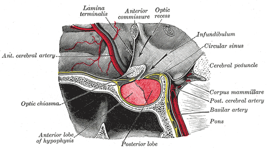
The pituitary gland is located within a depression of the sphenoid bone, called the pituitary (hypophyseal) fossa or sella turcica. It is connected to the hypothalamus via the pituitary stalk (infundibulum), which serves not only to connect them physically but also allowing passage of nerve fibres from the hypothalamus.3
Due to the pituitary gland’s location, it has important and complex anatomical relations:
- Inferiorly: sphenoid bone and the sphenoid sinus
- Superiorly: hypothalamus, pituitary stalk, optic chiasm, circle of Willis, diaphragma sellae (a fold of dura matter that covers the pituitary stalk)
- Laterally: cavernous sinus containing the internal carotids, oculomotor nerve, trochlear nerve, ophthalmic and maxillary divisions of the trigeminal nerve, and abducens nerve
- Anteriorly: anterior intercavernous sinus
- Posteriorly: posterior intercavernous sinus, dorsum sellae
Clinical significance: pituitary adenoma
Anterior lobe adenomas comprise about 10% of all intracranial tumours (posterior lobe adenomas do not occur).
These tumours can manifest clinically secondary to the secretion of excess hormone and compression of local structures.
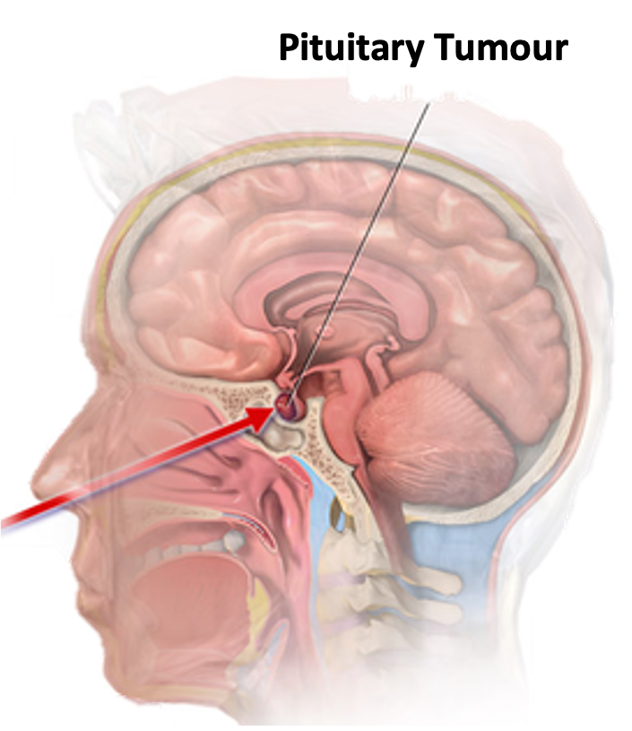
As these tumours grow, they can compress the optic chiasm which lies superior to the pituitary gland. This can cause visual field defects, notably bitemporal hemianopia, which if left untreated can lead to permanent visual field loss.5
One method of treatment involves surgery, approaching through the nose, and through the sphenoid sinus and sphenoid bone, a transsphenoidal approach (Figure 3). This can be performed with a microscope or endoscope.6
Posterior lobe
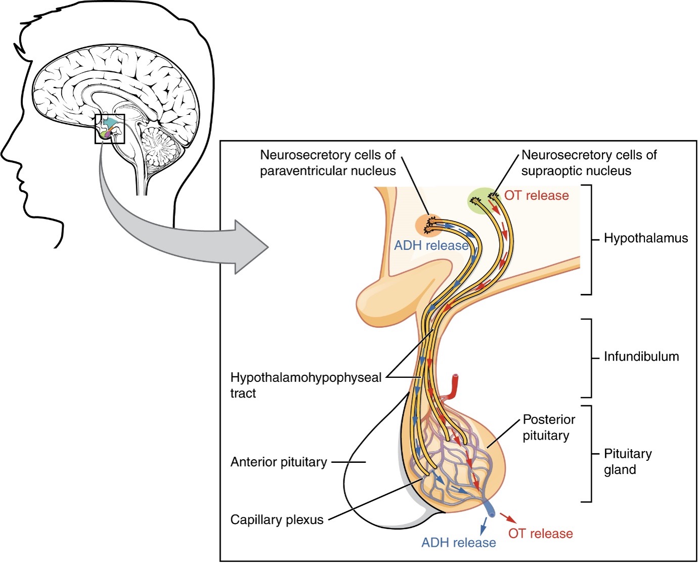
The posterior lobe (neurohypophysis) is formed from neural tissue and is an extension of the neurosecretory fibres of the paraventricular and supraoptic nuclei of the hypothalamus.
The posterior pituitary does not manufacture hormones. Instead, the posterior pituitary stores and secretes the hormones produced by the hypothalamus, specifically oxytocin (produced by the paraventricular nuclei) and antidiuretic hormone (produced by the supraoptic nuclei). In response to signals from the hypothalamus, these hormones are released into the bloodstream.1
Oxytocin stimulates uterine contractions and dilation of the cervix with a positive feedback loop from cervical stretching. It is also necessary for the milk ejection reflex and thought to contribute to parent-neonate bonding.1
Antidiuretic hormone (ADH) helps to control homeostasis of fluid in the body via increasing water reabsorption in the kidneys, this forms a negative feedback loop.1
Anterior lobe
The anterior lobe (adenohypophysis) is formed of glandular tissue split into three regions:
- Pars distalis
- Pars intermedia
- Pars tuberalis
The pars distalis is found in the most anterior portion of the anterior lobe and forms much of the anterior lobe.
The pars intermedia lies between the pars distalis and the posterior lobe, whilst the pars tuberalis wraps the pituitary stalk.4
The anterior lobe manufactures its hormones via endocrine cells. These cells are controlled via hypothalamic hormones that stimulate or inhibit release via secretion into a capillary plexus in the infundibulum.
The anterior lobes hormones are listed below, along with the hypothalamic hormones that control their release.1
Table 1. Hormones in relation to the hypothalamus and anterior pituitary.1,3
|
Hypothalamic releasing/inhibiting hormones (RH/IH) |
Anterior lobe hormone |
Target |
Effect |
|
Corticotropin-RH |
Adrenocorticotropic hormone (corticotropin) |
Adrenal glands |
Stimulate production of glucocorticoids, which regulate metabolism and stress response |
|
Gonadotropic RH |
Follicle-stimulating hormone (FSH) and luteinising hormone (LH) |
Reproductive system |
FSH stimulates maturation/production of sex cells LH stimulates sex hormones by the gonads |
|
Growth hormone-RH and Growth hormone-IH (somatostatin) |
Growth hormone (somatotropin) |
Liver, bone, muscles |
Induce production of insulin-like growth factors, which stimulate body growth and higher metabolic rate |
|
Prolactin-RH and prolactin-IH (dopamine) |
Prolactin |
Mammary glands |
Promotes milk production |
|
Thyrotropin-RH |
Thyrotropin (thyroid-stimulating hormone) |
Thyroid gland |
Stimulates the release of thyroid hormones which regulate metabolism |
Vasculature
The pituitary gland is supported by a rich vascular supply due to the high energy needs of the endocrine cells, with capillary beds and veins acting as the ductal system for hormones.3
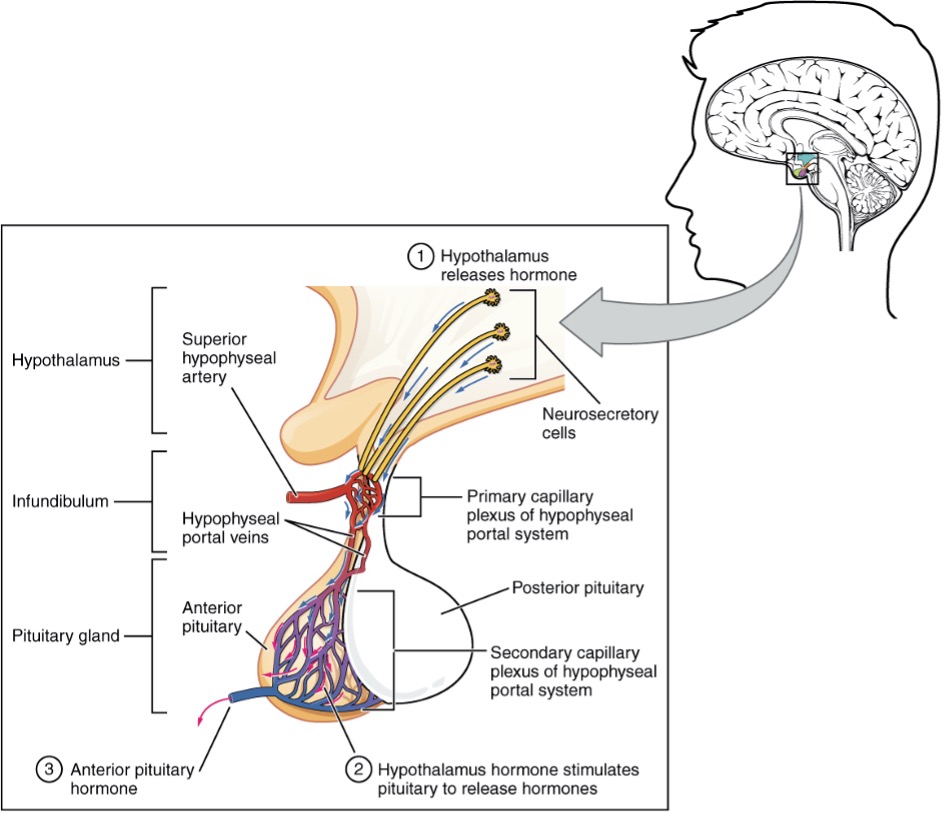
The arterial supply of the pituitary gland originates from the internal carotids, which give out hypophyseal branches.
One set of branches, including the superior hypophyseal artery, supplies a primary capillary bed in the wall of the pituitary stalk, which drains into portal vessels. These portal vessels drain and form a second capillary bed in the anterior lobe bathing the endocrine cells. This secondary capillary bed eventually drains into the cavernous sinus.3
The posterior lobe receives a direct supply from the inferior hypophyseal arteries, draining into the cavernous sinus.
The cavernous sinus transports the hormones secreted by both the anterior and posterior pituitary away and towards the target cells.3
Key points
- The pituitary gland is an endocrine organ with vital roles in homeostasis. It sits within the hypophyseal fossa of the sphenoid bone.
- The pituitary gland is split into two parts: anterior lobe and posterior lobe.
- The anterior lobe produces, stores, and secretes its own hormones: adrenocorticotropic hormone (ACTH), follicle-stimulating hormone (FSH), luteinising hormone (LH), growth hormone (GH), prolactin (PRL), thyrotropin (TSH).
- The anterior lobe is mediated by the hypothalamus via inhibiting and releasing hormones, which travel via the capillary bed and portal system.
- The posterior lobe stores and secretes hormones produced by neuron fibres from the supraoptic and paraventricular nuclei of the hypothalamus, specifically oxytocin and antidiuretic hormone.
- The blood supply originates from the internal carotids giving rise to hypophyseal branches with the arterial supply of the anterior lobe involving the superior hypophyseal artery and the posterior pituitary receiving a direct supply from the inferior hypophyseal arteries.
- The hormones and vasculature drain into the cavernous sinus.
Reviewer
Mr Ian Coulter
Consultant Neurosurgeon
Royal Victoria Infirmary
Editor
Dr Chris Jefferies
References
- OpenStax. 17.3 The Pituitary Gland and Hypothalamus – Anatomy and Physiology. 2021. Available from: [LINK]
- OpenStax. Hypothalamic–Pituitary Complex. Licence: [CC-BY]
- Mtui, Estomih, et al. Fitzgerald’s Clinical Neuroanatomy and Neuroscience. Eighth ed., 2021
- Gray’s Anatomy. Pituitary fossa. Licence: [Public domain]
- Xiu, Philip., and Atul, Anand. Pathology. 4th ed., Elsevier Mosby, 2012
- Laws, Edward R., and Giuseppe. Lanzino. Transsphenoidal Surgery. 1st ed., Saunders, 2010
- BruceBlaus. Adapted by Geeky Medics. Pituitary Tumour Removal. License: [CC BY-SA]
- OpenStax. Posterior Pituitary. Licence: [CC-BY]
- Mancall, Elliott L., et al. Gray’s Clinical Neuroanatomy: the Anatomic Basis for Clinical Neuroscience. Saunders, 2011
- OpenStax. Anterior Pituitary. Licence: [CC-BY]
