
- 📖 Geeky Medics OSCE Book
- ⚡ Geeky Medics Bundles
- ✨ 1300+ OSCE Stations
- ✅ OSCE Checklist PDF Booklet
- 🧠 UKMLA AKT Question Bank
- 💊 PSA Question Bank
- 💉 Clinical Skills App
- 🗂️ Flashcard Collections | OSCE, Medicine, Surgery, Anatomy
- 💬 SCA Cases for MRCGP
To be the first to know about our latest videos subscribe to our YouTube channel 🙌
Overview
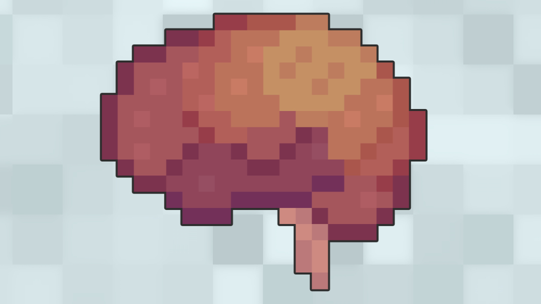
The cerebellum is a portion of the central nervous system (CNS) that is only concerned with the control of movement.
This article describes the gross anatomy of the cerebellum, the functional regions of the cerebellum and some of the associated CNS loops/pathways. The cerebellum is a complicated structure and so this article only highlights key anatomy and afferent/efferent connections.
For more information, watch the Geely Medics anatomy tutorial on the cerebellum to see the gross anatomy of the cerebellum in 3D and to hear tips on how to remember cerebellar signs.
Location, relations and structure
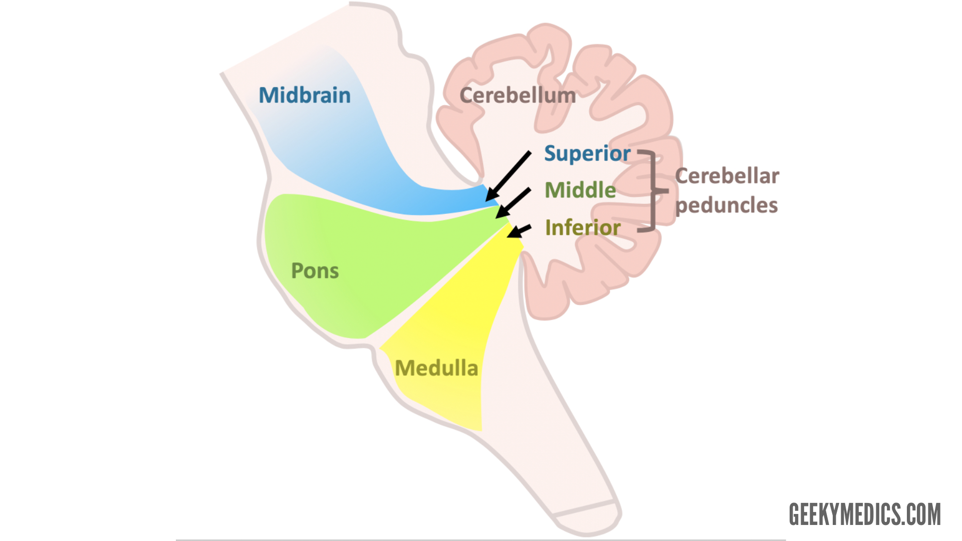
The cerebellum is a portion of the hindbrain located within the posterior cranial fossa of the skull, caudal to the cerebrum and tentorium cerebelli, and dorsal to the brainstem.
The cerebellum is directly connected to the midbrain, pons and medulla by the superior, middle and inferior cerebellar peduncles respectively (Figure 1).
Structure
Gross features
The cerebellum is divided into left and right hemispheres by a midline vermis. The external surface consists of a series of folds (folia) and fissures that gives the cerebellum a convoluted appearance.
Some of the fissures are used as landmarks to divide the cerebellum into lobes:
- The primary fissure divides the smaller anterior and larger posterior lobes
- The posterolateral fissure demarcates the flocculonodular lobe
- The horizontal fissure divides the cerebellum into superior and inferior portions (this division is not thought to be functionally significant)
Internal structure
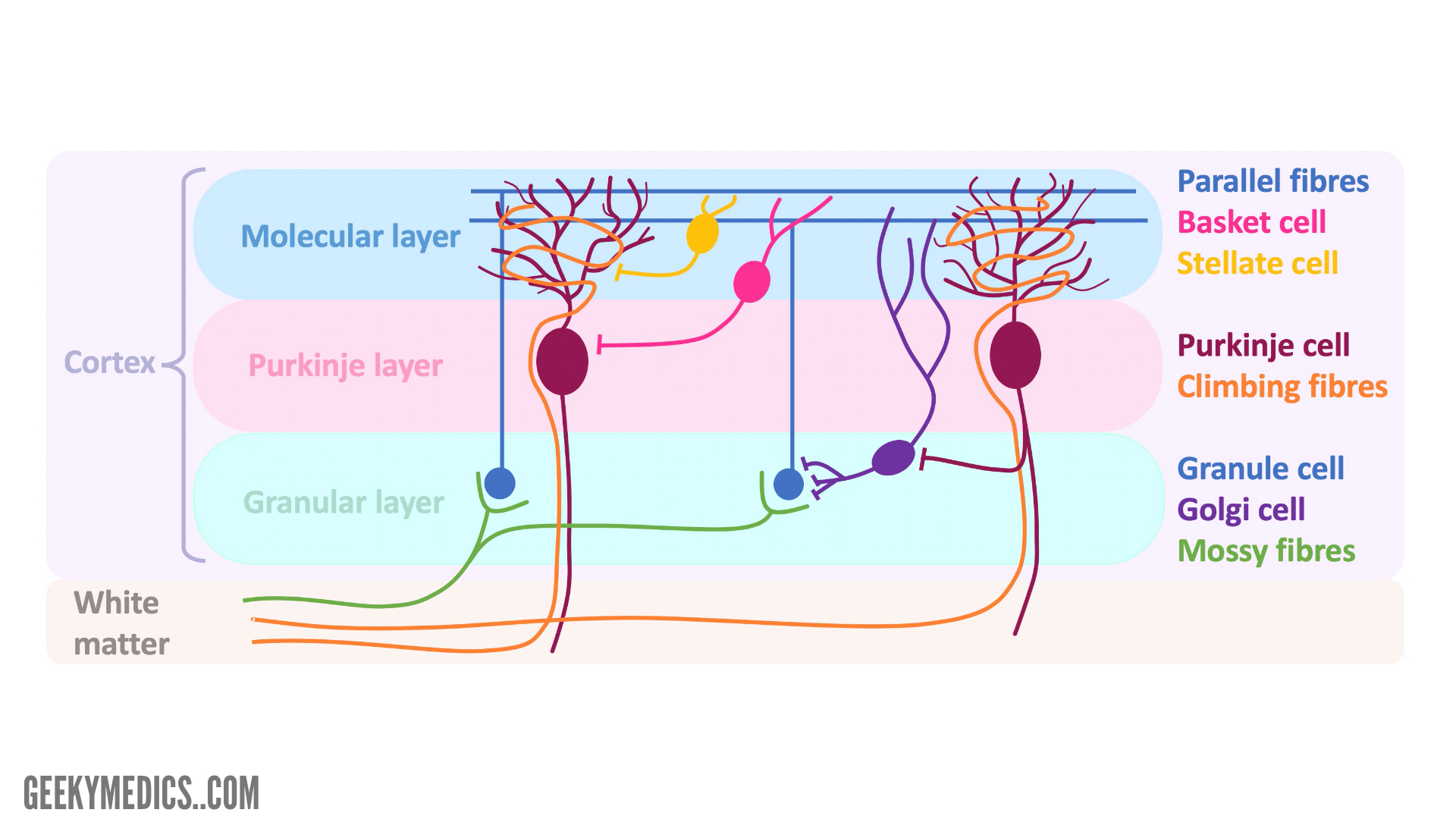
The arrangement of the internal structure of the cerebellum is not too dissimilar from the cerebrum (i.e. outer layer of grey matter that forms the cortex and an inner mass of white matter formed by fibre tracts).
The cerebellar nuclei are islands of grey matter located deep within the white matter that have important connections with the cerebellar cortex and some brainstem nuclei. The cerebellar nuclei include the fastigial, globose and emboliform (often described collectively as the interposed nuclei), and dentate.
Before looking at the histological layers of the cortex, it is worth studying the table and diagram below to gain an understanding of the cells and fibres associated with the cerebellum.
Table 1. The cells and fibres associated with the cerebellum.
| Cell/fibres | Location | Connections/functions |
| Granule cells/parallel fibres |
|
|
| Purkinje cells |
|
|
| Golgi cells |
|
|
| Stellate cells |
|
|
| Basket cells |
|
|
| Mossy fibres |
|
|
| Climbing fibres |
|
|
Summary of cerebellar cortical layers
There are three cortical layers in the cerebellum.
Granular layer
The granular layer is comprised of granule cells that receive afferent mossy fibres originating from the spinal cord and brainstem. The Golgi cells are also located in this layer and synapse onto the dendrites of granule cells. Golgi cells act to limit the output of mossy fibres and therefore granule cells.
However, Golgi cells also receive input from strongly stimulated Purkinje cells to limit the inhibitory activity of Golgi cells. This allows for prolonged excitation of strongly stimulated Purkinje cells.
Purkinje layer
The purkinje layer consists of the cell bodies of the Purkinje cells. The axons of Purkinje cells are the only axons to leave the cerebellar cortex to synapse at the deep cerebellar and brainstem nuclei. These cells receive input from climbing fibres from the contralateral inferior olivary nucleus or parallel fibres.
Molecular layer
The molecular layer primarily consists of the dendrites of the Purkinje fibres and axons (i.e. parallel fibres) of the granule cells. Within the molecular layer, the axons of the granule cells divide in a T-like fashion to form the parallel fibres that run parallel to the cerebellar fissures. The parallel fibres synapse onto the dendrites of the Purkinje cells. Other cell types included within this layer include stellate and basket cells.
Functional anatomy of the cerebellum
There is little agreement on the discrete functional regions of the cerebellum and the associated functions, and so there is often overlap. Therefore, the table and images below provide broad overviews that account for the variety often described.
Table 2. Functional anatomy of the cerebellum.
| Division | Possible/identified functional regions | Cerebellar nuclei | Peduncles | Functions |
|
Vestibulocerebellum
|
|
|
|
|
|
Spinocerebellum
|
|
|
|
|
|
Cerebrocerebellum
|
|
|
|
|
Cerebellar loops
This portion of the article will focus on the loops associated with the functional portions of the cerebellum to demonstrate how they influence movement.
The cerebellum typically acts on the ipsilateral side of the body, though the vestibulocerebellum has bilateral influence. Consequently, fibres associated with the cerebellar loops decussate a number of times, which can be confusing.
Vestibulocerebellum
The vestibulocerebellum is concerned with conjugate eye movements and balance control:
- The vestibular nerve synapses on the vestibular nuclei. Fibres project to the flocculonodular lobe and vermis via the inferior cerebellar peduncle.
- Cortical efferent fibres (Purkinje fibres) project to the fastigial nucleus.
- Fibres pass to the reticular and vestibular nuclei and the gaze centres within the brainstem via the inferior cerebellar peduncle. A significant portion of fastigial efferent fibres decussate to the contralateral brainstem and therefore have a bilateral influence.
- Information associated with gaze control is mediated by the medial longitudinal fasciculus.
- Information associated with balance control descends in the spinal cord via reticulospinal and vestibulospinal tracts to extensor motor neurons in the spinal cord.

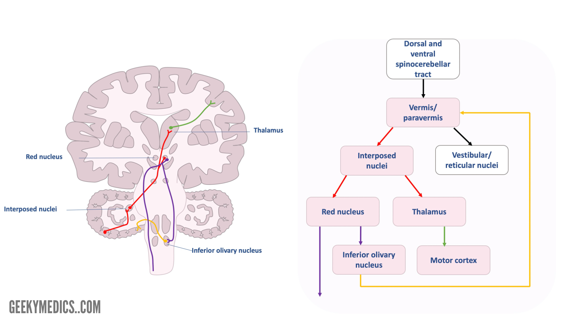
Spinocerebellum
The spinocerebellum is concerned with posture and the monitoring and correcting of the motor activity of the limbs:
- Afferents from dorsal and ventral spinocerebellar tracts, which carry information from muscle and joint receptors, enter the cerebellum via inferior and superior peduncles respectively.
- Both tracts project to the ipsilateral vermis and paravermal cortex.
- Information regarding posture control passes to ipsilateral reticular and vestibular nuclei via the superior cerebellar peduncle. Fibres then descend the spinal cord as reticulospinal and vestibulospinal tracts.
- Purkinje fibres also project to interposed nuclei and then pass within the superior cerebellar peduncle towards the contralateral red nucleus and thalamus.
- Fibres from the red nucleus pass caudally within the brainstem. Some fibres project to the ipsilateral inferior olivary nucleus and others decussate in the midbrain as the rubrospinal tract. Connections to the inferior olivary nucleus travel as olivocerebellar fibres to the contralateral cerebellar cortex via inferior cerebellar peduncle.
- Fibres that project to the thalamus continue to the motor cortex as thalamocortical fibres.
Cerebrocerebellum
The cerebrocerebellum is concerned with the planning and initiation of movement and motor learning.
The loop described below is associated with motor planning and initiation. Motor learning occurs as a consequence of ongoing activity of the cerebellar parallel fibres (known as depression):
- Corticopontine fibres from the motor cortices project to the deep pontine nuclei.
- Pontocerebellar fibres decussate and enter the cerebellum via the middle cerebellar peduncle to terminate in the cortex of cerebellar hemispheres.
- Purkinje cells project to the dentate nucleus.
- Fibres leave the cerebellum via the superior cerebellar peduncle and project to the contralateral red nucleus and thalamus. Fibres from the thalamus then pass back to the motor cortices.

Illustrator
Dr Hannah Hubbard
