- 📖 Geeky Medics OSCE Book
- ⚡ Geeky Medics Bundles
- ✨ 1300+ OSCE Stations
- ✅ OSCE Checklist PDF Booklet
- 🧠 UKMLA AKT Question Bank
- 💊 PSA Question Bank
- 💉 Clinical Skills App
- 🗂️ Flashcard Collections | OSCE, Medicine, Surgery, Anatomy
- 💬 SCA Cases for MRCGP
To be the first to know about our latest videos subscribe to our YouTube channel 🙌
Assessment of colour vision using Ishihara plates can sometimes appear in OSCEs. This guide provides a brief step-by-step guide to using Ishihara plates to assess colour vision, with an included video demonstration.
Introduction
Wash your hands and don PPE if appropriate.
Introduce yourself to the patient including your name and role.
Confirm the patient’s name and date of birth.
Briefly explain what the examination will involve using patient-friendly language.
Gain consent to proceed with the examination.
Position the patient sitting on a chair.
Ask if the patient has any pain before proceeding.
Colour vision assessment
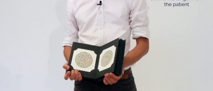
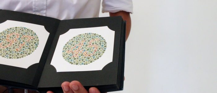
Colour vision can be assessed using Ishihara plates, each containing a coloured disc of dots. Within the pattern of each disc are dots which form a number or shape visible to those with normal colour vision. Those with a red-green colour vision defect may see some plates differently, be unable to read certain plates, or even see numbers on plates that appear blank to those with normal colour vision.
Although the test was originally designed for diagnosing and subtyping individuals with red-green colour blindness, these plates can be used in the acute setting to identify acute optic neuropathy (such as optic neuritis).
The Ishihara plates come in the original 36-plate version or the shortened 24-plate version. Only the first 17 plates of the 36-plate version and the first 13 plates of the 24-plate version should be used in most assessments. Further plates are meant for individuals who cannot read numbers or determine the extent of red-green blindness.
An easy way to remember this is to stop after the last plate with a number (which reads “73”).
How to use Ishihara plates
If the patient usually wears glasses for reading, ensure these are worn for the assessment.
1. Ask the patient to cover one of their eyes.
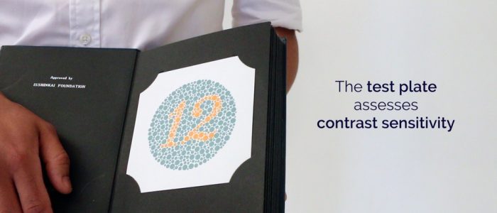
2. Then, ask the patient to read the numbers on the Ishihara plates. The first page is usually the ‘test plate’, which does not test colour vision but assesses contrast sensitivity. If the patient cannot read the test plate, you should document this.
3. If the patient can read the test plate, you should move through the Ishihara plates, asking the patient to identify the number on each. Stop at the plate that reads “73” (the last plate with a number).
4. Once the test is complete, document the number of plates the patient identified correctly, including the test plate (e.g. 17/17). Also, make a note of the reading speed for each eye.
5. Repeat the assessment on the other eye.
To complete the examination…
Explain to the patient that the examination is now finished.
Thank the patient for their time.
If mydriatic drops were instilled, remind the patient they cannot drive for the next 3-4 hours until their vision has returned to normal.
Dispose of PPE appropriately and wash your hands.
Summarise your findings
Suggest further assessment and investigations
All of the following further assessments and investigations are dependent on the patient’s presenting complaint, and in most cases, none of them would need to be performed:
- Complete visual assessment: including visual acuity, visual fields and fundoscopy.
- Cranial nerve examination: to further assess for evidence of cranial nerve pathology (e.g oculomotor nerve).
- Retinal photography: to better visualise any abnormalities noted on fundoscopy.
- Optical Coherence Tomography (OCT): measures the thickness of the retinal nerve fibre layer in microns. Used for baseline assessment and monitoring of optic nerve head swelling.


