
- 📖 Geeky Medics OSCE Book
- ⚡ Geeky Medics Bundles
- ✨ 1300+ OSCE Stations
- ✅ OSCE Checklist PDF Booklet
- 🧠 UKMLA AKT Question Bank
- 💊 PSA Question Bank
- 💉 Clinical Skills App
- 🗂️ Flashcard Collections | OSCE, Medicine, Surgery, Anatomy
- 💬 SCA Cases for MRCGP
To be the first to know about our latest videos subscribe to our YouTube channel 🙌
Introduction
The foot is anatomically defined as the distal part of the lower extremity and encompasses all structures below the ankle joint.
The muscles of the foot can be split into two groups, the extrinsic and intrinsic muscles. The extrinsic foot muscles are found in the lower leg and act to dorsiflex, plantarflex, invert and evert the foot. The intrinsic foot muscles are entirely contained within it, and primarily act to move the toes.
This article will cover the intrinsic muscles of the foot. For the extrinsic muscles, see the Geeky Medics article on the muscles of the lower leg.
The intrinsic muscles of the foot
All intrinsic muscles of the foot originate and insert within it. They have two main actions. The first is to stabilise the foot and support the arches to maintain foot structure. The second is to aid the actions of the muscles of the lower leg to produce fine movements of the toes.
All intrinsic muscles of the foot are innervated by branches of the tibial nerve except for extensor digitorum brevis, which is innervated by the deep fibular nerve. Blood supply is from branches of the posterior tibial and dorsalis pedis arteries.
These muscles can be further subdivided into two groups, the dorsal and plantar muscles of the foot.
Dorsal group muscles
The dorsal group consists of two muscles, extensor digitorum brevis and extensor hallucis brevis. Both muscles act to extend the toes.
They originate on the superolateral surface of the calcaneus and pass underneath the tendons of extensor digitorum longus as they pass over the dorsal aspect of the foot. Both muscles are innervated by the deep fibular nerve (S1, S2).
Extensor digitorum brevis
Extensor digitorum brevis originates on the superolateral surface of the calcaneus.
The muscle belly then divides into three and runs along the dorsal surface of the foot before forming three tendons.
These tendons insert onto the lateral aspect of the tendons of extensor digitorum longus, which in turn insert on the base of the proximal phalanx of the second, third and fourth toes.
Extensor hallucis brevis
Similar to extensor digitorum brevis, extensor hallucis brevis originates on the superolateral surface of the calcaneus and runs along the centre of the dorsal surface of the foot before inserting onto the base of the proximal phalanx of the big toe.
Plantar group muscles
The plantar group consists of all the other intrinsic muscles of the foot and can be subdivided into four layers, going from superficial (plantar) to deep (dorsal) within the foot.
First layer
The first layer is the most superficial and consists of abductor hallucis, flexor digitorum brevis and abductor digiti minimi. They lie immediately deep to the plantar aponeurosis in the sole of the foot.
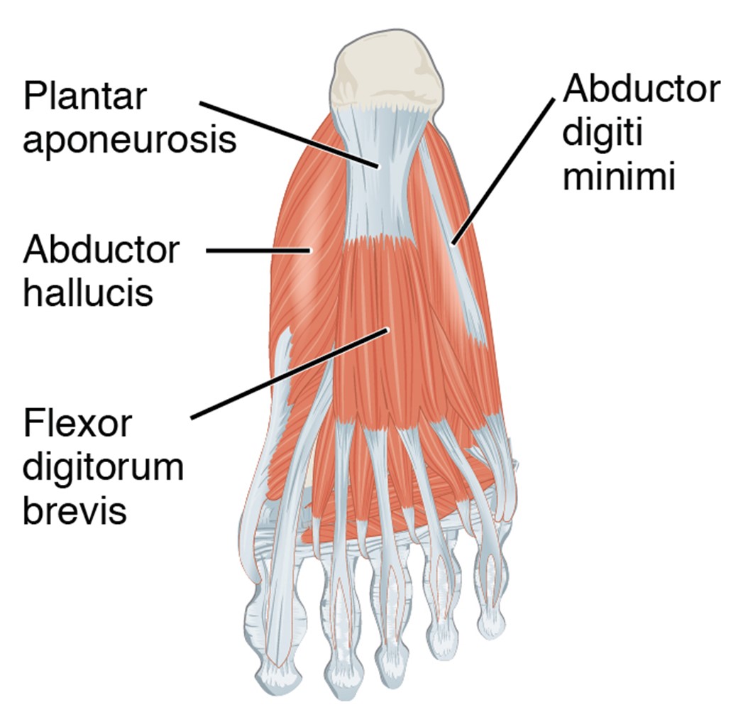
Abductor hallucis
Abductor hallucis is the most medial muscle of the first layer and forms the medial border of the foot.
It originates from the medial process of the calcaneal tuberosity and inserts as a tendon on the medial side of the base of the proximal phalanx of the big toe.
As its name suggests, it acts to abduct and additionally flexes the big toe at the metatarsophalangeal joint.
It is innervated by the medial plantar nerve, a branch of the tibial nerve derived from nerve roots S1-3.
Flexor digitorum brevis
Flexor digitorum brevis lies centrally in the first layer. It originates as a tendon on the medial process of the calcaneal tuberosity before becoming a muscle that runs centrally over the plantar aponeurosis. It then divides into four tendon branches.
These tendons split into two before running around the lateral aspects of each tendon of flexor digitorum longus and inserting on the margins of the middle phalanx.
It acts to flex the lateral four toes at the proximal interphalangeal joint and is innervated by the medial plantar nerve, a branch of the tibial nerve derived from nerve roots S1-3.
Abductor digiti minimi
Abductor digiti minimi is the most lateral of the muscles in the first layer.
It originates from several places: the medial and lateral aspects of the calcaneal tuberosity, and a fibrous band of connective tissue which connects calcaneus to metatarsal V.
Abductor digiti minimi forms a tendon that runs along the surface of metatarsal V before inserting onto the lateral side of the base of the proximal phalanx on the little toe.
It acts to abduct the little toe at the metatarsophalangeal joint and is innervated by the lateral plantar nerve, a branch of the tibial nerve derived from nerve roots S1-3.
Clinical relevance: Plantar fasciitis
The plantar aponeurosis is a thickening of the deep fascia that runs along the sole of the foot. It acts to support the longitudinal arch of the foot and protects deep foot structures.
Plantar fasciitis is a common injury in runners and is often presents in primary care.
Clinical features include pain in the heel and the sole of the foot, typically worse after long periods of rest or immediately after exercising.
Most cases of plantar fasciitis will improve with conservative management, such as rest, non-steroidal anti-inflammatories and stretching.
Second layer
The second layer of the plantar group consists of two muscle groups, the lumbricals and the quadratus plantae. These lie immediately deep to the first layer and act on toes two to five.
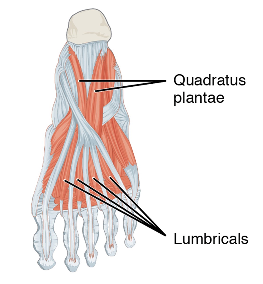
Quadratus plantae
The quadratus plantae muscle has two origins, one head originates from the medial surface, and the second from the lateral surface of the calcaneal tuberosity.
It inserts onto the lateral side of each tendon branch of flexor digitorum longus near where the tendons originate in the proximal half of the foot.
Along with the flexor digitorum longus, it acts to flex toes two to five and is innervated by the lateral plantar nerve.
Lumbricals
The lumbricals are four muscles that originate from the tendons of flexor digitorum longus and insert onto the medial aspect of the extensor hoods of toes two to five.
First lumbrical:
- Origin: medial side of the tendon of flexor digitorum longus associated with the second toe
- Insertion: medial side of the extensor hood of the second toe
Second lumbrical:
- Origin: medial side of the tendon of flexor digitorum longus associated with the third toe
- Insertion: medial side of the extensor hood of the third toe
Third lumbrical:
- Origin: medial side of the tendon of flexor digitorum longus associated with the fourth toe
- Insertion: medial side of the extensor hood of the fourth toe
Fourth lumbrical:
- Origin: medial side of the tendon of flexor digitorum longus associated with the fifth toe
- Insertion: medial side of the extensor hood of the fifth toe
These muscles act to resist excessive extension of the metatarsophalangeal joints and flex the interphalangeal joints during walking.
The first lumbrical is innervated by the medial plantar nerve, whilst the second, third and fourth lumbricals are innervated by the lateral plantar nerve.
Third layer
The third muscle layer of the plantar group lies deep to the second layer, and consists of three muscles: flexor hallucis brevis, adductor hallucis and flexor digiti minimi brevis.
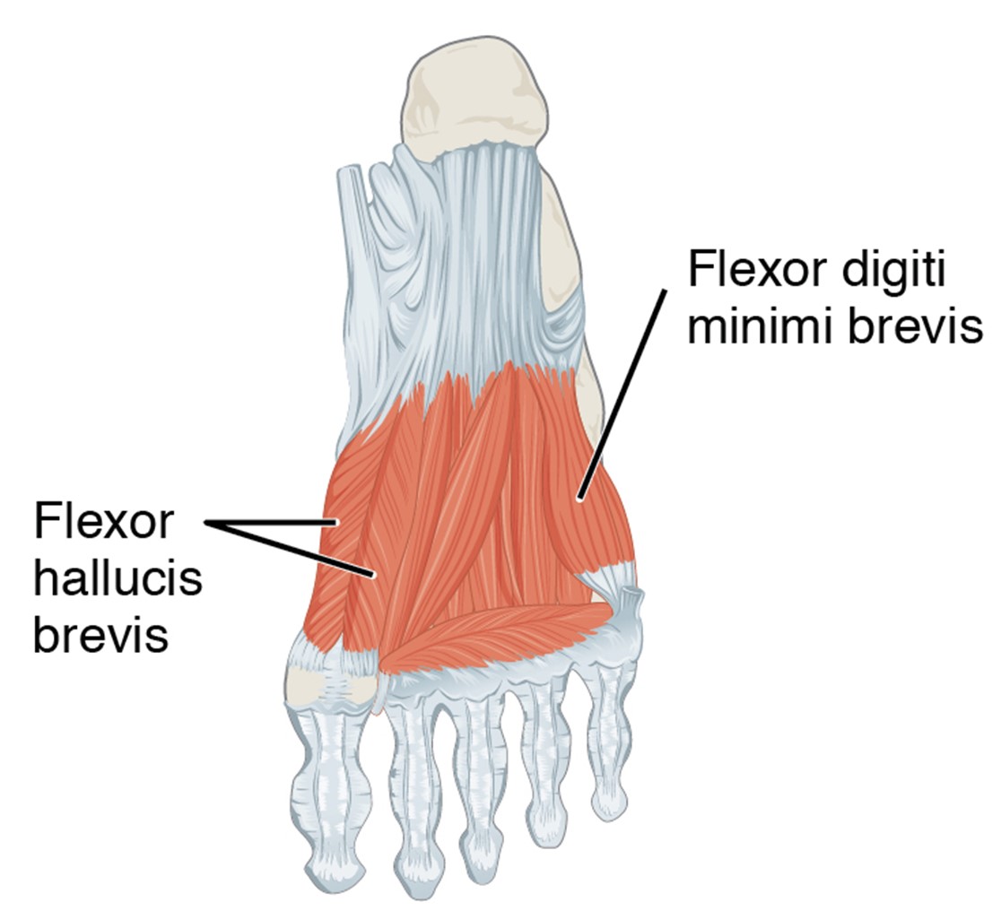
Flexor hallucis brevis
Flexor hallucis brevis has two heads of origin:
- Medial head: originates from the tendon of tibialis posterior
- Lateral head: originates from the plantar surface of the cuboid bone
These heads converge to form the belly of the muscle, which in turn divides forming two tendons for insertion on the medial and lateral side of the base of the proximal phalanx of the big toe.
As its name would suggest, the muscle acts to flex the big toe at the metatarsophalangeal joint and is innervated by the medial plantar nerve.
Adductor hallucis
Adductor hallucis also has two heads of origin, the transverse head and the oblique head.
The transverse heads originate separately on the metatarsophalangeal joints of the lateral three toes and run medially before converging and inserting onto the lateral side of the base of the proximal phalanx of the big toe, along with the oblique head.
The oblique head originates from the plantar surfaces of the bases of the second to fourth metatarsals as well as the fibularis longus sheath. It runs anterolaterally along the foot before converging with the transverse heads and inserting onto the lateral side of the base of the proximal phalanx of the big toe.
These muscles act to adduct the big toe at the metatarsophalangeal joint and are innervated by the lateral plantar nerve.
Flexor digiti minimi brevis
Flexor digiti minimi brevis originates from the base of the fifth metatarsal and the adjacent sheath of the fibularis longus tendon. It inserts onto the lateral side of the base of the proximal phalanx of the little toe.
It acts to flex the little toe at the metatarsophalangeal joint and is innervated by the lateral plantar nerve.
Fourth layer
The fourth layer is the deepest of the plantar group and comprises the dorsal and plantar interossei. Both groups act to resist extension of the metatarsophalangeal joints and flex the interphalangeal joints, however have opposite actions on the movement of the toes in the long axis.
Dorsal interossei
There are four dorsal interossei that run along with the metatarsal bones of the second to fifth toes. They all act to abduct the toes.
The second toe is associated with two dorsal interossei and can therefore be abducted to either side of its long axis.
The third and fourth toes are only associated with dorsal interossei on their lateral border, and so can only be abducted in this direction.
Origins: sides of the adjacent metatarsals
Insertions: extensor hoods and bases of the proximal phalanges of the second to fifth toes
Actions:
- Abduction of the third, fourth and fifth toes at the metatarsophalangeal joints
- Resist extension of the metatarsophalangeal joints
- Flexion of the interphalangeal joints
Innervation:
- All are innervated by the lateral plantar nerve
- The first and second dorsal interossei are also innervated by the deep fibular nerve (S2, S3)
Plantar interossei
The plantar interossei consist of four muscles that act together to adduct the third, fourth and fifth toes.
Origin: medial sides of the third to fifth metatarsals
Insertion: extensor hoods and base of the proximal phalanx of the third, fourth and fifth toes
Actions:
- Adduction of the third, fourth and fifth toes at the metatarsophalangeal joints
- Resist extension of the metatarsophalangeal joints
- Flexion of the interphalangeal joints
Innervation: lateral plantar nerve (S2, S3)
Clinical relevance: Morton’s neuroma
Morton’s neuroma is an enlargement of the common plantar nerve. It is usually found in the space between the third and fourth toes (the site where the medial and lateral plantar nerves merge).
Typical clinical features include sharp or dull pain in the space between the third and fourth toes, worsened by wearing shoes or walking.
Management of Morton’s neuroma includes non-steroidal anti-inflammatory injections and surgical removal.
Key points
- The extrinsic muscles of the foot originate in the lower leg, whilst the intrinsic muscles are contained within the foot itself.
- The intrinsic foot muscles act to stabilise the foot and support the arches, as well as to produce fine movement of the toes.
- The intrinsic foot muscles can be divided into two main groups, plantar and dorsal.
- The dorsal group consists of extensor digitorum brevis and extensor hallucis brevis which both act to extend the toes.
- The plantar group consists of four muscle layers, going from superficial (plantar) to deep (dorsal) within the foot.
- All of the muscles are innervated by branches of the tibial nerve (nerve roots S1-3), except extensor digitorum brevis, which is innervated by the deep fibular nerve.
- Vascular supply is derived from branches of the dorsalis pedis and posterior tibial arteries.
Editor
Dr Chris Jefferies
References
- OpenStax. Anatomy and Physiology. Intrinsic muscles of the foot. Licence: [CC-BY].
- Anatomy, Foot & Ankle: Musculoskeletal Medicine. OrthoPaedia. 2020.
- Drake R, Vogl W, Mitchell A, Gray H. Gray’s Anatomy for Students. 3rd Ed. Elselvier; 2015.
- OpenStax. Anatomy and Physiology. April 2013. Available from: [LINK]
- James Thing, Mahiben Maruthappu and John Rogers. Diagnosis and management of plantar fasciitis in primary care. British Journal of General Practice 2012; 62 (601): 443-444. Available from: [LINK]
