

- 📖 Geeky Medics OSCE Book
- ⚡ Geeky Medics Bundles
- ✨ 1300+ OSCE Stations
- ✅ OSCE Checklist PDF Booklet
- 🧠 UKMLA AKT Question Bank
- 💊 PSA Question Bank
- 💉 Clinical Skills App
- 🗂️ Flashcard Collections | OSCE, Medicine, Surgery, Anatomy
- 💬 SCA Cases for MRCGP
To be the first to know about our latest videos subscribe to our YouTube channel 🙌
Introduction
The muscles of the posterior compartment of the thigh are the biceps femoris, semitendinosus and semimembranosus, more commonly referred to as the hamstrings. The term ‘hamstring’ came about as it was common to use the long tendons of these muscles to tie up hams or pork thighs for curing.
The actions of the hamstrings include hip extension, knee flexion and medial/lateral rotation at both joints. Collectively the hamstrings are innervated by the sciatic nerve (L5, S1, S2) and their arterial supply is derived from a combination of the inferior gluteal artery and the perforating arteries of profunda femoris. The perforating arteries supply the majority of the hamstring muscles whereas the inferior gluteal artery only supplies the superior portion.
In this article, we will explore the anatomy of each of the hamstrings looking at their action, attachments, innervation and arterial supply. At the end of this article, we will also look at the clinical relevance of this anatomy in hamstring injuries.
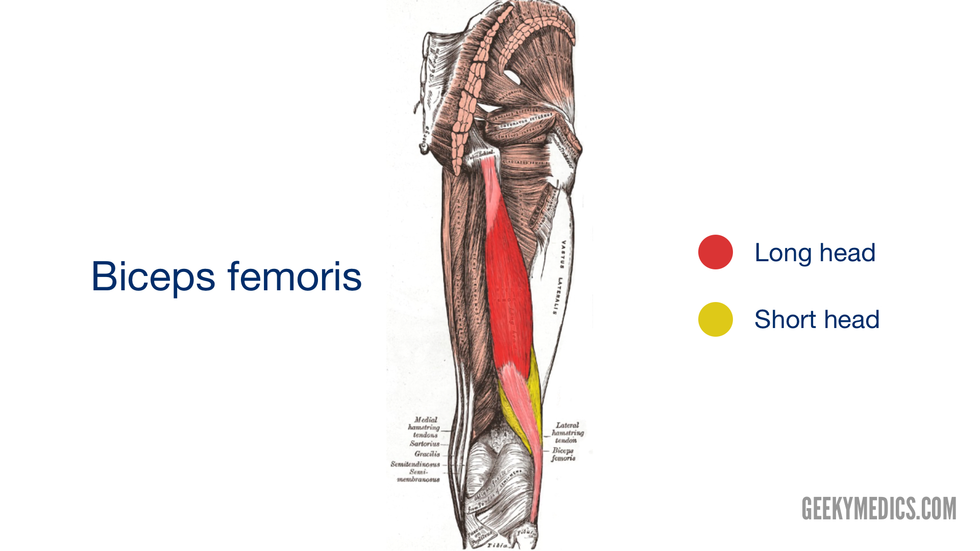
Biceps femoris
The biceps femoris is the most lateral muscle in the posterior compartment of the thigh and, as the term bicep suggests, has two heads: the long head and the short head.
These muscle heads have different origins but join to form a palpable tendon on the lateral distal thigh inserting on to the head of the fibula. The long head of biceps femoris protects the sciatic nerve as it passes from the gluteal region into the posterior compartment of the thigh.
Origin
- Long head: inferomedial part of the ischial tuberosity
- Short head: lateral lip of the linea aspera
Insertion
- Head of the fibula
Action
- Knee flexion
- Hip extension
- Lateral rotation at the hip and knee joint
Innervation
- Long head: tibial division of the sciatic nerve
- Short head: common fibular division of the sciatic nerve
Blood supply
- Inferior gluteal artery supplies the superior portion of the muscle
- Perforating arteries of profunda femoris supply the majority of the muscle
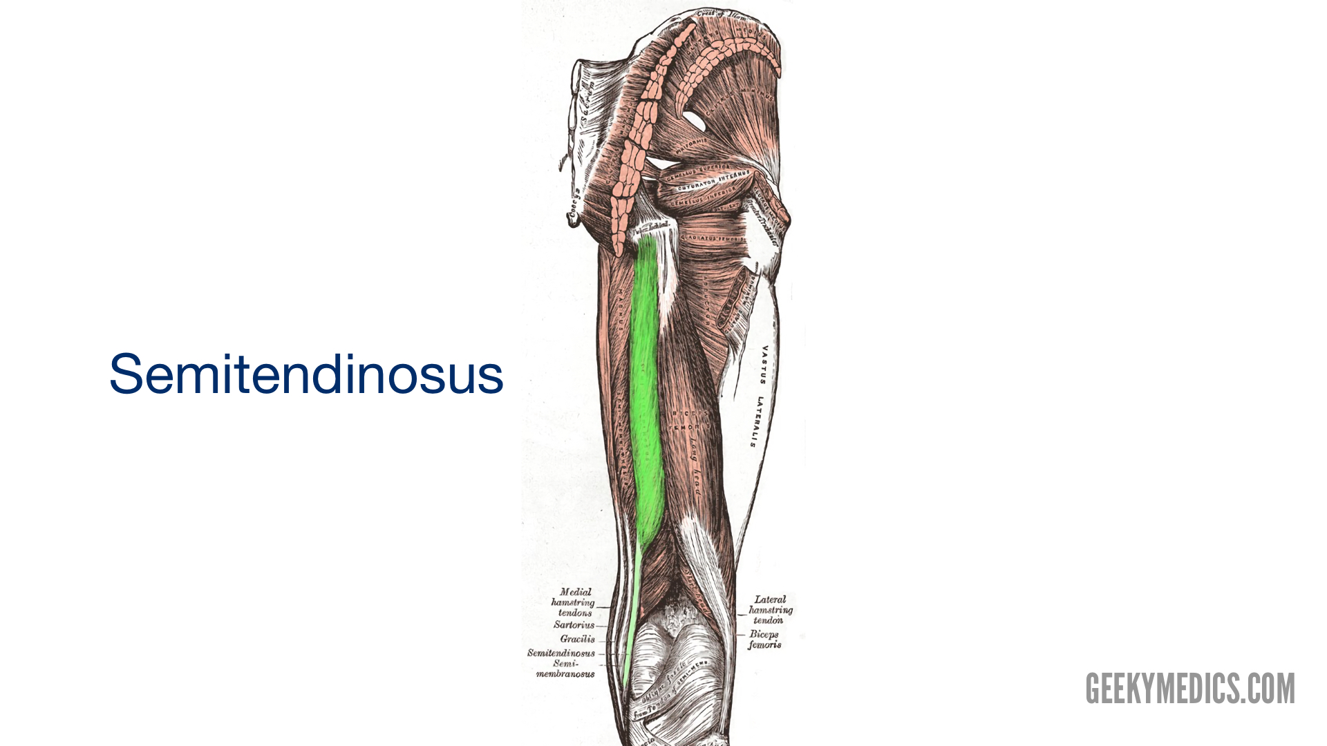
Semitendinosus
Aptly named, semitendinosus is half tendinous. It lies medial to biceps femoris and shares the same origin as its long head.
Semitendinosus lies superficial to semimembranosus and forms its tendon approximately two-thirds of the way down the thigh. This tendon inserts to the medial surface of the tibia as part of the ‘pes anserinus’, that is also made up of the tendons of the sartorius and gracilis muscles.
Origin
- Inferomedial part of the ischial tuberosity
Insertion
- Medial surface of the tibia
Action
- Knee flexion
- Hip extension
- Medial rotation of the thigh at the hip joint
- Medial rotation of the lower leg at the knee
Innervation
- Tibial division of the sciatic nerve
Blood supply
- Inferior gluteal artery supplies the superior portion of the muscle
- Perforating arteries of profunda femoris supply the majority of the muscle
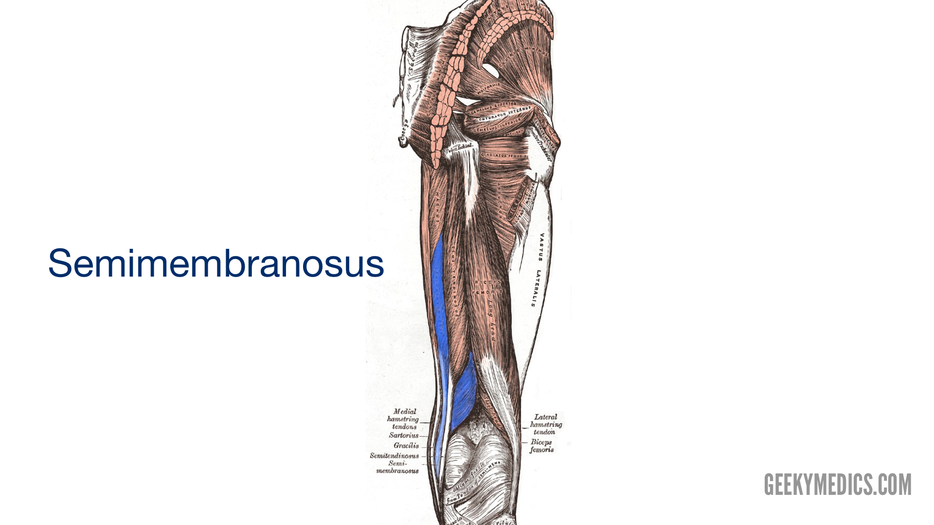
Semimembranosus
Again another aptly named muscle, semimembranosus is named because of the flat membranous form of its proximal attachment. It also attaches to the ischial tuberosity but more superiorly compared to semitendinosus and the long head of biceps femoris.
Distally, the semimembranosus tendon extends to contribute to the fascia and ligaments of the knee joint. It is the most medial muscle of the hamstrings and can be found underneath semitendinosus.
Origin
- Superolateral part of the ischial tuberosity
Insertion
- Posterior surface of the medial tibial condyle
Action
- Knee flexion
- Hip extension
- Medially rotation of the thigh at the hip joint
- Medial rotation of the lower leg at the knee joint
Innervation
- Tibial division of the sciatic nerve
Blood supply
- Inferior gluteal artery supplies the superior portion of the muscle
- Perforating arteries of profunda femoris supply the majority of the muscle
Clinical relevance: Hamstring injuries
Injuries to the hamstrings are common for those competing in sports such as sprinting and football due to excessive stretching of the muscles. An injury is most likely to occur with a sudden change in speed or direction. The level of injury can vary from a mild strain, where the muscles are excessively stretched, to a complete tear of the muscle itself.
In adults, the most injured point is the muscle-tendon junction. Whereas, in adolescents, an avulsion fracture of the ischial tuberosity is more likely. This is because the ischial apophysis (the site where the tendons attach) is the weakest part of the hamstrings in this age group. In an avulsion fracture the tendons of the muscle tear-off taking a fragment of bone with them.
MRI and ultrasound can be used to assess an injured hamstring, with MRI being able to provide more information about the extent of the injury.
References
Reference texts
- Richard L. Drake, A. Wayne Vogyl, Adam W.M. Mitchell. Gray’s Anatomy for Students 4th Elsevier, 2020.
- Keith L. Moore, Arthur F. Dalley, Anne M. R. Agur. Moore Clinically Orientated Anatomy 7th Lippincott Williams & Wilkins, 2013.
Reference images
- Henry Vandyke Carter. Licence: [Public domain]. Available from: [LINK].
