
- 📖 Geeky Medics OSCE Book
- ⚡ Geeky Medics Bundles
- ✨ 1300+ OSCE Stations
- ✅ OSCE Checklist PDF Booklet
- 🧠 UKMLA AKT Question Bank
- 💊 PSA Question Bank
- 💉 Clinical Skills App
- 🗂️ Flashcard Collections | OSCE, Medicine, Surgery, Anatomy
- 💬 SCA Cases for MRCGP
To be the first to know about our latest videos subscribe to our YouTube channel 🙌
Introduction
The ventricular system is the interconnecting network of fluid-filled cavities within the brain, comprising two lateral ventricles, the third ventricle and the fourth ventricle. The ventricles contain cerebrospinal fluid (CSF), which is produced by the choroid plexus within the ventricles.1
The ventricular system helps protect the brain by cushioning against head trauma, and the CSF within the ventricles provides nutrients to and removes waste products from the brain tissue.2,3
This article will discuss the structure of the brain’s ventricular system, the production of CSF by the choroid plexus and the reabsorption of CSF back into the systemic circulation.
Lateral ventricles
The left and right lateral ventricles are the largest cavities within the ventricular system and are highlighted in red in Figure 1.
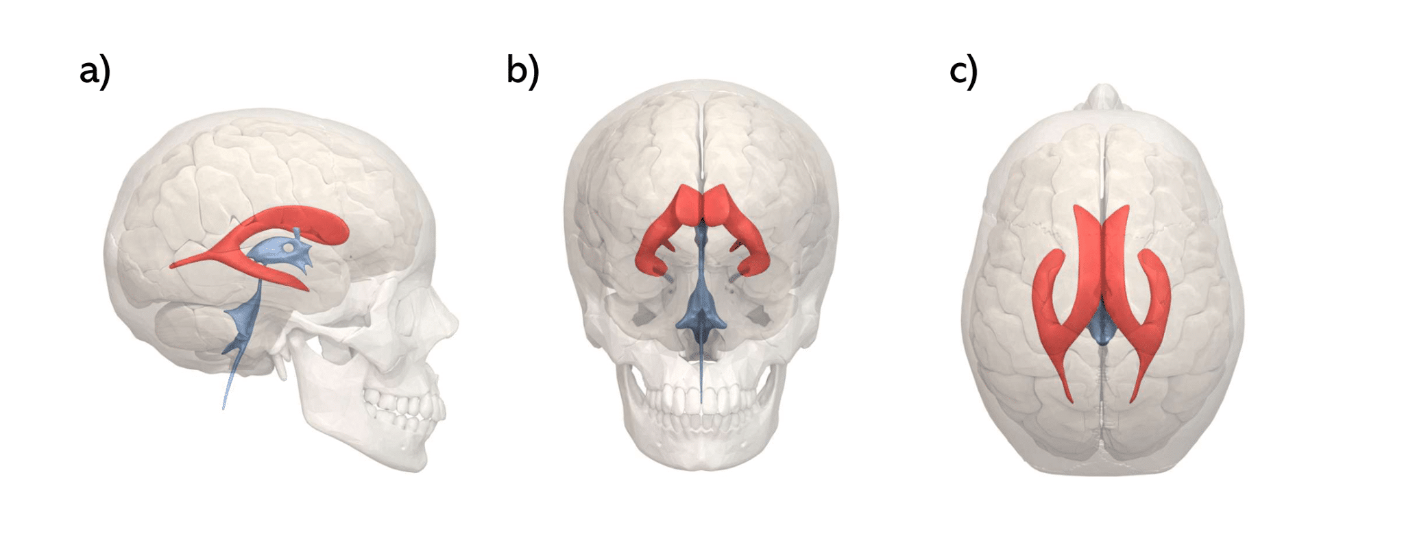
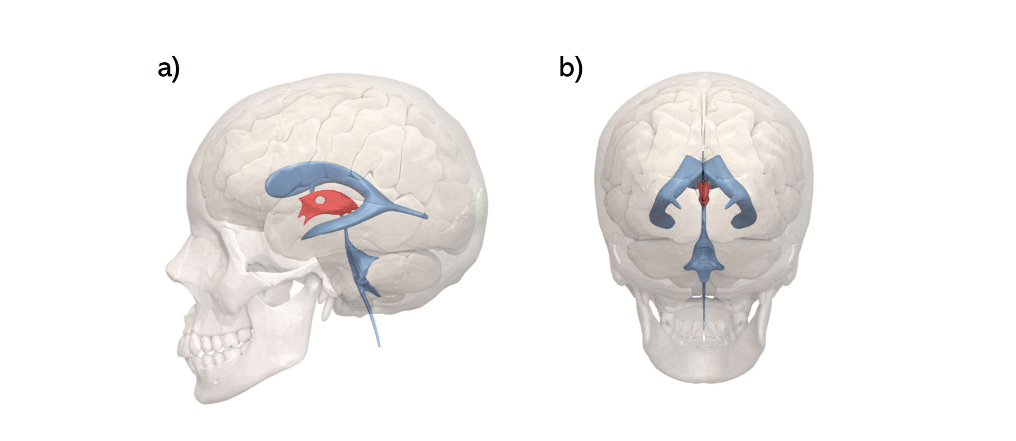
Each of the lateral ventricles can be subdivided into the body, trigone, frontal horn, temporal horn and occipital horn (Figure 2).4
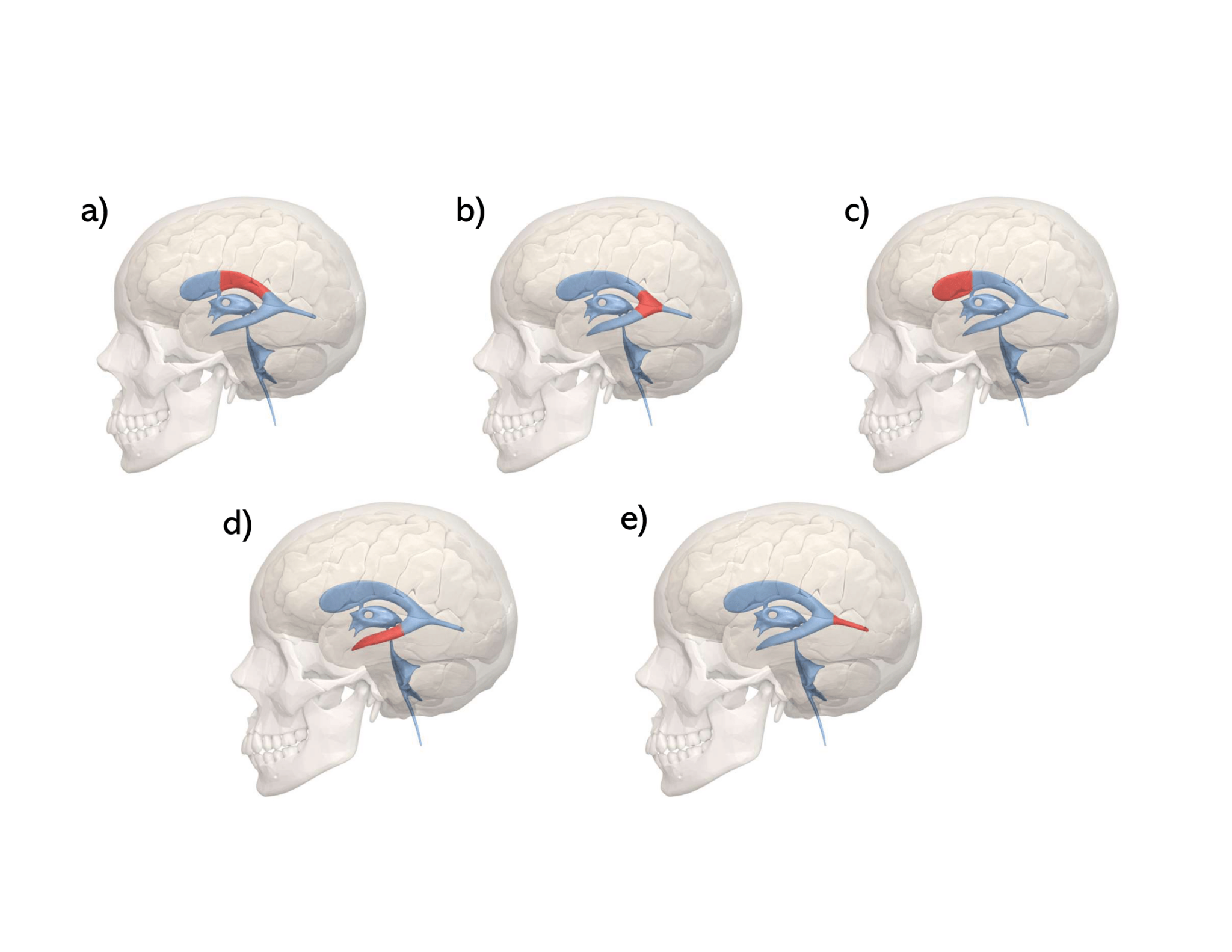
The body (Figure 2a) is found between the trigone (posteriorly) and the frontal horn (anteriorly). It is bounded superiorly by the corpus callosum and inferiorly by the thalamus and caudate nucleus. Medially, the bodies of the left and right lateral ventricles are separated by a thin membrane called the septum pellucidum.4
The trigone (Figure 2b, also known as the atrium) takes its name from its triangular cross-section. This is the point where the temporal horn, occipital horn and body of the lateral ventricles meet.4 The trigone contains an enlarged choroid plexus called the choroid glomus.5
The frontal horn (or anterior horn, Figure 2c) is the extension of the lateral ventricle into the frontal lobe. Like the body, it is bounded superiorly by the corpus callosum and medially by the septum pellucidum. It is bounded inferiorly mainly by the head of the caudate nucleus, and the anterior boundary is formed by the genu and rostrum of the corpus callosum.5
The temporal horn (or inferior horn, Figure 2d) is located anteriorly and inferiorly to the trigone and is the largest part of the lateral ventricle. It is bounded superiorly by the tapetum of the corpus callosum and inferiorly by the hippocampus and fimbria of the hippocampus. The amygdala is found at the anterior extremity of the temporal horn.5
The occipital horn (or posterior horn, Figure 2e) is the extension of the lateral ventricle into the occipital lobe. It is bounded both superiorly and laterally by the tapetum of the corpus callosum, and medially by the white matter of the occipital lobe.5
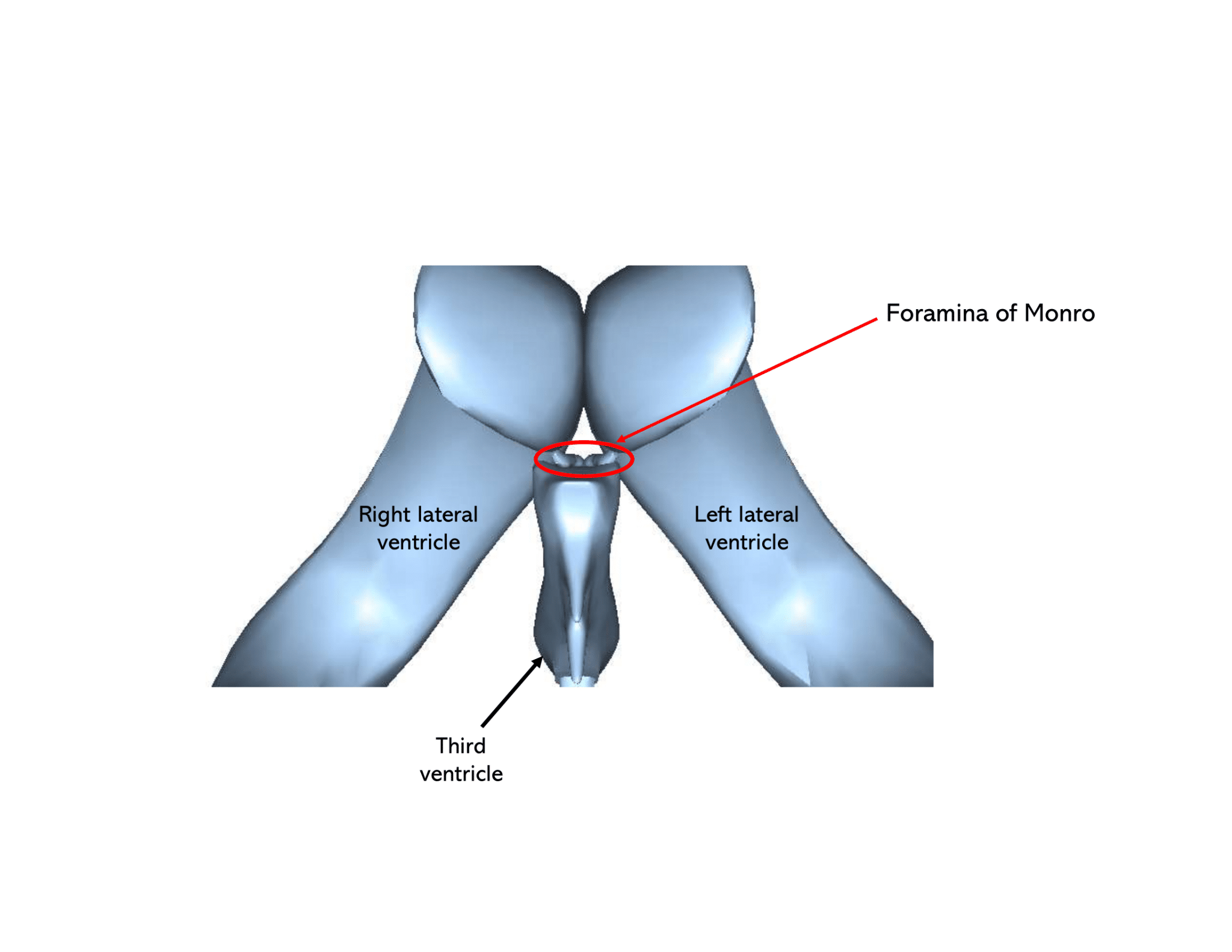
The interventricular foramina (or foramina of Munro) connect the lateral ventricles (specifically the anterior horns) to the third ventricle (Figure 3).6
Third ventricle
The third ventricle (seen in Figure 4) is a narrow, CSF-filled space located between the left and right thalami.4
In addition to the main cavity, there are also four recesses of the third ventricle:4
- Supraoptic recess (located superior to the optic chiasm)
- Infundibular recess (projects into the infundibulum of the pituitary gland)
- Pineal recess (projects posteriorly into the pineal gland)
- Suprapineal recess (located superior to the pineal gland)
The third ventricle connects to the fourth ventricle via the cerebral aqueduct (Figure 5), a very narrow channel running through the midbrain.1

Fourth ventricle
The fourth ventricle is a CSF-filled cavity located within the hindbrain, with a characteristic diamond shape (seen in Figure 6).4

Located anterior to the fourth ventricle is the pons and upper medulla, and posterior is the cerebellum.
The roof of this diamond-shaped cavity consists of the two cerebellar peduncles and the thin layer of white matter that connects the two peduncles (the superior medullary velum). The rhomboid fossa (a diamond-shaped depression on the posterior surface of the pons) forms the floor of the fourth ventricle.4
A small amount of CSF from the fourth ventricle enters the central canal of the spinal cord, but most CSF exits the fourth ventricle via three foramina (the medial foramen of Magendie and two lateral foramina of Luschka) into the cisterna magna, one of the subarachnoid cisterns.1
Subarachnoid cisterns
The subarachnoid cisterns are spaces in the brain where there is a gap between the subarachnoid and pia mater, leading to a collection of CSF within the enlarged subarachnoid space.7
Examples of subarachnoid cisterns include:7
- Cisterna manga: the largest subarachnoid cistern, located posterior to the medulla oblongata and inferior to the cerebellum
- Pontine cistern: found anterior to the pons, containing the basilar artery
- Interpeduncular cistern: a collection of CSF found between the two cerebral peduncles (on the anterior midbrain)
Cerebrospinal fluid (CSF)
Reabsorption of CSF
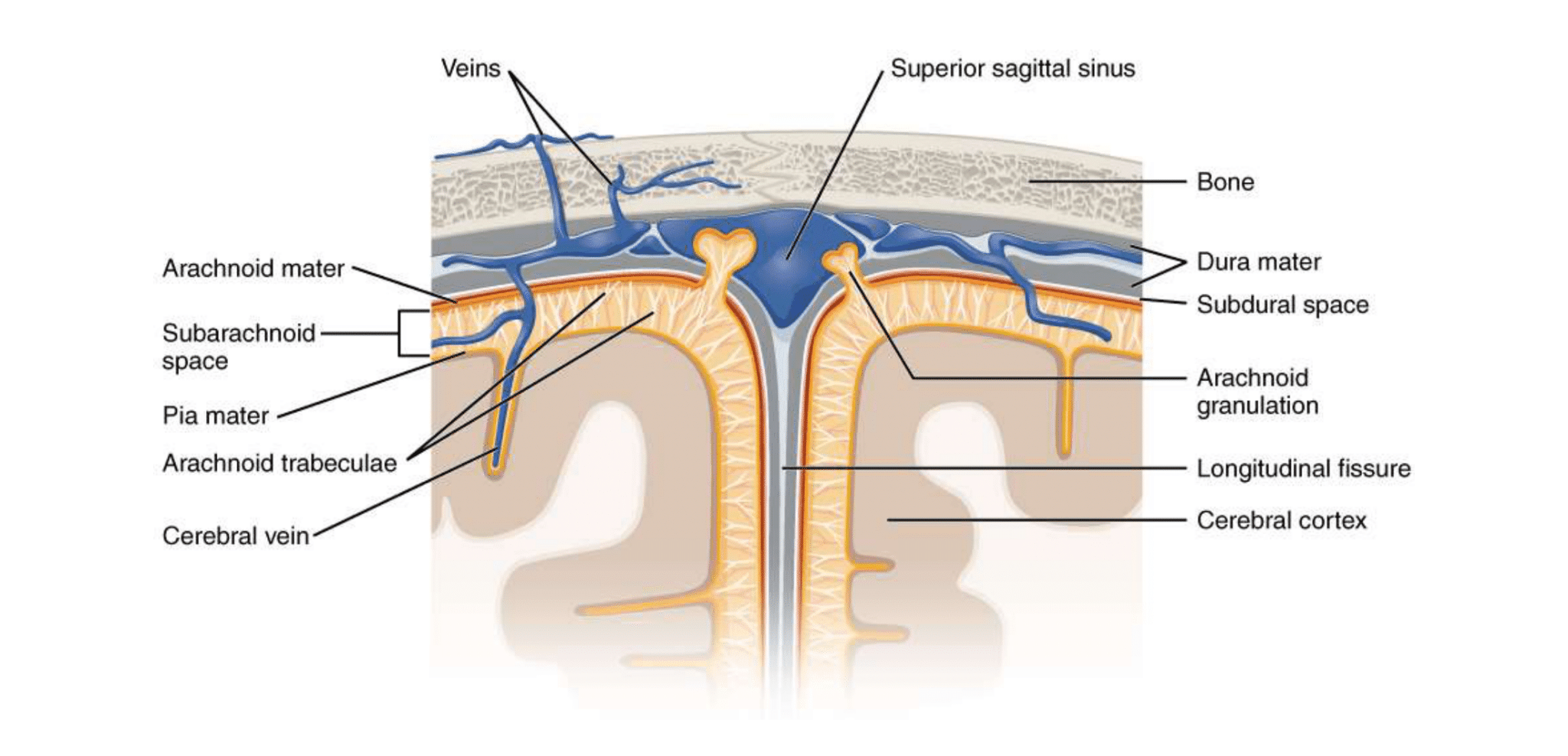
After CSF has exited the ventricular system, it enters the venous system via arachnoid granulations (extensions of arachnoid mater into the dural venous sinuses, shown in Figure 7), and thus enters the systemic circulation.8
Choroid plexus
The choroid plexus is the tissue responsible for the production of CSF and is found in each of the ventricles. Approximately 500ml of CSF is produced each day.9
Microscopically, the choroid plexus consists of cuboidal epithelial cells, beneath which is connective tissue containing capillaries. The formation of CSF begins with plasma leaving the capillaries and entering the interstitial space of the connective tissue. CSF is then produced from the plasma by the choroid epithelium.9
Clinical relevance: hydrocephalus
Hydrocephalus refers to an abnormally increased volume of CSF within the ventricular system.10 A CT scan of a patient with hydrocephalus is shown in Figure 8 (note the significantly enlarged lateral ventricles).

The two main types of hydrocephalus are communicating and non-communicating hydrocephalus:10
- Non-communicating: occurs when the flow of CSF between the ventricles is blocked (e.g. by a tumour in the base of the third ventricle)
- Communicating: occurs when there is a blockage to the flow of CSF after it has left the ventricular system (e.g. meningitis, subarachnoid haemorrhage)
Symptoms of hydrocephalus may include vomiting, irritability and seizures. In addition, infants with hydrocephalus may present with increased head size and/or a swollen fontanelle on the top of the skull.10
Hydrocephalus is usually treated by surgery and may involve a shunt being inserted into one of the ventricles, or a small hole being made in the base of the third ventricle (endoscopic third ventriculostomy).10
Key points
- The ventricular system is an interconnecting network of CSF-filled cavities within the brain
- The ventricular system comprises two lateral ventricles, the third ventricle and the fourth ventricle
- CSF is produced by tissue called choroid plexus, which is found in each of the ventricles
- After exiting the fourth ventricle, CSF is reabsorbed into the systemic venous circulation via arachnoid granulations
- Excess build-up of CSF within the ventricular system leads to a condition called hydrocephalus
Reviewer
Dr Kasra Bahadori
IMT 3
Editor
Dr Chris Jefferies
References
- Grow WA. Development of the Nervous System. Elsevier; 2018. p. 72-90.e1.
- Shenoy SS, Lui F. Neuroanatomy, Ventricular System. StatPearls. Treasure Island (FL): StatPearls Publishing Copyright © 2022, StatPearls Publishing LLC.; 2022.
- Fame RM, Cortés-Campos C, Sive HL. Brain Ventricular System and Cerebrospinal Fluid Development and Function: Light at the End of the Tube. BioEssays. 2020;42(3):1900186.
- Stratchko L, Filatova I, Agarwal A, Kanekar S. The Ventricular System of the Brain: Anatomy and Normal Variations. Seminars in Ultrasound, CT and MRI. 2016;37(2):72-83.
- Scelsi CL, Rahim TA, Morris JA, Kramer GJ, Gilbert BC, Forseen SE. The Lateral Ventricles: A Detailed Review of Anatomy, Development, and Anatomic Variations. American Journal of Neuroradiology. 2020;41(4):566-72.
- Tubbs RS, Oakes P, Maran IS, Salib C, Loukas M. The foramen of Monro: a review of its anatomy, history, pathology, and surgery. Child’s Nervous System. 2014;30(10):1645-9.
- Altafulla J, Bordes S, Jenkins S, Litvack Z, Iwanaga J, Loukas M, et al. The Basal Subarachnoid Cisterns: Surgical and Anatomical Considerations. World Neurosurg. 2019;129:190-9.
- Harris C, Khasawneh A, Garling R. Cerebrospinal fluid circulation: What do we know and how do we know it? Brain Circulation. 2018;4(1):14.
- Lun MP, Monuki ES, Lehtinen MK. Development and functions of the choroid plexus–cerebrospinal fluid system. Nature Reviews Neuroscience. 2015;16(8):445-57.
- Koleva M, De Jesus O. Hydrocephalus. StatPearls. Treasure Island (FL): StatPearls Publishing Copyright © 2022, StatPearls Publishing LLC.; 2022.
Image references
- BodyParts3D by DBCLS. Lateral ventricle. Licence: [CC BY-SA 2.1 JP]
- BodyParts3D by DBCLS. Lateral ventricle subsections. Licence: [CC BY-SA 2.1 JP]
- BodyParts3D by DBCLS. Anterior view of ventricular system showing foramina of Monro. Licence: [CC BY-SA 2.1 JP]
- BodyParts3D by DBCLS. Third ventricle. Licence: [CC BY-SA 2.1 JP]
- BodyParts3D by DBCLS. Cerebral aqueduct. Licence: [CC BY-SA 2.1 JP]
- BodyParts3D by DBCLS. Fourth ventricle. Licence: [CC BY-SA 2.1 JP]
- Meningeal layers showing arachnoid granulations. Licence: [CC BY 4.0]
- Reza Akhavan. Head CT scan of a patient with hydrocephalus. Licence: [CC BY-NC-SA 2.0]
