

- 📖 Geeky Medics OSCE Book
- ⚡ Geeky Medics Bundles
- ✨ 1300+ OSCE Stations
- ✅ OSCE Checklist PDF Booklet
- 🧠 UKMLA AKT Question Bank
- 💊 PSA Question Bank
- 💉 Clinical Skills App
- 🗂️ Flashcard Collections | OSCE, Medicine, Surgery, Anatomy
- 💬 SCA Cases for MRCGP
To be the first to know about our latest videos subscribe to our YouTube channel 🙌
Overview
The forearm is the portion of the arm distal to the elbow and proximal to the wrist. There are 20 muscles separated into two compartments.
In this article, we will discuss the posterior compartment of the forearm in the setting of their attachment points, function, innervation and vascular supply. In addition, we’ll also be providing clinical examples to reinforce this information.
For the anterior compartment, see the Geeky Medics guide to the muscles of the anterior forearm.
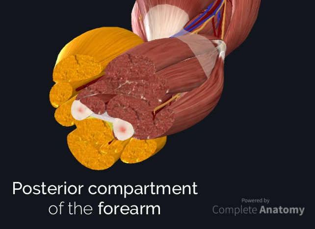
Summary: muscles of the posterior compartment of the forearm
The muscles of the posterior compartment of the forearm produce wrist and/or finger extension and thumb abduction. They are located posterior to the interosseous membrane and are arranged as twelve muscles in two layers (superficial and deep).
The muscles can be also divided functionally into four groups:
- Forearm fixator
- Elbow extension and supination
- Wrist and finger extension
- Thumb abduction
They are mainly supplied by the posterior interosseous branch of the radial nerve. Blood supply is from the radial recurrent, posterior, anterior interosseous and interosseous recurrent arteries. The deep veins mimic the arteries,
When identifying the function of the forearm muscles, it is important to note that any forearm compartment muscle that crosses the elbow joint will act at this joint. Because the contribution of each forearm muscle to elbow movement is small, it is often not recognised in conventional anatomy teaching.
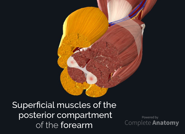
Superficial layer of the posterior compartment
The superficial layer of the posterior compartment contains seven muscles that have a common origin of the supracondylar ridge and laterally epicondyle of the humerus (the common extensor tendon):
- Brachioradialis
- Extensor carpi radialis longus
- Extensor carpi radialis brevis
- Extensor digitorum
- Extensor digiti minimi
- Extensor carpi ulnaris

Brachioradialis
Brachioradialis produces minimal flexion at the elbow, but becomes active as a powerful elbow fixator (like when holding a box in front of you). It is most efficient in partial pronation and is the only muscle of the posterior forearm compartment that produces flexion at a joint.
Brachioradialis forms the lateral boundary of the cubital fossa, and the radial nerve bifurcates before diving deep to this muscle in this region.
- Function: weak elbow flexion; primary elbow fixator
- Origin: the proximal third of the lateral supracondylar ridge
- Insertion: at or immediately proximal to the radial styloid process
- Innervation: radial nerve
- Arterial supply: radial recurrent

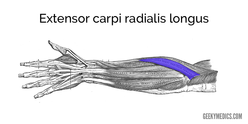
Extensor carpi radialis longus
The extensor carpi radialis longus has a relatively short muscle belly and longer tendon. It is this long tendon, and its superficial appearance, that identifies it from the extensor carpi radialis brevis.
- Function: wrist extension and abduction
- Origin: lateral supracondylar ridge and lateral epicondyle
- Insertion: dorsal surface of metacarpal II
- Innervation: radial nerve (before bifurcating into superficial and deep branches)
- Arterial supply: radial recurrent artery
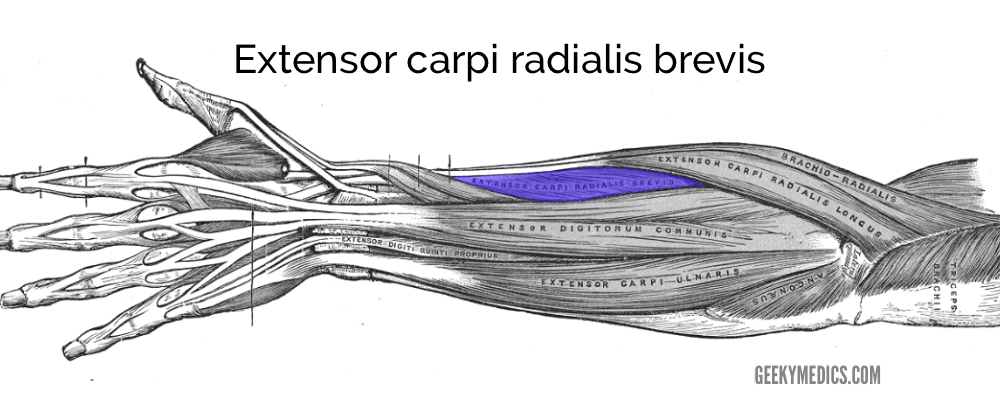
Extensor carpi radialis brevis
The little sibling of the extensor carpi radialis longus, the extensor carpi radialis brevis is distinguishable because of its seemingly larger muscle belly, shorter tendon, and deep appearance to the extensor carpi radialis longus. It is the last superficial muscle of the posterior forearm supplied by the radial nerve before it passes through the supinator.
- Function: extension and abduction of the wrist
- Origin: lateral epicondyle
- Insertion: base of metacarpals II and III
- Innervation: radial nerve
- Arterial supply: radial artery
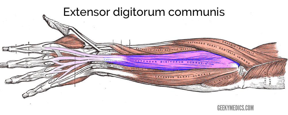
Extensor digitorum (communis)
The extensor digitorum is the major extensor muscle of digits II-V. This muscle arises from a very short common muscle belly that diverges into four individual muscle bellies, each giving rise to a single tendon. Each tendon inserts into the interconnected triangular aponeurosis (dorsal hood, extensor expansion) on the dorsal surface of each digit.
- Function: extension of digits II-V
- Origin: lateral epicondyle
- Insertion: extensor expansion of the dorsal hand
- Innervation: posterior interosseous nerve
- Arterial supply: posterior interosseous artery
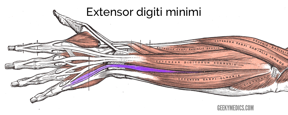
Extensor digiti minimi (extensor digiti quinti proprius)
Extensor digiti minimi is an accessory extensor of the little finger.
- Function: extension of digit V
- Origin: lateral epicondyle
- Insertion: extensor expansion, immediately medial to extensor digitorum
- Innervation: posterior interosseous nerve
- Arterial supply: posterior interosseous artery
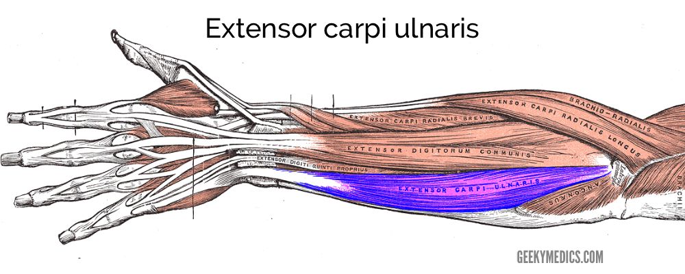
Extensor carpi ulnaris
The extensor carpi ulnaris is an important extensor and adductor of the wrist. It has a relatively short muscle belly with a long, flat tendon that converges distally into a round tendon.
- Function: extension and adduction of the wrist
- Origin: lateral epicondyle
- Insertion: medial aspect of the base of metacarpal V
- Innervation: posterior interosseous nerve
- Arterial supply: posterior interosseous artery
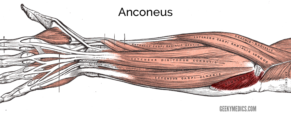
Anconeus
Named for its triangular shape, the anconeus is a small muscle that provides accessory support to other posterior forearm muscles.
- Function: extension; in pronation, abducts ulna
- Origin: lateral epicondyle
- Insertion: the dorsolateral surface of the olecranon process and dorsal ulna
- Innervation: radial nerve
- Arterial supply: recurrent interosseous artery
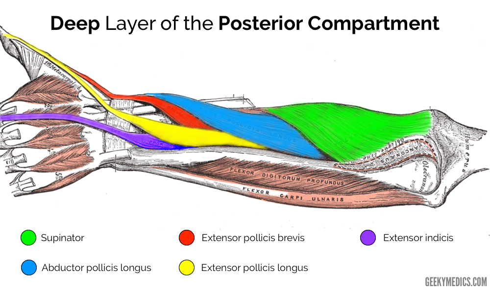
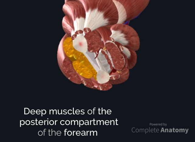
Deep layer of the posterior compartment
The deep layer of the posterior compartment contains five muscles, all of which are innervated by the posterior interosseous nerve:
- Supinator
- Abductor pollicus longus
- Extensor pollicus longus
- Extensor pollicus brevis
- Extensor indicis
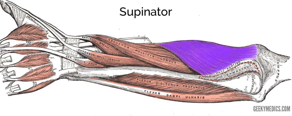
Supinator
The supinator muscle has superficial and deep heads that help to supinate the forearm. Passing between these two heads is the deep branch of the radial nerve. As the superficial head forms the ‘roof’ of this tunnel, it is termed the Arcade of Frohse.
- Function: supination of the forearm
- Origin:
- Superficial head: lateral epicondyle
- Deep head: supinator crest (dorsolateral ulna)
- Insertion: proximal lateral radius, at or superior to the oblique line
- Innervation: deep branch of the radial nerve
- Arterial supply: radial recurrent artery
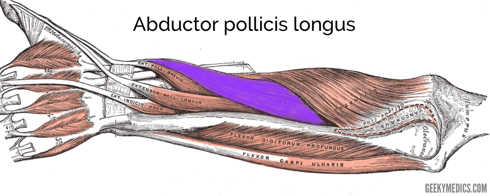
Abductor pollicis longus
The abductor pollicis longus not only contributes to thumb abduction between metacarpal I and the (os) trapezium, but assists in the abduction of the wrist, too. The abductor pollicis longus assists the extensor pollicis brevis in forming the inferior margin of the anatomical snuffbox.
- Function: abduction of the thumb (and assists in abduction at the wrist)
- Origin: proximal dorsal radius, ulna and interosseous membrane
- Insertion: lateral aspect of metacarpal I
- Innervation: posterior interosseous nerve
- Arterial supply:
- Proximal aspect: posterior interosseous artery
- Distal aspect: anterior interosseous artery
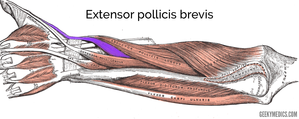
Extensor pollicis brevis
Extensor pollicis brevis is the little sibling to extensor pollicis longus. The extensor pollicis brevis takes a short-cut to reach the thumb; extensor pollicis longus takes the long way to the thumb, passing through the extensor retinaculum. The extensor pollicis brevis assists the abductor pollicis longus in forming the inferior margin of the anatomical snuffbox.
- Function: extension of the carpometacarpal and metacarpophalangeal joints of digit I (assists abduction of the wrist)
- Origin: dorsal radius, ulna and interosseous membrane
- Insertion: dorsal surface of the proximal phalanx of digit I
- Innervation: posterior interosseous nerve
- Arterial supply: posterior interosseous artery
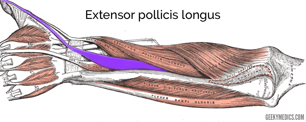
Extensor pollicis longus
The extensor pollicis longus takes a longer course to reach the thumb than its little sibling extensor pollicis brevis. The extensor pollicis longus passes around the dorsal radial tubercle (Lister’s tubercle) before coursing to the thumb. The extensor pollicis longus forms the superior border of the anatomical snuff box.
- Function: extension of the carpometacarpal and metacarpophalangeal joints of digit I (assists abduction of the wrist)
- Origin: dorsal ulna and interosseous membrane
- Insertion: dorsal surface of the distal phalanx of digit I
- Innervation: posterior interosseous nerve
- Arterial supply: posterior interosseous artery
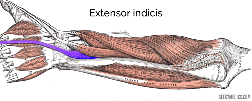
Extensor indicis
The extensor indicis is an accessory extensor of the index finger, allowing the index finger finer, more dexterous movement.
- Function: mainly extension of digit II at the metacarpophalangeal joint
- Origin: dorsal ulna
- Insertion: extensor expansion of digit II (adjacent to the digit II tendon of extensor digitorum)
- Innervation: posterior interosseous nerve
- Arterial supply:
- Proximal aspect: posterior interosseous artery
- Distal aspect: anterior interosseous artery
Clinical relevance: intersection syndrome versus De Quervain’s syndrome
Intersection syndrome
Intersection syndrome is a painful inflammatory condition that occurs at the intersection of the abductor pollicis longus and extensor pollicis brevis muscle bellies as they cross over extensor carpi radialis longus and brevis on the dorsolateral forearm.
Classically, this inflammatory disease presents as a result of repetitive movements, such as rowing or weight lifting.
Intersection syndrome is best managed with rest from aggravating activities and ice or cool compresses, but steroid injections may help to reduce inflammation.
It is often confused with De Quervain’s syndrome.
De Quervain’s syndrome
De Quervain’s syndrome is an inflammatory condition affecting the extensor pollicis brevis and abductor pollicis longus muscles. These are the only two muscles involved in this condition.
It is usually due to overuse and is referred to as one of the repetitive strain injuries. Pain is felt down the lateral side of the distal forearm, radiating to the thumb and region of the dorsal interossei.
It is usually diagnosed clinically with Finkelstein’s test (Figure 18), but MRI can confirm the presence of inflammation and local oedema around the tendons of extensor pollicis brevis and abductor pollicis longus.
Treating De Quervain’s syndrome can be a lengthy process, and is best achieved with the reduction of aggravating injuries, NSAIDs and cool compresses. There is contention about the use of steroid injections, but plastic surgery can offer definitive therapy.
Exam tips
- The radial nerve supplies all muscles of the extensor compartment
- The order of tendons running down the lateral aspect of the forearm can provide a simple basis for learning the muscles, or help you out in a spot of trouble in anatomy exams:
- Brachioradialis
- Extensor carpi radialis longus
- Extensor carpi radialis brevis
- Abductor pollicis longus
- Extensor pollicis brevis
- Extensor pollicis longus
- If you get stuck on an exam and forget the name of a muscle, break down the name into logical components:
- Are you in the flexor or extensor compartment?
- Does this muscle move the wrist (carpi), fingers (digitorum) or thumb (pollicis)?
- Does this muscle run down the radial (radialis) or ulnar (ulnaris) aspect of the forearm?
- Does this muscle have a longer (longus) or shorter (brevis) sibling?
Editor
William Maish
Geeky Medics Anatomy Lead
References
Reference texts
- Richard L. Drake, A. Wayne Vogyl, Adam W.M. Mitchell. Gray’s Anatomy for Students. s.l. : Elsevier, 2015.
- Steven F Morris, G. Ian Taylor. Vascular territories. [book auth.] Peter C. Neligan Geoffrey C. Gurtner. Plastic Surgery: Volume 1. s.l. : Elsevier, 2018.
Reference images
- Complete Anatomy 2019. 3D4 Medical. Available from: [LINK]
- Henry Gray. Modified by Geeky Medics. Published in 2019.
- James Heilman, MD. License: [CC BY-SA]
