

- 📖 Geeky Medics OSCE Book
- ⚡ Geeky Medics Bundles
- ✨ 1300+ OSCE Stations
- ✅ OSCE Checklist PDF Booklet
- 🧠 UKMLA AKT Question Bank
- 💊 PSA Question Bank
- 💉 Clinical Skills App
- 🗂️ Flashcard Collections | OSCE, Medicine, Surgery, Anatomy
- 💬 SCA Cases for MRCGP
To be the first to know about our latest videos subscribe to our YouTube channel 🙌
Overview
The arm, anatomically known as the brachium, forms the connection between the antebrachium (forearm) and the bones of the shoulder girdle. It contains anterior and posterior compartments, that functionally contain muscles of flexion and extension respectively.
In this article, we discuss the anatomy of the arm and learn anatomy in the context of clinical examples.
The terms agonist and synergist will be used throughout this article. An agonist is a muscle with a primary function at that joint; it is a term usually used to describe muscles crossing only a single joint. A synergist is an assisting muscle and is a term usually reserved for muscles that serve functions at more than one joint. There may be single or multiple agonists and/or synergists at any joint in the body.
Anterior compartment
The anterior compartment contains three muscles:
- Biceps brachii
- Coracobrachialis
- Brachialis
The major actions of the muscles of the anterior arm are flexion of the elbow (agonists) and shoulder (synergists).
The anterior compartment derives its innervation from the musculocutaneous nerve (C5, C6, C7).
Most muscles of the anterior arm compartment are supplied by the brachial artery, with proximal contributions from the axillary artery.
Biceps brachii
The biceps brachii has both long and short heads both of which converge to insert in the forearm as a single tendon. This muscle is biarticular, crossing both the shoulder and the elbow, and has important functions in not only flexion, but supination of the forearm as well.
- Function:
- Shoulder: flexion
- Elbow: flexion and supination
- Origin:
- Short head: lateral aspect of the coracoid process of the scapula
- Long head: supraglenoid tubercle of the scapula
- Insertion: (proximal, ventral) radial tuberosity and the deep fascia of the forearm via the bicipital aponeurosis
- Innervation: musculocutaneous nerve

Clinical relevance: biceps brachii tears
Biceps brachii tears can be classified as either partial or complete.
A complete tear involves complete separation of the biceps brachii tendon from its insertion at the radial tuberosity. Partial tears occur either at this same region, but to a lesser severity, or occur at the junction of the long and short heads of the biceps brachii.
Tears are often the result of athletic activities and are precipitated by either rapid onset of high forces or repetitive trauma.
Biceps brachii tears are painful and present with weakness in forearm flexion and supination. Tears may also present with significant localised oedema.
Treatment of biceps brachii tears initially involves a conservative approach of rest from aggravating activities, ice or cold compress and non-steroidal anti-inflammatory medications. Physical therapy is often required to return to normal or near-normal strength, while surgery is usually reserved for more significant tears in athletes.
Coracobrachialis
Functionally, the coracobrachialis is a weak agonist of shoulder flexion.
- Function: shoulder flexion and stabilisation
- Origin: the tip of the coracoid process
- Insertion: medial aspect of humeral midshaft
- Innervation: musculocutaneous nerve

Brachialis
The brachialis muscle is relatively short, thick muscle that crosses the elbow joint. It is an agonist of elbow flexion in both forearm supination and pronation; compared to the biceps brachii, which is an elbow flexor only during forearm supination.
- Function: elbow flexion
- Origin: anterior and lateral aspect of the distal half of the humerus
- Insertion: ulnar tuberosity
- Innervation: musculocutaneous nerve (and minor lateral section by the radial nerve)
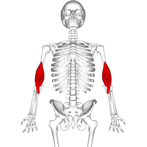
Posterior compartment
The posterior compartment is formed by the triceps brachii.
This three-headed muscle is responsible for both elbow (agonist) and shoulder (synergist) extension.¹
While the proximal triceps brachii derives some vascular supply from the axillary artery, most supply is from the profunda brachii (deep brachial artery).
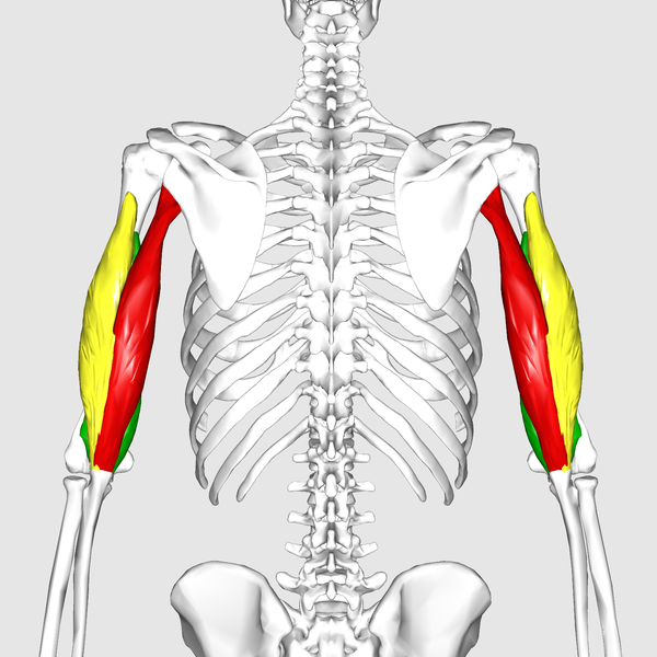
Triceps brachii
The triceps brachii has three heads: long, medial, and lateral.
The three heads of triceps brachii have different origins, one of which is on the scapula, and all converge into a single tendon that inserts onto the ulna.
- Function: elbow extension (agonist) and shoulder extension (synergist)
- Origin:
- Long head: infraglenoid tubercle of the scapula
- Lateral head: postero-lateral humerus superior to the radial (spiral) groove
- Medial head: postero-lateral humerus inferior to the radial (spiral) groove
- Insertion: olecranon process of the ulna
- Innervation: radial nerve
Editor
William Maish
Geeky Medics Anatomy Lead
References texts
- Richard L. Drake, A. Wayne Vogyl, Adam W.M. Mitchell. Gray’s Anatomy for Students. s.l. : Elsevier, 2015.
- Javed O, Ashmyan R. Anatomy, Shoulder and Upper Limb, Muscles. Treasure Island: StatPearls, 2019.
Reference images
- Anatomography. License: [CC BY-SA]
- Anatomography. License: [CC BY-SA]
- Anatomography. License: [CC BY-SA]
- Anatomography. License: [CC BY-SA]
