

- 📖 Geeky Medics OSCE Book
- ⚡ Geeky Medics Bundles
- ✨ 1300+ OSCE Stations
- ✅ OSCE Checklist PDF Booklet
- 🧠 UKMLA AKT Question Bank
- 💊 PSA Question Bank
- 💉 Clinical Skills App
- 🗂️ Flashcard Collections | OSCE, Medicine, Surgery, Anatomy
- 💬 SCA Cases for MRCGP
To be the first to know about our latest videos subscribe to our YouTube channel 🙌
Introduction
An ulcer is defined as a break in the skin or mucous membrane which fails to heal leading to a loss of surface tissue, disintegration and epithelial necrosis.1
Oral ulceration can be idiopathic, infectious, autoimmune or malignant in aetiology. Diagnosis of oral ulceration remains a challenge for clinicians due to the overlap in clinical presentation.2
This article provides a brief overview of the common causes of oral ulceration.
History and examination
History
Along with a comprehensive medical, social, dental and family history, key points to cover in the history include:
- Number of ulcers present in the oral cavity
- Duration of ulcer(s)
- Previous episodes of oral ulceration/frequency of ulceration
- Any recent changes in medication
- Associated symptoms such as fever or malaise
- Any extra-oral changes which may be indicative of an autoimmune disorder
Clinical examination
Important points to address in the clinical examination include:
- Number of ulcers present
- Site of the ulceration: on keratinised or non-keratinised mucosa
- Appearance, margins and base of ulcer(s)
- Texture of ulcer(s)
- Mucosal scarring
Recurrent aphthous ulcers
Recurrent aphthous stomatitis (RAS)
RAS is a common oral disorder affecting up to 25% of the population.3
It can be idiopathic or due to an underlying systemic condition.
Aetiology of RAS
The aetiology of RAS remains unknown however a number of factors, including stress, trauma, smoking cessation, hormonal imbalance, food hypersensitivity and genetics can predispose an individual to RAS.4
Clinical presentation
RAS has three typical clinical presentations which are discussed below.
Minor RAS
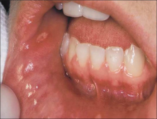
Minor RAS is the most common clinical presentation. Ulcers appear in clusters of 1-6 on non-keratinised mucosa. Minor RAS is self-limiting and usually heals within 7-10 days with no scarring.
Minor RAS presents clinically as ulcers 2-4mm in diameter with an erythematous margin and yellowish floor (figure 1).4
Major RAS
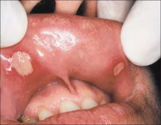
Major RAS can present on keratinised and non-keratinised oral mucosa. These ulcers are usually greater than 10mm in diameter, occurring in groups of 1-6 (figure 2). Major RAS is self-limiting but can last for several months. These ulcers usually heal with scarring. It is important for clinicians to be aware that these ulcers can mimic a malignant ulcer.4
Herpetiform RAS
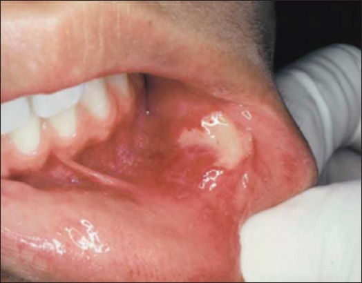
Herpetiform RAS is extremely painful. Lesions present as multiple small vesicles usually less than 5mm in diameter coalesce together to form large widespread erythematous areas (figure 3). They usually appear on non-keratinised mucosa, lasting 10-14 days.4
Nutritional deficiencies
Several nutritional deficiencies are linked with the oral manifestation of RAS. The common nutritional deficiencies seen in patients with RAS are iron, vitamin B12 and folate.
It is therefore important to screen for underlying haematinic deficiencies. Investigations to consider include a full blood count (FBC), serum ferritin, serum B12 and folate.
Behcet’s disease
This is a multisystem inflammatory disorder, which commonly presents in the oral cavity as aphthous ulceration. Other defining features of Bechet’s disease include genital ulceration, uveitis, erythema nodosum, arthritis, gastrointestinal lesions, vascular lesions and central nervous system involvement.8
Aetiology
The aetiology Bechet’s is unknown, but a combination of genetic and environmental factors plays a role, affecting all age groups. The HLA-B51 gene is found in the majority of the patients with this disease.8
Clinical presentation
Bechet’s disease presents in the oral cavity as minor, major or herpetiform recurrent aphthous ulceration, recurring at least three times over a 1- year period. Oral ulceration is the hallmark of this disease.8
Crohn’s disease
Crohn’s disease is an autoimmune disorder affecting the gastrointestinal tract.
Aetiology
The aetiology of Crohn’s disease is unknown, however, genetic and environmental factors can predispose an individual to Crohn’s.
Clinical presentation
Approximately 80% of the individuals with Crohn’s disease exhibit oral lesions. These include persistent lip swelling, cobblestone appearance of the oral mucosa, mucogingivitis, deep linear ulceration, aphthous ulcers along with angular cheilitis and perioral dermatitis.9
Ulcerative colitis and coeliac disease
Ulcerative colitis is characterised by a diffuse inflammation of the colonic mucosa, whilst coeliac disease is caused by an abnormal immune reaction to gluten.10
In both, an oral manifestation of RAS is common.
Acute solitary ulcers
Traumatic ulceration
Aetiology
Traumatic ulcers are usually caused by physical, thermal or chemical trauma resulting in damage to the oral mucosa.
These ulcers usually heal within 7-10 days after the cause has been eliminated.
Clinical presentation
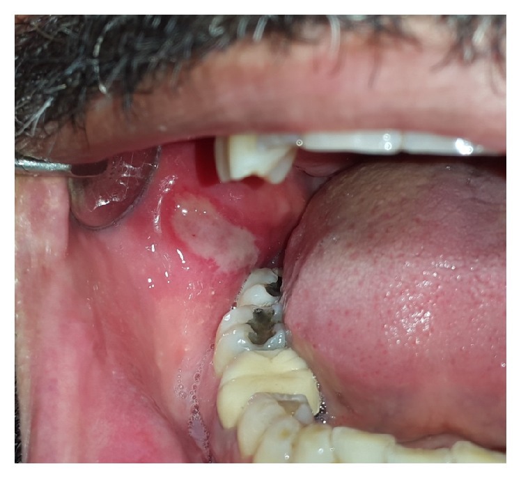
The most common sites are buccal mucosa, tongue and lower lip.
Traumatic ulcers usually present as a raised lesion with an erythematous border and yellowish-white necrotic pseudomembrane (figure 4).11
Acute multiple ulcers
Primary herpetic gingivostomatitis
Aetiology
Primary herpetic gingivostomatitis is a common oral manifestation of systemic herpes simplex virus (HSV) infection.11
Clinical presentation
These ulcers can present on keratinised and non-keratinised mucosa.
Typical clinical features of primary herpetic gingivostomatitis include:11
- Pyrexia
- Malaise
- Lymphadenopathy
- Halitosis
- Difficulty swallowing
- Intraoral pinhead vesicles which coalesce into large painful ulcers
Hand-foot mouth disease
Aetiology
This is a highly infectious viral disease caused by the Coxsackie virus affecting infants and children under 5 years of age.13
Site of ulceration
- Hands
- Feet
- Oral cavity: tongue, palate, buccal mucosa, gums, lips
Clinical presentation
Intra-oral lesions present as vesicles which ulcerate resulting in erythematous halos or macules approximately 2mm in diameter.13
Erythema multiforme
Erythema multiforme (EM) is an immunological vesiculobullous disorder affecting the skin and mucous membranes.
It is further categorised into:14
- EM major: has the involvement of one or several mucous membranes.
- EM minor: has no mucosal membrane involvement.
Aetiology
The aetiology of erythema multiforme is obscure. Infectious agents such as herpes simplex virus or use of certain drugs (nonsteroidal anti-inflammatories, antibiotics and anticonvulsants) can precipitate erythema multiforme.14
Clinical presentation
EM lesions are acute with mild or no prodromal symptoms. Oral lesions occur on the lips and anterior oral mucosa. The lesions present as asymmetrical, erythematous, lesions. These lesions usually heal without scarring.14
Chronic solitary ulcers
Ulcerative squamous cell carcinoma (SCC)
Ulcerative squamous cell carcinoma often presents as a single persistent ulcer. It is usually asymptomatic in the initial stages.11
Risk factors
Smoking, tobacco chewing and excessive alcohol intake increase the risk of developing oral cancer.
Clinical presentation
These ulcers can present anywhere on the oral mucosa but the following areas are the high-risk sites, where SCC is more likely to develop:
- The floor of the mouth
- Lateral and ventral borders of the tongue
- Retromolar region
- Lips
An ulcerative SCC can present as a crater-like lesion with rolled out margins, an indurated border and a velvety base (figure 5).11

Chronic multiple ulcers
Pemphigus vulgaris
Pemphigus vulgaris is an autoimmune, life-threatening disease presenting as blisters and erosions of the skin and mucous membrane.13
Clinical presentation
In the oral cavity, the most affected sites are the buccal mucosa, palate and gingivae.
The clinical presentation of the oral lesions starts as a bulla, which breaks to form shallow ulcers. When rubbed, the thin layer of epithelium sheds leaving a denuded base (known as Nikolsky’s sign).13
Mucous membrane pemphigoid
Mucous membrane pemphigoid is an autoimmune blistering disease that affects the basement membrane. This disease can affect the eyes, oral cavity, nasopharynx, larynx, oesophagus, genitourinary tract and anus.13
Clinical presentation
This disease manifests orally as vesicles, erosions, and desquamative gingivitis.
General management of oral ulceration
Primary care management
Along with reassurance and advice, management in primary care may include:
- Removing the source of trauma: patient education, smoothening sharp teeth/restoration.
- Chlorhexidine mouthwash.
- Topical corticosteroids.
- Topical anaesthetic for symptomatic relief.
- Regular monitoring of the oral mucosa.
- Appropriate referral to secondary care.
A 2-week urgent referral to secondary care is required if malignancy is suspected. For all other oral ulcerations where malignancy is not suspected, referrals can be made as urgent or routine to establish a definitive diagnosis or for management, if the condition cannot be managed in primary care.
Investigations
Further investigations are required when the aetiology of oral ulceration is unclear.
Laboratory investigations
Laboratory investigations should aim to exclude underlying nutritional deficiencies and gastrointestinal tract pathology:
- Full blood count
- Serum B12
- Ferritin
- Folate
Other investigations
- Biopsy: indicated if there is a clinical suspicion of malignancy or a definitive diagnosis cannot be established clinically.
- Direct/indirect immunofluorescence: used to help diagnose autoimmune blistering diseases (e.g. pemphigus vulgaris or mucous membrane pemphigoid and connective tissue disorders such as lupus).
These investigations are performed in secondary care.
Secondary care management
Management will differ depending on the underlying condition but may include:
- Topical or systemic corticosteroids
- Surgical removal of persistent ulcer
- Monitoring of oral mucosa.
Reviewer
Dr Rajinder Dodd
OMFS Specialty doctor at Pinderfields General Hospital
Editor
Dr Chris Jefferies
References
- Edsberg, L.E., Black, J.M., Goldberg, M., McNichol, L., Moore, L. and Sieggreen, M. Revised National Pressure Ulcer Advisory Panel Pressure Injury Staging System. Published in 2016. Available from: [LINK]
- Minhas, S., Sajjad, A., Kashif, M., Taj, F., Waddani, H.A. and Khurshid, Z. (2019). Oral Ulcers Presentation in Systemic Diseases: An Update. Published in 2019. Available from: [LINK]
- Scully, C. and Shotts, R. Mouth ulcers and other causes of orofacial soreness and pain. Published in 2001. Available from: [LINK]
- Tarakji, B., Gazal, G., Al-Maweri, S.A., Azzeghaiby, S.N. and Alaizari, N. (2015). Guideline for the Diagnosis and Treatment of Recurrent Aphthous Stomatitis for Dental Practitioners. Published in 2015. Available from: [LINK]
- Tarakji, B., Gazal, G., Al-Maweri, S.A., Azzeghaiby, S.N. and Alaizari, N. Minor aphthous ulceration. License: [CC-BY]. Available from: [LINK]
- Tarakji, B., Gazal, G., Al-Maweri, S.A., Azzeghaiby, S.N. and Alaizari, N. Major aphthous ulceration. License: [CC-BY]. Available from: [LINK]
- Tarakji, B., Gazal, G., Al-Maweri, S.A., Azzeghaiby, S.N. and Alaizari, N. Herpetiform aphthous ulceration. License: [CC-BY]. Available from: [LINK]
- Nair, J.R. and Moots, R.J. (2017). Behcet’s disease. Published in 2017. Available from: [LINK]
- Lankarani, K.B., Sivandzadeh, G.R. and Hassanpour, S. (2013). Oral manifestation in inflammatory bowel disease: A review. Published in 2013 Available from: [LINK]
- Collins, P. and Rhodes, J. (2006). Ulcerative colitis: diagnosis and management. Published in 2006. Available from: [LINK]
- Mortazavi, H., Safi, Y., Baharvand, M. and Rahmani, S. (2016). Diagnostic Features of Common Oral Ulcerative Lesions: An Updated Decision Tree. Published in 2016 Available from: [LINK]
- Mortazavi, H., Safi, Y., Baharvand, M. and Rahmani, S. Traumatic ulcer coated with pseudomembrane and surrounded by inflammatory halo. License: [CC-BY]. Available from: [LINK]
- Sundharam, Sivapatha & Sundararaman, Preethi & Kannan, S Karthiga. (2018). Oral Ulcers – A Review. Published in 2018. Available from: [LINK]
- Hasan, S., Jangra, J., Choudhary, P. and Mishra, S. (2018). Erythema Multiforme: A Recent Update. Biomedical and Pharmacology Journal, 11(1), pp.167–170.
- Mortazavi, H., Safi, Y., Baharvand, M. and Rahmani, S. Ulcerative SCC of tongue. License: [CC-BY]. Available from: [LINK]
