

- 📖 Geeky Medics OSCE Book
- ⚡ Geeky Medics Bundles
- ✨ 1300+ OSCE Stations
- ✅ OSCE Checklist PDF Booklet
- 🧠 UKMLA AKT Question Bank
- 💊 PSA Question Bank
- 💉 Clinical Skills App
- 🗂️ Flashcard Collections | OSCE, Medicine, Surgery, Anatomy
- 💬 SCA Cases for MRCGP
To be the first to know about our latest videos subscribe to our YouTube channel 🙌
Introduction
The suboccipital muscles are a set of four paired muscles in the back of the neck.
The suboccipital muscles attach to the atlas (C1), axis (C2) and occipital bone, connecting the atlas to the axis and the two vertebrae to the base of the skull. They are deep to the trapezius, splenius and semispinalis muscles and superficial to the atlanto-occipital and atlanto-axial joints.
These small muscles contribute to the movements of the head. When the suboccipital muscles contract, they extend the head at the atlanto-occipital joint and rotate the head at the atlanto-axial joints.
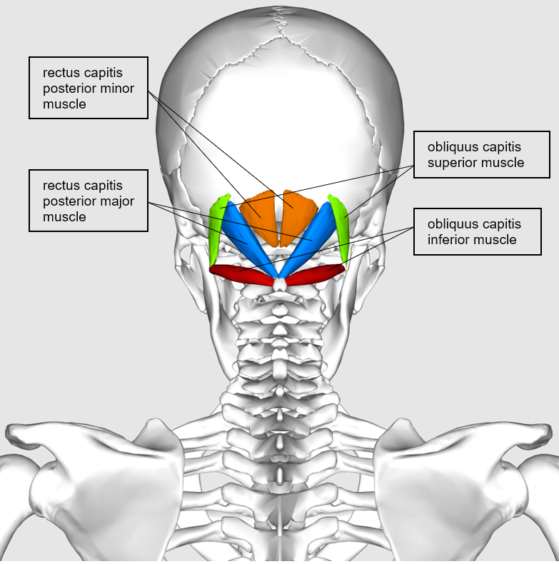
Rectus capitis posterior major
Origin
Spinous process of the axis
Insertion
Lateral aspect of the occipital bone, below the inferior nuchal line
Function
Extends the head and rotates the head to the ipsilateral side of the muscle
Innervation
Suboccipital nerve (posterior ramus of C1)
Blood supply
Vertebral artery and deep descending branches of the occipital artery
Rectus capitis posterior minor
Origin
Posterior tubercle of the atlas
Insertion
Medial aspect of the occipital bone, below the inferior nuchal line
Function
Extends the head
Innervation
Suboccipital nerve (posterior ramus of C1)
Blood supply
Vertebral artery and deep descending branches of the occipital artery
Obliquus capitis superior
Origin
Transverse process of the atlas
Insertion
Between the superior and inferior nuchal lines of the occipital bones
Function
Extends the head and bends it to the ipsilateral side (laterally)
Innervation
Suboccipital nerve (posterior ramus of C1)
Blood supply
Vertebral artery and deep descending branches of the occipital artery
Obliquus capitis inferior
Origin
Spinous process of the axis
Insertion
Transverse process of the atlas
Function
Rotates the head to the ipsilateral side
Innervation
Suboccipital nerve (posterior ramus of C1)
Blood supply
Vertebral artery and deep descending branches of the occipital artery
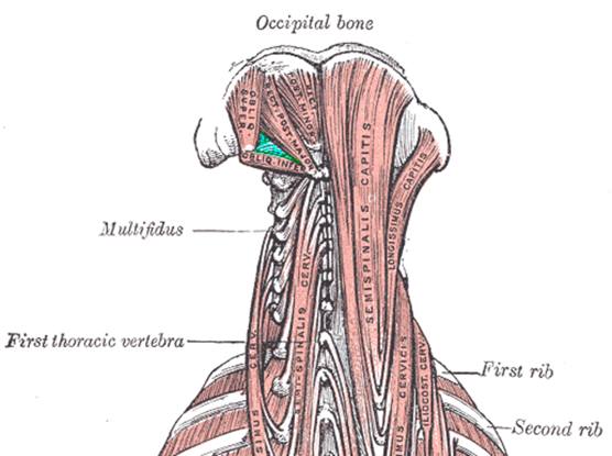
The suboccipital triangle
Three of the suboccipital muscles form the boundaries of the suboccipital triangle:
- The medial border of the triangle is formed by the rectus capitis posterior major muscle.
- The lateral border of the triangle is formed by the obliquus capitis superior muscle.
- The inferior border of the triangle is formed by the obliquus capitis inferior muscle.
- The roof of the triangle includes dense connective tissue.
- The floor of the triangle includes the posterior atlanto-occipital membrane and the posterior arch of the atlas.
The contents of the triangle include important structures such as:
- The vertebral artery
- The vertebral vein and suboccipital plexus
- The greater occipital nerve
- The suboccipital nerve
Key points
- The suboccipital muscles are a group of four paired deep muscles located at the base of the occipital bone.
- Their main actions are to extend and rotate the head.
- They consist of the rectus capitis posterior major, rectus capitis posterior minor, obliquus capitis superior and obliquus capitis inferior.
- They are innervated by the suboccipital nerve and their arterial supply comes from the vertebral artery and deep descending branches of the occipital artery.
Editor
Dr Chris Jefferies
References
- Richard L. Drake, A. Wayne Vogyl, Adam W. M. Mitchell. Gray’s Anatomy for Students. 4th Elsevier. Published in 2019.
- John T. Hansen. Netter’s Clinical Anatomy. 4th Elsevier. Published in 2018.
- Susan Standring. Gray’s Anatomy. 42nd Elsevier. Published in 2020.
- Donald A. Neumann. Kinesiology of the Musculoskeletal System. 3rd Elsevier. Published in 2016.
- Neil S. Norton. Netter’s Head and Neck Anatomy for Dentistry. 3rd Elsevier. Published in 2016.
Image references
- Anatomography. The suboccipital muscles. License: [CC BY-SA]
- Häggström, Mikael (2014). “Medical gallery of Mikael Häggström 2014”. WikiJournal of Medicine 1 (2). License: [Public domain]
