

- 📖 Geeky Medics OSCE Book
- ⚡ Geeky Medics Bundles
- ✨ 1300+ OSCE Stations
- ✅ OSCE Checklist PDF Booklet
- 🧠 UKMLA AKT Question Bank
- 💊 PSA Question Bank
- 💉 Clinical Skills App
- 🗂️ Flashcard Collections | OSCE, Medicine, Surgery, Anatomy
- 💬 SCA Cases for MRCGP
To be the first to know about our latest videos subscribe to our YouTube channel 🙌
Introduction
Tooth wear is the loss of hard dental tissue through non-carious causes. It is a multifactorial process encompassing both chemical and mechanical aetiologies and is characterised by the loss of natural tooth surface morphology.
Tooth wear is a prevalent condition and tends to accumulate with age.1 21st century lifestyle changes including increasingly acidic diets and overenthusiastic brushing in pursuit of the perfect smile, have led to increased prevalence of tooth wear in younger generations. As these individuals are likely to retain their teeth throughout life, it is imperative that tooth wear is identified swiftly and managed appropriately.
Aetiology
There are three main processes which contribute to the aetiology of tooth wear: erosion, abrasion and attrition.
Erosion
Erosion is the chemical dissolution of hard dental tissue without bacterial involvement. Acids are divided into two major categories:
- Intrinsic acid: gastric acid (hydrochloric acid) brought back up the alimentary tract into the oral cavity via regurgitation, vomiting or involuntary gastro-oesophageal reflux disease
- Extrinsic acid: dietary acids, acidic medicines, occupational exposure to acidic fumes
Erosion is recognised as the dominant factor in tooth wear with acid softening the surface layer of the tooth making it more susceptible to physical damage from abrasion or attrition.2 Erosive tooth wear is a frequently used term referring to tooth wear where dental erosion is the primary aetiological factor.3
Abrasion
Abrasion is the wearing of hard dental tissue by something other than the teeth. It can be due to foreign objects or substances repeatedly contacting the teeth. Most frequently abrasive tooth wear is associated with traumatic toothbrushing, although other habits such as holding objects between the teeth can also cause abrasion.
Attrition
Attrition is physical wear caused by tooth-to-tooth contact. Tooth clenching and grinding are linked to attrition and are habits that the patient may not be aware of.
Previously, abfraction was also suggested to play a contributory role in the aetiology of tooth wear and referred to wedge-shaped defects at the cementoenamel junction. Abfraction is theorised to be caused by occlusal loading resulting in tooth flexure and mechanical microfractures, causing loss of dental tissue at the cementoenamel junction. However, a recent consensus workshop has discouraged the use of this terminology as there is insufficient supporting evidence to justify abfraction as a separate process.3
Risk factors
Saliva is the main protective biological factor for the prevention of erosion. It acts to dilute, neutralise and clear acids whilst also providing a reservoir of calcium, phosphate and fluoride ions to enhance remineralisation.
Compromised salivary flow increases the risk of erosive tooth wear. The main causes are:
- Ageing
- Radio or chemotherapy
- Systemic diseases
- Drug-induced xerostomia
Other risk factors which increase the risk of tooth wear include:
- Frequent consumption of dietary acids: soft drinks, fresh fruit, fruit juices, pickles, sports drinks etc.
- Gastro-oesophageal reflux disease
- Alcoholism
- Anorexia nervosa/ bulimia nervosa
- Antidepressants/ sleeping medications
- Occupational: competitive swimmers, wine tasters
- Bruxism
Diagnosis
Tooth wear can result from a range of processes, as described above, and these tend to act synergistically rather than in isolation. The morphology of defects varies depending on the predominant cause. Due to this multifactorial nature of tooth wear, a thorough history of patient diet and lifestyle habits is also necessary to help inform the diagnosis.
Clinical characteristics
Enamel erosion tends to give tooth surfaces a smooth, glazed or dull appearance. For smooth surfaces, convex areas appear flattened and concavities can form. Initial lesions are located coronally to the cementoenamel junction with a preserved enamel ‘halo’ along the gingival margin. This is suggested to be due to the neutralising effect of gingival crevicular fluid.4
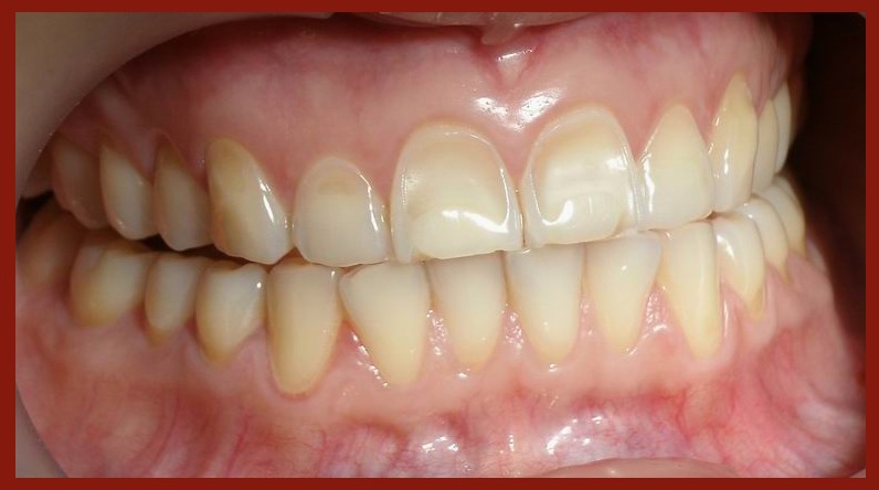
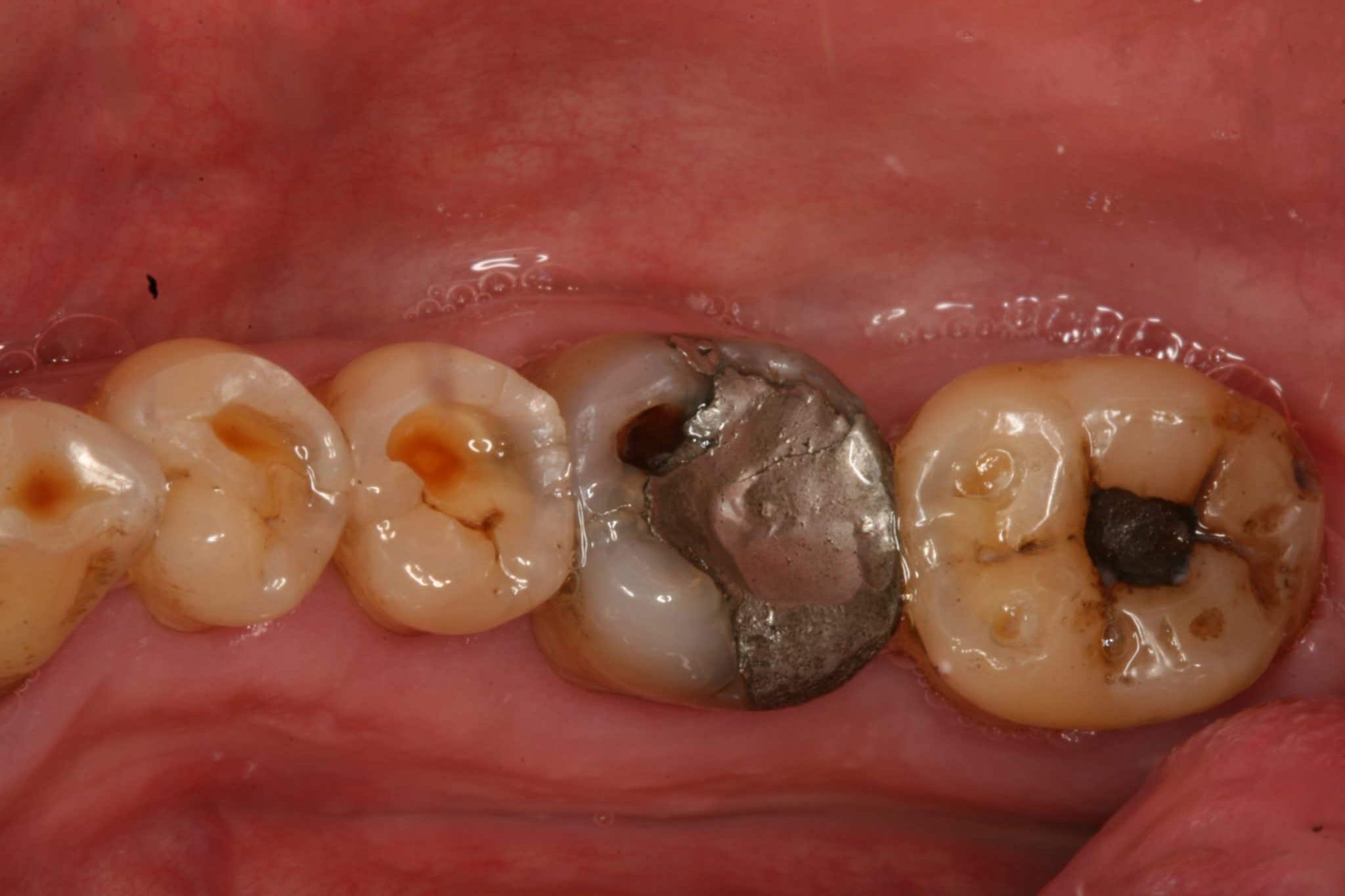
Occlusally, characteristic features of erosion are cupped occlusal surfaces and proud-standing restorations. Incisal edges become progressively more translucent and chipping may occur.
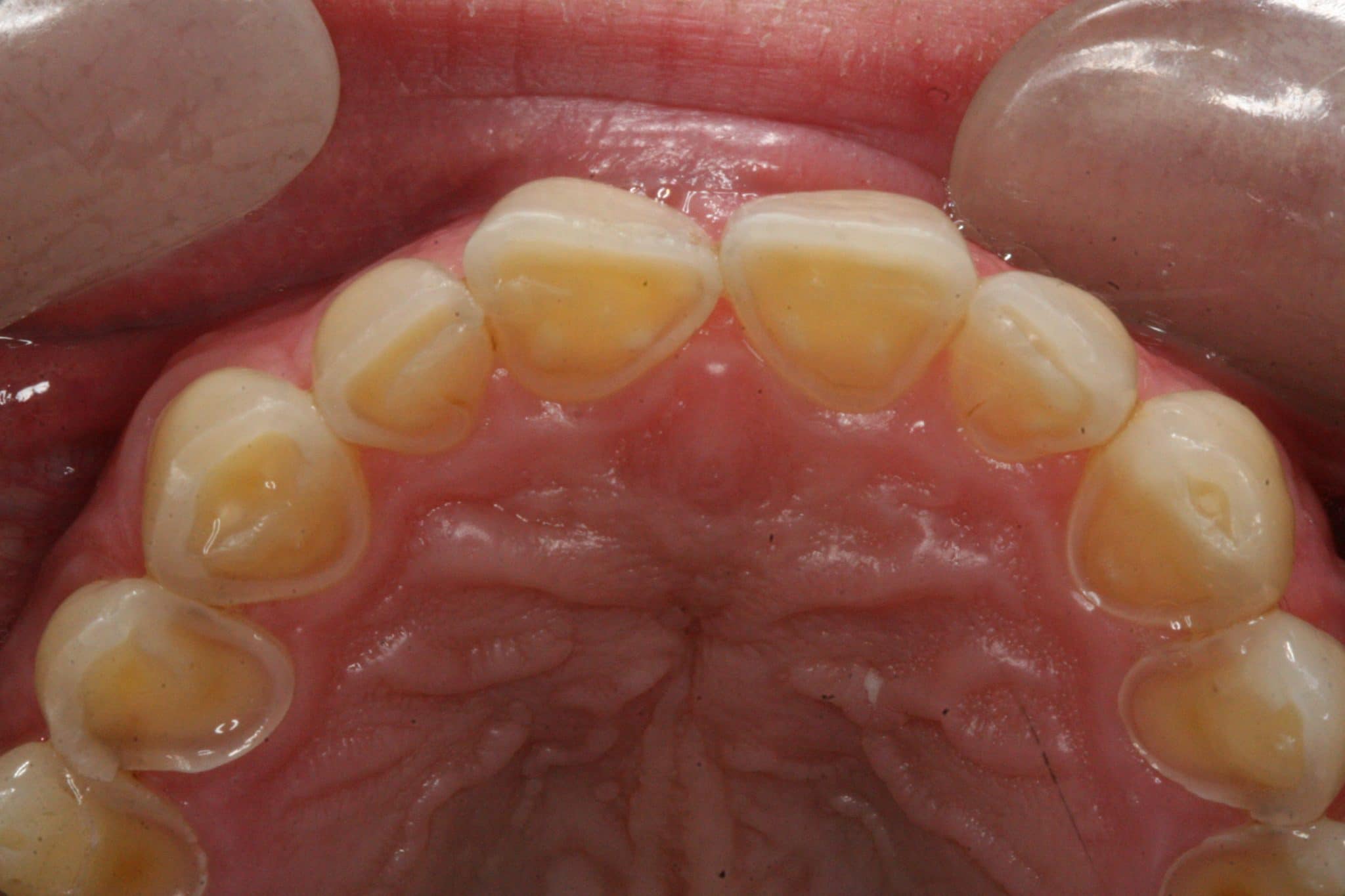
Intrinsic acids erode the palatal surfaces of the upper anterior teeth initially.
Non-carious cervical lesions tend to result from a combination of erosive and abrasive processes.
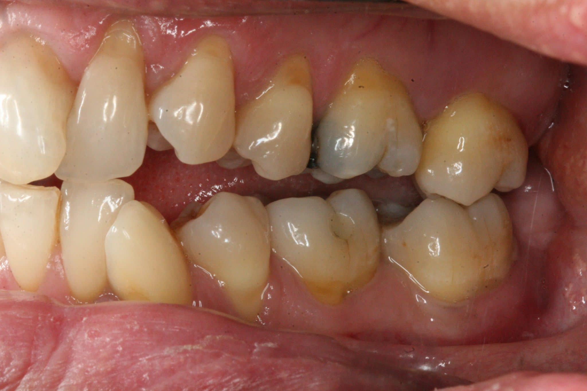
Stained worn surfaces suggest historic tooth wear, whereas glassy unstained surfaces suggest that wear may still be ongoing.
How to distinguish between occlusal erosion and attrition
Key points on differentiating occlusal erosion and attrition include:
- Attrition tends to result in flat lesions with sharp margins, whereas occlusal erosion leads to rounding of cusps and incisal edges.
- Generally, attrition creates corresponding wear facets on opposing teeth.
- Look for other signs of attritive wear, such as frictional keratosis or fracture lines in teeth/ restorations.
Why tooth wear is an issue for patients
Tooth wear is an irreversible process, except in the very early stages, and therefore accumulates with age.
For many patients a worn dentition can present the following problems:
- Pain: dentine hypersensitivity may be experienced if dentine is exposed. Severe tooth wear may also result in pulpal inflammation requiring endodontic treatment.
- Aesthetic concerns: yellowing due to reparative dentine deposition, dissatisfaction with the morphology of teeth
- Functional impairment
Severe tooth wear has also been shown to affect patient quality of life, with the negative impact comparable to that reported by edentulous patients.6
Management of tooth wear
Prevention
Prevention is the essential first-line management of tooth wear. This includes counselling and educating the patient to help them identify and reduce aetiological factors: 7
- Diet diary and analysis with individually focused advice: highlight dietary acids, recommend decrease frequency of consumption and keep to mealtimes.
- Explore intrinsic acid sources: vomiting, reflux, eating disorders – refer to GP if required.
- Recommend fluoridation measures to increase the resistance of tooth surface to erosion: use of fluoride mouth rinse during the day, stannous fluoride toothpaste reinforcing spit don’t rinse.
- Address any traumatic toothbrushing habits, ensure brushing is not straight after an acidic intake. Consider brushing before breakfast.
Preventative agents can be applied in the surgery or at home but the need for frequent application makes home care products a more desirable option. Stannous fluoride has been clinically proven to reduce erosive tooth wear and this is readily available in over-the-counter toothpaste.8 Professionally applied high fluoride varnishes also provide some protection against tooth wear but they need to be applied regularly making them a less cost-effective solution.
Monitoring
The regular monitoring of tooth wear with study models and intra-oral photographs allows for the detection of tooth wear progression. Tooth wear indices are also a useful tool to diagnose, grade and monitor wear.
The Basic Erosive Wear Examination (BEWE)
The BEWE is currently the gold standard tooth wear index and has both a grading and management section.9 General dental practitioners are encouraged to use the BEWE as part of their routine examinations to ensure any tooth wear is recorded.
Table1. BEWE criteria for grading erosive wear9
|
Score |
Clinical appearance |
|
0 |
No erosive tooth wear |
|
1 |
Initial loss of surface texture |
|
2 |
Distinct defect, hard tissue loss <50% of surface area |
|
3 |
Hard tissue loss ≥50% of surface area |
Similarly to the Basic Periodontal Examination (BPE), the BEWE examination is completed for all teeth but only the surface with the highest score is recorded for each sextant. Once all sextants have been assessed, the scores are added to give a cumulative score which can be used to guide clinical management.
Table 2. BEWE risk levels to guide clinical management9
|
Risk level |
Cumulative score of all sextants |
Management |
|
None |
Less than or equal to 2 |
Routine maintenance and observation Repeat at 3-year intervals |
|
Low |
Between 3 and 8 |
Oral hygiene and dietary assessment, and advice, routine maintenance and observation Repeat at 2-year intervals |
|
Medium |
Between 9 and 13 |
Oral hygiene and dietary assessment, and advice, identify the main aetiological factor(s) for tissue loss and develop strategies to eliminate respective impacts Consider fluoridation measures or other strategies to increase the resistance of tooth surfaces Ideally, avoid the placement of restorations and monitor erosive wear with study casts, photographs, or silicone impressions Repeat at 6–12-month intervals |
|
High |
14 and over |
Oral hygiene and dietary assessment, and advice, identify the main aetiological factor(s) for tissue loss and develop strategies to eliminate respective impacts Consider fluoridation measures or other strategies to increase the resistance of tooth surfaces Ideally, avoid restorations and monitor tooth wear with study casts, photographs, or silicone impressions Especially in cases of severe progression consider special care that may involve restorations Repeat at 6–12-month intervals |
Restorative intervention
Restorations should be avoided if possible, particularly in the cervical region as they can act as a plaque trap. If restorations are required for aesthetics, function or to manage symptoms of pain, it is important to stabilise the causative factors prior to restorative rehabilitation to ensure longevity. In general, bonded restorations should be used where possible to minimise the need to heavily prepare teeth resulting in further tissue loss.
Key points
- Tooth wear is the loss of hard tissue through non-cariogenic causes.
- The three main types of tooth wear are erosion, abrasion and attrition.
- Most frequently tooth wear is caused by a combination of these processes acting synergistically, although erosion is often the dominant factor.
- Stained worn surfaces suggest tooth wear is historic, whereas glassy unstained surfaces suggest that wear may still be ongoing.
- A worn dentition can cause pain, functional impairment and aesthetic concerns, all of which can impact on a patient’s quality of life.
- The BEWE is the gold standard tooth wear index used to grade and monitor tooth wear in general dental practice.
- Management of tooth wear involves identifying the main aetiological factor(s), instigating preventative measures and monitoring for progression.
- If restorative intervention is required it should involve minimum preparation of the tooth and utilise bonding agents.
References
- Van’t Spijker A, Rodriguez JM, Kreulen CM, Bronkhorst EM, Bartlett DW, Creugers NH. Prevalence of tooth wear in adults. Int J Prosthodont. 2009;22(1):35-42. Available from: [LINK].
- Shellis RP, Addy M. The interactions between attrition, abrasion and erosion in tooth wear. Monogr Oral Sci. 2014;25:32-45. Available from: [LINK].
- Schlueter N, Amaechi BT, Bartlett D, et al. Terminology of Erosive Tooth Wear: Consensus Report of a Workshop Organized by the ORCA and the Cariology Research Group of the IADR. Caries Res. 2020;54(1):2-6. Available from: [LINK].
- Ganss C, Lussi A. Diagnosis of erosive tooth wear. Monogr Oral Sci. 2014;25:22-31. Available from: [LINK].
- Финитор. Dental erosion. Licence: CC BY-SA. Available from: [LINK].
- Papagianni CE, van der Meulen MJ, Naeije M, Lobbezoo F. Oral health-related quality of life in patients with tooth wear. J Oral Rehabil. 2013;40(3):185-90. Available from: LINK
- Griffith LJ, Newcombe RG, Daly S, Seong J, Davies M, West NX. A novel cervical tooth wear and recession index, The Cervical Localisation Code, and its application in the prevention and management of dentine hypersensitivity. J Dent. 2020;103432. Available from: [LINK].
- West NX, Seong J, Hellin N, Eynon H, Barker ML, He T. A clinical study to measure anti-erosion properties of a stabilized stannous fluoride dentifrice relative to a sodium fluoride/triclosan dentifrice. Int J Dent Hyg. 2017;15(2):113-119. Available from:[LINK].
- Bartlett D, Ganss C, Lussi A. Basic Erosive Wear Examination (BEWE): a new scoring system for scientific and clinical needs. Clin Oral Investig. 2008;12 Suppl 1:S65-8. Available from: [LINK].
