

- 📖 Geeky Medics OSCE Book
- ⚡ Geeky Medics Bundles
- ✨ 1300+ OSCE Stations
- ✅ OSCE Checklist PDF Booklet
- 🧠 UKMLA AKT Question Bank
- 💊 PSA Question Bank
- 💉 Clinical Skills App
- 🗂️ Flashcard Collections | OSCE, Medicine, Surgery, Anatomy
- 💬 SCA Cases for MRCGP
To be the first to know about our latest videos subscribe to our YouTube channel 🙌
Introduction
Flexor sheath tenosynovitis is inflammation of the tendon sheath and has non-infective and infective causes. Bacterial infections are the most common cause of flexor sheath infections, causing 2.5% to 9.4% of hand infections and are often secondary to penetrating trauma.1
Causative organisms include Staphylococcus aureus, Staphylococcus epidermis, B-haemolytic Streptococcus, and gram-negative Pseudomonas aeruginosa.
Infective flexor sheath tenosynovitis is a surgical emergency as the raised pressure in the tendon sheath can impair blood flow, leading to necrosis of the tendons and devitalisation of the fingers.2
The focus of this article will be the management of infective flexor sheath tenosynovitis.
Aetiology
Common mechanisms of flexor sheath infection include:1,3,4
- Penetrating trauma (e.g. animal bites)
- Direct spread from felons, septic joints and deep space infections
- Haematogenous spread
In the hand, flexor sheaths are closed continuous systems that contain the flexor digitorum profundus and the flexor digitorum superficialis tendons. The infection travels around the synovial sheath, which surrounds the flexor tendon and contributes to protecting and nourishing the tendon.
The infection begins as exudative fluid, followed by the development of purulent fluid. Bacterial overgrowth increases the inflammatory response and purulent fluid production. This increases the pressure between the layers of the flexor sheath. As the vascular supply to the tendons is from the surrounding digital arteries, the increased pressure interferes with this, leading to tendon ischaemia and necrosis.
The most common organism in infective flexor sheath tenosynovitis is Staphylococus aureus, however other skin commensals are commonly involved Staphylococcus epidermidis, beta-hemolytic Streptococcus, Pseudomonas aeruginosa. Eikenella and Pasturella multocida are involved in human and animal bites, respectively. Mixed flora and gram-negative organisms are common in patients who are immunocompromised.5
Risk factors
Risk factors for flexor sheath infections include:6
- Diabetes
- Use of intravenous drugs
- Immunocompromised patients (e.g. transplant)
Clinical features
History
Symptoms include pain and swelling of the palmar aspect of the affected digit. Patients usually present with symptoms of 24-48 hours duration.6
Penetrating injury in the preceding two to five days may help to establish the causative organism involved.
Other important areas to cover in the history include:6
- Past medical history: diabetes, renal failure, and peripheral vascular disease have poorer outcomes
- Medication history: immunosuppressants
- Use of illicit substances and intravenous drug use
- Occupation and hand dominance: this guides rehabilitation requirements and support postoperatively
Clinical examination
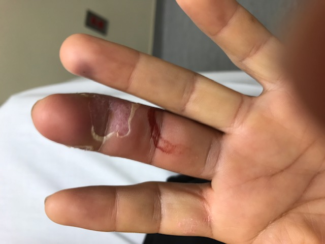
There are four cardinal features known as Kanavel’s signs of flexor sheath infection:7
- Pain on passive extension of the digit
- Tenderness on palpation along the flexor sheath
- Fusiform swelling of the affected digit
- Flexed finger posture
The earliest of these signs is pain on passive extension. However, the signs can be less evident in patients who are immunocompromised.8
The hand should also be examined for any penetrating wound and erythema, swelling or tenderness of the thenar or hypothenar eminences, which may indicate concurrent infected radial or ulnar bursa, respectively.9
Differential diagnoses
The differential diagnosis for infective flexor sheath tenosynovitis includes other common hand infections. However, flexor sheath tenosynovitis may also have a non-infective aetiology (autoimmune, overuse and idiopathic).
Table 1. Differential diagnoses for flexor sheath tenosynovitis6,10
| Differential diagnosis | Clinical features |
| Infective causes of flexor sheath tenosynovitis | |
| Felon | A closed space infection of the distal finger pulp. Throbbing abscess over the distal finger phalanx, usually due to penetrating injury. |
| Cellulitis | Diffuse inflammation without underlying pus collection. |
| Septic arthritis of the interphalangeal or metacarpophalangeal joint | Symptoms are localised to the affected joint with restricted range of movement and pain. |
| Deep space infection and collar button infections | Usually affecting the webspace between fingers (collar button infection), thenar or hypothenar spaces. Pain on flexion of the affected fingers. |
| Gout | Often presents with monoarticular inflamed, red and swollen joint. Look for gouty tophi. |
| Non-infective causes of flexor sheath tenosynovitis | |
| Autoimmune tenosynovitis | Rheumatoid arthritis: may have associated rheumatoid nodules or hand deformity Psoriatic arthritis: skin changes may be present. |
| Overuse tenosynovitis | Repetitive movements such as prolonged computer work. Findings may be non-elucidative |
| Idiopathic tenosynovitis | Flexor sheath inflammation with no clear cause |
Investigations
Laboratory investigations
Relevant laboratory investigations include:
- Full blood count: typically raised white cell count
- Inflammatory markers (CRP): usually raised
- Serum urate: useful when considering gout as a differential
- Blood cultures: should be obtained if there are signs of sepsis
Imaging
Plain X-rays may be useful to exclude a foreign body or osteomyelitis.
Diagnosis
Flexor sheath infection is a clinical diagnosis based on the history and examination.
The presence of all four of Kanavel’s signs predicts flexor sheath tenosynovitis, but not all four signs are needed to make a diagnosis. Detection of the four signs has a high sensitivity for diagnosis of 91.4 to 97.1% but a low specificity of 51.3% to 69.2%.11
Management
Immediate management
Immediate measures included hand elevation and broad-spectrum antibiotics. Patients should be urgently referred to the hand surgery department (plastic surgeons or orthopaedic surgeons, depending on the hospital).
Operative management
Patients should be seen immediately with an aim to undergo operative management as early as possible, within 24 hours.12
Operative management consists of a thorough surgical flexor sheath washout using an open or closed approach in a formal operating theatre environment. Samples of the fluid inside the flexor sheath should be sent to the microbiology laboratory to tailor antibiotic therapy.
Strict elevation and close monitoring of the hand is essential post-operatively. Patients may return to theatres for a re-look and further washout, especially if clinical features do not resolve or infections are particularly purulent.
Non-operative management
A trial of non-operative management, including hospital admission, intravenous antibiotics, splinting and hand elevation, may be useful in very early and mild presentations. However, a senior surgeon should make this decision. If no improvement is seen within 24 hours, surgery would be indicated.
Non-infective tenosynovitis
Non-infective causes of tenosynovitis vary in treatment based on the cause (e.g. non-steroidal anti-inflammatory drugs). Senior input is required, and rheumatology involvement may be indicated.11
Follow-up
Patients should be followed up with hand therapy weekly for at least six weeks.12
Complications
Complications if a flexor sheath infection is not diagnosed or treated promptly can include:
- Stiffness
- Tendon or pulley rupture
- Spread of infection
- Loss of soft tissue
- Necrotising fasciitis
- Finger amputation
- Osteomyelitis
- Horseshoe abscess
Key points
- Flexor sheath tenosynovitis is inflammation of the tendon sheath
- This may be non-infective or infective
- Non-infective causes include autoimmune conditions, tendon overuse and idiopathic
- Infective tenosynovitis is often caused by a bacterial infection (i.e. Staphylococcus aureus, Staphylococcus epidermidis and Beta-haemolytic streptococcus), following penetrating trauma
- Kanavel’s signs of pain on passive digit extension, a flexed digit with fusiform swelling and pain on palpation along the flexor sheath can help with diagnosis
- Diagnosis of infective flexor sheath tenosynovitis is mainly clinical
- Patients with an infective cause will need hand elevation, antibiotics and urgent referral to a hand surgery team for consideration of surgery, which almost always will entail a thorough flexor sheath washout
- If flexor sheath tenosynovitis is treated as a surgical emergency some of the negative sequelae can be mitigated
Reviewer
Professor Benjamin Miranda
Consultant Plastic Surgeon
Editor
Dr Chris Jefferies
References
- Abrams, R. A., & Botte, M. J. (1996). Hand infections: treatment recommendations for specific types. JAAOS-Journal of the American Academy of Orthopaedic Surgeons, 4(4), 219-230.
- Pang, H. N., Teoh, L. C., Yam, A. K., Lee, J. Y. L., Puhaindran, M. E., & Tan, A. B. H. (2007). Factors affecting the prognosis of pyogenic flexor tenosynovitis. JBJS, 89(8), 1742-1748.
- Harris, P. A., & Nanchahal, J. (1999). Closed continuous irrigation in the treatment of hand infections. The Journal of Hand Surgery: British & European Volume, 24(3), 328-333.
- Schaefer, R. A., Enzenauer, R. J., Pruitt, A., & Corpe, R. S. (1992). Acute gonococcal flexor tenosynovitis in an adolescent male with pharyngitis: a case report and literature review. Clinical Orthopaedics and Related Research®, 281, 212-215.
- Flevas, D. A., Syngouna, S., Fandridis, E., Tsiodras, S., & Mavrogenis, A. F. (2019). Infections of the hand: an overview. EFORT Open Reviews, 4(5), 183-193.
- Hermena S, Tiwari V. Pyogenic Flexor Tenosynovitis. StatPearls Publishing; 2023. Available from: [LINK]
- Kanavel, A. B. (1921). Infections of the Hand. Lea & Febiger.
- Draeger, R. W., & Bynum Jr, D. K. (2012). Flexor tendon sheath infections of the hand. JAAOS-Journal of the American Academy of Orthopaedic Surgeons, 20(6), 373-382.
- Chovanec, K., Arsene, C., Gomez, C., Brixey, M., Tolles, D., Galliers, J. W., … & Goodwin, L. (2021). Association of CLABSI with hospital length of stay, readmission rates, and mortality: A retrospective review. Worldviews on Evidence‐Based Nursing, 18(6), 332-338.
- Ray G, Sandean DP, Tall MA. Tenosynovitis. StatPearls Publishing; 2023. Available from: [LINK]
- Kennedy, C. D., Lauder, A. S., Pribaz, J. R., & Kennedy, S. A. (2017). Differentiation between pyogenic flexor tenosynovitis and other finger infections. Hand, 12(6), 585-590.
- The British Society for Surgery of the Hand. BSSH standards of care in hand trauma. Available from: [LINK]
Image references
- Figure 1. Jonrako. Infective tenosynovitis. License: [CC BY-SA]
