

- 📖 Geeky Medics OSCE Book
- ⚡ Geeky Medics Bundles
- ✨ 1300+ OSCE Stations
- ✅ OSCE Checklist PDF Booklet
- 🧠 UKMLA AKT Question Bank
- 💊 PSA Question Bank
- 💉 Clinical Skills App
- 🗂️ Flashcard Collections | OSCE, Medicine, Surgery, Anatomy
- 💬 SCA Cases for MRCGP
To be the first to know about our latest videos subscribe to our YouTube channel 🙌
Introduction
Malignant spinal cord compression (MSCC) is a medical emergency caused by compression of the spinal cord or cauda equina. It can result from direct pressure, vertebral collapse or instability due to metastatic or local spread of tumours.
It occurs in 5-10% of cancer patients and will be the presenting feature of cancer in 20% of patients with MSCC.1
Aetiology
MSCC occurs when there is compression of the spinal cord or cauda equina related to malignancy. This can occur through several mechanisms:2,3
- Direct compression from tumour expansion into the epidural space: most commonly due to metastases
- Direct compression from collapsed vertebral bodies: most commonly due to metastases; the most common region of bony metastases is the thoracic spine (around 70%), followed by the lumbosacral (20%) and cervical (10%)
- Direct extension of an intra-abdominal or intra-thoracic primary malignancy
- Direct compression from a primary malignancy of the vertebral body
- Compression from intradural spinal cord malignancies
The compression leads to oedema, venous congestion and demyelination. This leads to loss of neurological function, which is variable depending on the level of the lesion(s).
Risk factors
While any malignancy has the potential to metastasise to the spine, it is more common in certain types of cancer:2
- Prostate cancer (20%)
- Lung cancer (20%)
- Breast cancer (17%)
- Renal cancer (12%)
- Multiple myeloma
The risk of MSCC is also related to the duration of the disease. The median age of patients at the time of MSCC diagnosis is 65.
Clinical features
History
The most common presenting complaint in MSCC is back pain, and it may have been present for several weeks before MSCC is diagnosed.
Red flag features of back pain that may suggest MSCC (particularly in patients with a known cancer diagnosis) include:
- Thoracic or cervical pain
- Progressive lumbar pain
- Spinal pain aggravated by straining
- Localised spinal tenderness
- Nocturnal pain preventing sleep
Other symptoms can vary depending on the level of compression but commonly include:
- Limb weakness (e.g. the limb feeling “heavy” or “stiff”, difficulty climbing stairs)
- Loss of coordination
- Sensory disturbance
- Autonomic dysfunction (e.g. urinary retention, faecal incontinence due to loss of anal tone, constipation)
- Cauda equina syndrome (bladder/bowel dysfunction, saddle anaesthesia, leg weakness, gait disturbance, back pain)
Patient education
The Christie NHS Foundation Trust have produced a range of MSCC patient information resources, including alert cards which list the red flag symptoms of MSCC.
Clinical examination
Patients with suspected MSCC should have a full neurological examination.
Clinical findings will vary depending on the level of compression, but there is often poor concordance between the spinal level and symptoms. Additionally, over 20% of patients will have compression at multiple levels.
Typical examination findings below the level of compression include:
- Limb weakness or paralysis (assess using the MRC muscle power scale)
- Sensory disturbance
- Brisk reflexes
- Increased muscle tone
- Clonus
- Upgoing plantars
- Reduced anal tone
- Palpable, distended bladder
Investigations
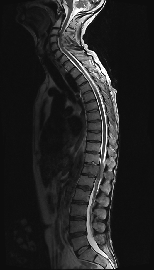
Any patient with suspected MSCC should be referred for an urgent MRI of the spine within 24 hours. If an MRI is contraindicated (e.g.pacemakers and metallic foreign bodies), the next best imaging modality is CT.
Once imaging has been obtained, further investigations can help to guide treatment.
Bedside investigations
Relevant bedside investigations include:
- Bladder scan: to assess for urinary retention
Laboratory investigations
Relevant laboratory investigations include:
- Baseline blood tests (FBC, U&Es, LFTs): to assess general fitness and for imaging
- Bone profile: to assess for hypercalcaemia
- Clotting and group & save: if the patient is likely to require surgery
- LDH: higher levels are associated with poor prognosis
- Myeloma screen: if the patient does not have a known cancer diagnosis
- Tumour markers: may help with the assessment of cancer stage and suitability for treatment or diagnosis if MSCC is the initial presentation
An up-to-date staging CT is needed to help assess the cancer stage and suitability for treatment.
Diagnosis
MSCC is diagnosed by imaging (spinal MRI or CT). The scan should also be reported within 24 hours of patient presentation to enable rapid treatment.
If MSCC is the presenting feature, then patients will require further investigation to diagnose the primary tumour, but this should not delay the assessment and treatment of the cord compression.
Management
The management of MSCC can be split into initial management of suspected MSCC, definitive management of confirmed MSCC and ongoing rehabilitation.
Initial management (suspected MSCC)
Initial management of suspected MSCC includes:
- Dexamethasone (16mg OD or 8mg BD): to reduce inflammation and oedema
- Omeprazole (or alternative PPI) for stomach protection
- Low molecular weight heparin (or alternative) for thromboprophylaxis
- Contact the local MSCC coordinator to arrange admission and MRI spine (follow local referral guidelines)
- Immobilise the spine if instability is suspected
- Analgesia
Definitive management (confirmed MSCC)
Management options for confirmed MSCC include surgery +/- radiotherapy, radiotherapy alone or best supportive care.
The management choice depends on various factors, including the patient’s performance status, the extent of their malignancy, other comorbidities, level(s) of compression and neurological deficit at presentation. If the patient has had paraplegia and loss of sphincter control for >48 hours, treatment is unlikely to improve neurological function.
Surgery
Surgical decompression can be considered in patients with a life expectancy >6 months, some preserved neurological function and limited levels of compression. This would usually be followed by radiotherapy to reduce the recurrence risk.
Radiotherapy alone
Radiotherapy alone is generally the management of choice for most patients, particularly those with multiple medical comorbidities, rapidly progressive neurological deficits and radiosensitive tumours (e.g. myeloma).
In these patient groups, radiotherapy is given to improve function. However, it can also be given for palliative pain relief in those with a life expectancy of <6 months, low-performance status and established paraplegia >24 hours.
Best supportive care
Patients who are frail, unwell or have a short life expectancy require palliative management. This focuses on managing symptoms and considering referral to specialist palliative care teams for advice.
Rehabilitation
Ongoing rehabilitation is essential, and the patient’s exact needs will vary depending on the neurological deficit and treatment given. Common considerations include:
- Pain control
- Thromboprophylaxis
- Weaning of dexamethasone
- Breathing exercises and forced expiratory techniques to aid chest clearance
- Prevention of contractures/spasticity
- Continence management
- Prevention of pressure ulcers
- Communication assistance
- Mobility aids
- Psychological care
Complications
Complications of MSCC include:
- Pressure ulcers
- Autonomic dysreflexia
- Deep vein thrombosis
- Pulmonary embolism
- Falls
- Urinary tract infections
- Pneumonia
- Muscle contractures
- Psychological complications
Key points
- MSCC occurs when there is compression of the spinal cord or cauda equina related to malignancy and can lead to significant disability
- It affects 5-10% of cancer patients and is most commonly associated with prostate, lung, breast and renal cancer
- Symptoms vary depending on the level of compression, but the most common presenting symptoms are back pain, limb weakness and sensory disturbances
- Diagnosis is made by spinal MRI, which should be performed within 24 hours of the initial presentation
- High-dose oral steroids (dexamethasone) should be given once MSCC is suspected
- Definitive management consists of surgical decompression, radiotherapy and symptom management, with the exact choice depending on the patient’s fitness, the extent of the underlying disease and neurological deficits
Editor
Dr Chris Jefferies
References
- Scottish Palliative Care Guidelines. Malignant spinal cord compression. Published 2021. Available from: [LINK]
- BMJ Best Practice. Malignant Spinal Cord Compression. Published 2023. Available from: [LINK]
- NHS Greater Manchester, Lancashire and South Cumbria Strategic Clinical Networks. Guidelines for the management of malignant spinal cord compression. Published 2014. Available from: [LINK]
Image references
- Figure 1. Case courtesy of Andrew Murphy, Radiopaedia.org, rID: 47515
