

- 📖 Geeky Medics OSCE Book
- ⚡ Geeky Medics Bundles
- ✨ 1300+ OSCE Stations
- ✅ OSCE Checklist PDF Booklet
- 🧠 UKMLA AKT Question Bank
- 💊 PSA Question Bank
- 💉 Clinical Skills App
- 🗂️ Flashcard Collections | OSCE, Medicine, Surgery, Anatomy
- 💬 SCA Cases for MRCGP
To be the first to know about our latest videos subscribe to our YouTube channel 🙌
Introduction
A haemorrhoid is an enlargement of the vascularity of the anal cushions in the anal canal. As a result, the anal cushions increase in size and can project distally, both within the anal canal and externally.
Haemorrhoids are common in the adult population and are seen in acute and outpatient settings.
Aetiology
The sliding anal canal lining theory, first described in 1975, is a widely accepted theory to explain the development of haemorrhoids.1 This is where the tissues supporting the anal cushions deteriorate, resulting in the downward displacement of the internal anal cushions and subsequently causing venous dilatation.
If you imagine the anus as a clock, with 12 o’clock situated towards to perineum, the major anal cushions lie at:
- 3 o’clock
- 7 o’clock
- 11 o’clock
Classification of haemorrhoids
Haemorrhoids are classified using Goligher’s classification based on the appearance, characteristics and degree of the prolapse:
- First degree: anal cushions bleed but remain in the rectum (no prolapse)
- Second degree: prolapse of haemorrhoids on defaecation or straining (spontaneously reduces)
- Third degree: prolapse of haemorrhoids on defaecation or straining (requires manual reduction)
- Fourth degree: prolapse remains at all times and is irreducible
Risk factors
Risk factors for haemorrhoids include:
- Constipation: this will predispose the patient to increase time straining on the toilet
- Increased age
- Increased abdominal pressure, such as in pregnancy and labour
- Diarrhoea
- High BMI
Clinical features
History
Patients with haemorrhoids can be asymptomatic or only have intermittent symptoms.
Typical symptoms of haemorrhoids may include
- Pruritis ani (itching peri-anally)
- Rectal bleeding: this is usually bright red, fresh blood seen on the tissue paper upon wiping
- Palpable lump in or around the anus: this may or may not be reducible
- Pain: most haemorrhoids are painless; however, they become acutely tender and painful if the haemorrhoid thromboses
- Discomfort around the anus, fullness or feeling of incomplete defaecation (known as tenesmus)
Clinical examination
As haemorrhoids are vascular structures, you cannot always palpate them on internal examination.
Visualisation on external examination is arguably the most important examination step for diagnosing haemorrhoids.
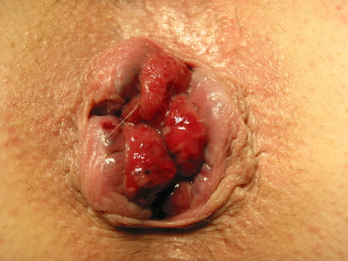
On external examination, there may be a swelling, which may be reducible or irreducible and is usually non-tender. However, if there is suspicion the haemorrhoid has thrombosed, it will be painful to touch, purple or blue in colour and oedematous.
Differential diagnoses
Differential diagnoses to consider in the context of suspected haemorrhoids include:
- Anorectal or colorectal cancer: usually a craggy, irregular mass with contact bleeds on rectal examination. If there is new rectal bleeding, it is important to rule out malignancy; this is dependent on red flag symptoms such as age, change in bowel habit, abdominal pain or mass or weight loss.
- Adenomatous polyp: a rectal mass may be visualised on proctoscopy; it can present with rectal bleeding, diarrhoea and constipation
- Perianal abscess: this is a local collection of pus around the anus; it is usually painful, increases in size over a shorter timeframe, is fluctuant and will have signs of local infection such as erythema and warmth. If the patient is systemically unwell, they can present with signs of sepsis.
- Portal hypertension: this can cause varices in multiple locations, such as the anal canal.
Investigations
Beside investigations
Relevant bedside investigations in the context of haemorrhoids include:
- Proctoscopy: to visualise the anal canal
Laboratory investigations
Relevant laboratory investigations in the context of haemorrhoids include:
- Full blood count (FBC): to identify anaemia (a red flag for malignancy)
- Faecal immunochemical test (FIT): a stool sample usually performed in the community if there is suspicion of colorectal cancer
Colonoscopy
A colonoscopy may be required to exclude other differential diagnoses (e.g. colorectal cancer). This can be limited to a flexible sigmoidoscopy (if suspecting of lower gastrointestinal tract pathology).
Imaging
Relevant imaging investigations in the context of haemorrhoids include:
- CT colonoscopy: if suspecting colorectal cancer and the patient is unfit for a colonoscopy
- MRI rectum: a good modality for defining complex anorectal pathology
Diagnosis
A diagnosis of haemorrhoids can be reached with a history and typical examination findings. Usually, imaging is not required.
Management
Acute management
Acutely thrombosed haemorrhoids can present acutely with pain and tenderness. Initial management includes:
- Analgesia
- Laxatives (to reduce straining)
- Sitz hot salt bath: this is a shallow bath to sit in to relieve discomfort in the perineal region
Conservative management
Most patients can be treated conservatively; this involves lifestyle advice and laxatives.
Lifestyle advice includes increasing fibre and fluid to ensure the stools are looser and avoid constipation. Regular laxative use is encouraged to keep stools loose. Medications which cause constipation should be avoided, such as opioids. If discomfort occurs, topical local anaesthetic gels can be applied (e.g. Instillagel®).
Rubber band ligation
Rubber band ligation is a common outpatient non-surgical treatment for haemorrhoids.
A suction gun draws up the haemorrhoid, followed by a firing of a rubber band to cut off the blood supply to the haemorrhoid (strangulate). This requires precision suction of the haemorrhoid and correct placement of the rubber band. If placed below the dentate line, it will cause the patient pain. Patients may require multiple sessions of banding.
Surgical management
There are two main surgical methods:
- Transanal haemorrhoid dearterialisation (THD): performed using a doppler attached to a proctoscope; the surgeon will identify the artery feeding into the anal cushion (typically located at 1 o’clock, 3 o’clock, 5 o’clock, 7 o’clock, 9 o’clock and 11 o’clock), these are tied off, the haemorrhoid then shrinks following this.
- Haemorrhoidectomy: this is usually via the Milligan Morgan haemorrhoidectomy technique, where the prolapsed haemorrhoids and/or anal cushions are excised (3 o’clock, 7 o’clock and 11 o’clock). These wounds are typically left open and are sore; hot Sitz baths aid healing.
Complications
Haemorrhoids do not typically cause significant complications. However, they can become acutely thrombosed. This will result in a purple or blue-appearing haemorrhoid, which is oedematous and painful. Thrombosed haemorrhoids are typically treated conservatively with analgesia, laxatives to keep the stools loose and Sitz hot water baths.
Key points
- A haemorrhoid is an enlargement of the vascularity of the anal cushions in the anal canal, projects distally and can be classified using Goligher’s classification
- Risk factors include chronic constipation, high BMI or increased intraabdominal pressures
- History typically includes pruritis ani, fresh red rectal bleeding and a palpable lump in the rectum
- Haemorrhoids can usually be diagnosed clinically, but further investigations (e.g. blood tests, colonoscopy) may be required to exclude other differential diagnoses
- Medical management includes lifestyle advice, such as increased dietary fibre and water intake with laxatives to keep stools soft.
- Rubber band ligation can be utilised in outpatient settings
- Surgical management consists of transanal haemorrhoid arterialisation or a haemorrhoidectomy.
Editor
Dr Chris Jefferies
References
- Lohsiriwat, V. (2012). Hemorrhoids: from basic pathophysiology to clinical management. World journal of gastroenterology: WJG, 18(17), 2009.
- Dr. K.-H. Günther, Klinikum Main Spessart, Lohr am Main. Piles of 3rd degree. License: [CC BY]
