

- 📖 Geeky Medics OSCE Book
- ⚡ Geeky Medics Bundles
- ✨ 1300+ OSCE Stations
- ✅ OSCE Checklist PDF Booklet
- 🧠 UKMLA AKT Question Bank
- 💊 PSA Question Bank
- 💉 Clinical Skills App
- 🗂️ Flashcard Collections | OSCE, Medicine, Surgery, Anatomy
- 💬 SCA Cases for MRCGP
To be the first to know about our latest videos subscribe to our YouTube channel 🙌
Introduction
The ankle joint is a synovial hinge joint that connects the lower leg with the foot. It is made up of three articulating points: the distal end of the tibia, the distal end of the fibula, and the talus of the foot.
The primary movements of the ankle joint include plantarflexion and dorsiflexion, with limited inversion and eversion. The joint is stabilised by several ligaments.
Movement in the ankle is produced by muscles located in the lower leg. These muscles are split into the anterior, lateral and posterior compartments of the lower leg and each compartment is responsible for a different movement at the ankle joint. For more detail, please see the Geeky Medics guide to the muscles of the lower leg.
Bony structure
The ankle joint consists of three bony surfaces: the distal end of the tibia, the distal end of the fibula, and the superior surface of the talus in the foot.
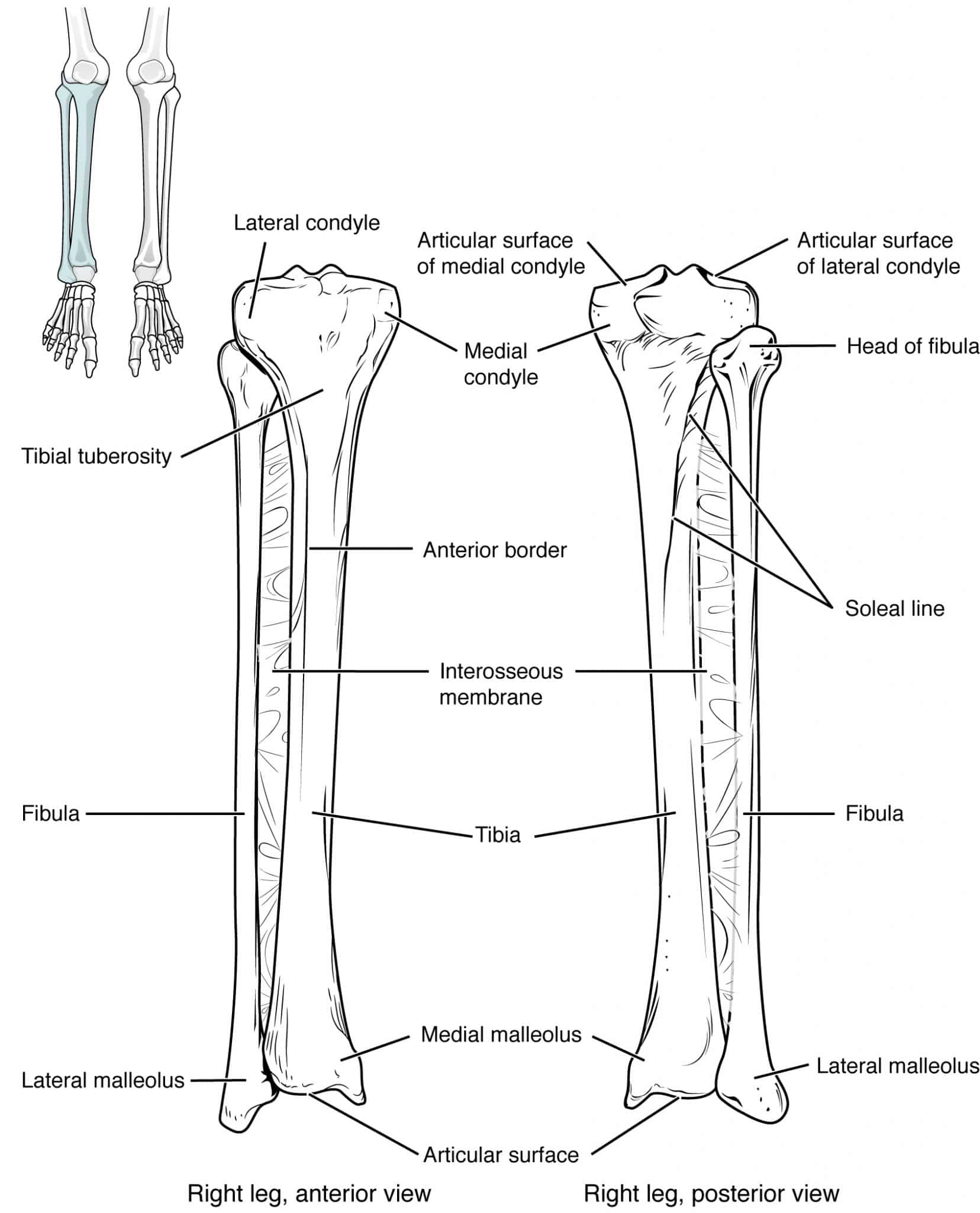
Tibia
The tibia lies on the medial side of the lower leg and is larger than the fibula. It is the main weight-bearing bone in the leg and has two articulating surfaces. At the proximal end, the tibial condyles articulate with the medial and lateral condyles of the femur to form the knee joint. At the distal end, the bone widens and forms the medial malleolus on the medial aspect of the ankle.
The inside aspect of the medial malleolus articulates with the talus to form the medial side of the ankle joint. On the lateral end of the tibia is the fibular notch, which articulates with the distal end of the fibula. This forms the distal tibiofibular joint.
Fibula
The fibula is the smaller of the two bones in the leg and lies on the lateral side. It is not weight-bearing, and its primary function is to act as a site of muscle attachment. The head of the fibula articulates proximally with the inferior lateral tibial condyle, forming the proximal tibiofibular joint.
Similarly to the tibia, the distal end of the fibula expands to form the lateral malleolus, the deep aspect of which articulates with the talus to form the lateral ankle joint.
Talus
The talus is part of the proximal group of tarsal bones in the foot. It lies superior to the calcaneus and posterior to the navicular bone. The superior surface is elevated in order to articulate with both the tibia and fibula within the ankle joint. The upper surface of this elevation articulates with the distal end of the tibia, the lateral surface articulates with the lateral malleolus of the fibula and the medial surface articulates with the medial malleolus of the tibia.
Clinical Relevance: ankle fractures
Ankle fractures are a common injury and are assessed using the Ottawa ankle rules. These are highly specific and designed to identify clinically significant injuries and reduce the use of unnecessary X-ray imaging. For more information on X-ray interpretation, see the Geeky Medics guide to ankle X-ray interpretation.
An ankle X-ray is indicated if there is bony tenderness in any of the following areas:
- The distal 6cm of the posterior tibia
- The distal 6cm of the posterior fibula
- Tip of the medial malleolus
- Tip of the lateral malleolus
AND an inability to weight bear immediately after the injury and in the emergency department.
A foot X-ray is indicated if there is bony tenderness in any of the following areas:
- The base of the fifth metatarsal
- The site of the navicular bone
AND an inability to weight bear immediately after the injury and in the emergency department.
Blood supply and innervation
The blood supply to the ankle joint is derived from the malleolar branches of the fibular artery and the anterior and posterior tibial arteries.
The joint is innervated by branches of the tibial nerve and the deep fibular nerve.
Ligaments
Distal tibiofibular joint
The distal aspects of the tibia and fibula are held together by a band of connective tissue called a syndesmosis. This is a type of fibrous joint that prevents the two bones from separating and therefore holds the talus in place within the ankle joint. The function of the syndesmosis is, therefore, to strengthen and stabilise the ankle joint and allow efficient weight-bearing.
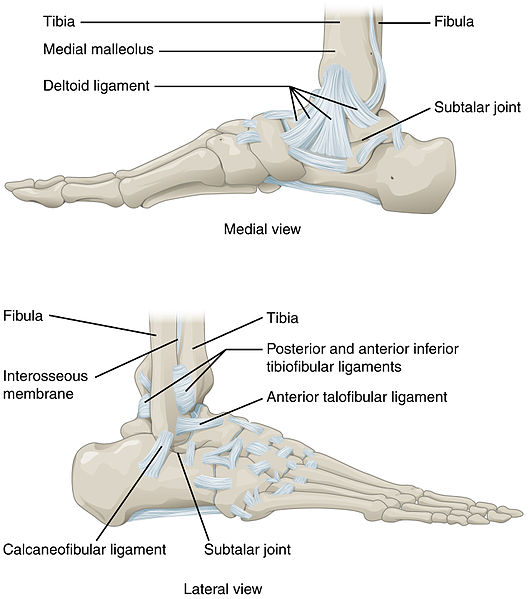
Medial (deltoid) ligament
The medial side of the joint is supported by the medial (deltoid) ligament. This is triangular in shape and originates just above the medial malleolus. It is sub-classified into four parts based on the four inferior points of insertion: tibionavicular, tibiocalcaneal, posterior tibiotalar and anterior tibiotalar.
Lateral ligament
Three ligaments form the lateral ligament of the ankle: anterior talofibular, posterior talofibular, and calcaneofibular.
Clinical Relevance: ankle sprains
Ankle sprains can be classified into either inversion or eversion sprains. Inversion sprains are much more common and occur when the lateral supporting ligaments are stretched or torn. The most commonly injured is the talofibular ligament. In severe sprains, the ligament can rupture and may be associated with distal fibular fractures.
Eversion sprains are less common and often occur following an awkward landing from a jump. These result in damage to the deltoid ligament on the medial aspect of the joint. In severe cases, this may lead to dislocation of the joint and a bimalleolar (Pott’s) fracture.
The mainstay of treatment for ankle sprains is RICE: rest, ice, compression and elevation. More severe injuries associated with fractures may require prolonged immobilisation or surgery.
Muscles
The movement of the ankle joint is produced by muscles of the lower leg. The table below summarises the muscles that act to move the ankle.
Table 1. Summary of the muscles that act to move the ankle joint.
| Compartment | Muscle | Action at ankle joint |
|
Anterior |
Tibialis anterior |
Dorsiflexion, inversion |
|
Extensor hallucis longus |
Dorsiflexion |
|
|
Extensor digitorum longus |
Dorsiflexion |
|
|
Fibularis tertius |
Dorsiflexion, eversion |
|
|
Posterior
|
Gastrocnemius |
Plantarflexion |
|
Soleus |
Plantarflexion |
|
|
Plantaris |
Plantarflexion |
|
|
Tibialis posterior |
Plantarflexion, inversion |
|
|
Lateral
|
Fibularis longus |
Plantarflexion, eversion |
|
Fibularis brevis |
Eversion |
Editor
Dr Chris Jefferies
References
- Drake, R. Gray’s Anatomy for Students (4thEdition). Feb 2019.
- OpenStax. Anatomy and Physiology. April 2013. Available from: [LINK]
- Keith L. Moore, Arthur F. Dalley, Anne M. R. Agur. Clinically Oriented Anatomy (6th Edition). 2010.
Image references
- OpenStax. Tibia and Fibula. Anatomy and Physiology. April 2013. License: [CC-BY 4.0]
- OpenStax. Ankle Joint. Anatomy and Physiology. April 2013. License: [CC-BY 4.0]
