
- 📖 Geeky Medics OSCE Book
- ⚡ Geeky Medics Bundles
- ✨ 1300+ OSCE Stations
- ✅ OSCE Checklist PDF Booklet
- 🧠 UKMLA AKT Question Bank
- 💊 PSA Question Bank
- 💉 Clinical Skills App
- 🗂️ Flashcard Collections | OSCE, Medicine, Surgery, Anatomy
- 💬 SCA Cases for MRCGP
To be the first to know about our latest videos subscribe to our YouTube channel 🙌
Introduction
A 58-year-old man presents to the emergency department with a cough and breathlessness. Work through the case to reach a diagnosis.
UK Medical Licensing Assessment (UKMLA)
This clinical case maps to the following UKMLA presentations:
- Breathlessness
- Cough
History
Presenting complaint
“I’ve really been struggling to catch my breath with this cough.”
History of presenting complaint
Cough history
When did the cough start?
“The cough started about 4 months ago, but it’s gotten worse over the last week”
Is the cough constant or does it come and go?
“It’s been on and off for a few months but this past week it’s been constant”
Is there anything that triggers the cough or anything that makes the cough better?
“Generally the cough is manageable and just gets worse when I exercise, but this past week it’s always there and nothing makes it better”
Do you bring up any sputum when you cough or is it dry/tickly?
“Normally I bring up a small amount of mucus, but I’ve been bringing up loads this past week”
What colour is the mucus you’re bringing up?
“Normally it’s clear, but this week it turned yellow and green”
Have you ever coughed up any blood?
“No”
For more information, see the Geeky Medics guide to cough history taking.
Shortness of breath history
When did you start getting short of breath?
“I’ve been getting more easily out of breath for about 6 months”
Has the shortness of breath been getting worse over time?
“Yes, it’s been impacting me at work for the last 6 months and this past week I haven’t been able to go in”
Is there anything that makes the shortness of breath worse?
“My job is quite physical and I get breathless after about 20 minutes at work or when I’m exercising”
Are you short of breath at rest or when lying down?
“No”
For more information, see the Geeky Medics guide to respiratory history taking.
Other parts of the history
Past medical and surgical history
- Do you have any medical problems?
- Have you had any problems with your breathing before?
- Have you ever been in hospital with your breathing before?#
“My GP diagnosed me with COPD about 6 months ago, but I’ve never been to hospital with it before”
Medication history/allergies
- Do you take any regular medication?
- Do you have any allergies?
“I have a salbutamol inhaler which I take about once a day, but this week I’ve had to take it more. I’m not allergic to anything”
Family history
- Do any medical problems run in the family?
“My dad passed away from lung cancer when he was about my age”
Social history
- Do you live with anyone?
- What do you do for work?
- How are you managing at home?
- Do you smoke? If yes, how many cigarettes do you smoke a day? And how long have you smoked?
- Do you drink alcohol? If yes, how many drinks would you say you have a week?
- Have you ever been exposed to asbestos?
“I live with my wife and work as a car mechanic. Normally I cope ok but I’ve really struggled the last few days. I’ve been smoking one or two packs a day for about 35 years, I know I should stop but haven’t yet. I probably drink about 6 pints a week of beer. I don’t think I’ve been exposed to any asbestos or anything like that”
Systems review
- Have you noticed any fevers?
- Have you had any night sweats?
- Have you been in contact with anyone with similar symptoms recently?
- Have you lost any weight recently?
- Have you had any nausea or vomiting?
- Have you had any changes to your sense of smell or taste?
“I’ve felt quite feverish, but haven’t had any night sweats. I don’t know anyone with anything like this. I haven’t lost any weight, had any nausea, and haven’t lost my sense of smell or taste.”
Clinical examination
Examination findings
Inspection
- Laboured, symmetrical breathing
- No pectus excavatum or carinatum
- No scars
Palpation
- Trachea central
- Full symmetrical expansion
- No palpable apex beat
Percussion
- Dullness to percussion in the right lower lobe
Auscultation
- Reduced breath sounds across lower lobes
- Coarse crackles in the right lower lobe
- Expiratory wheeze is present throughout
- Increased vocal resonance in the right lower lobe
Patient’s hands
An image of the patient’s hands is shown below.
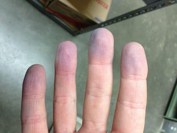
Peripheral cyanosis
Investigations
This patient is acutely unwell with signs of hypoxia with a background of COPD. An ABG is a rapid investigation that can be used to rapidly assess pH, hypoxia, and hypercapnia, which will be crucial for managing this patient.
ABG results
The patient’s ABG (taken on air) is shown below:
- pH: 7.23 (7.35-7.45)
- pO2: 7.8 (10–14)
- pCO2: 8.1 (4.5–6.0)
- HCO3: 28 (22-26)
- BE: +3 (-2 to +2)
This ABG shows type 2 respiratory failure and acute-on-chronic respiratory acidosis.
- Type 2 respiratory failure is seen as the oxygen is low (hypoxaemia) and the CO2 is high (hypercapnia).
- Respiratory acidosis is seen as the pH is low (acidosis), due to the raised CO2 (respiratory).
- The bicarbonate is raised as the body compensates for the high CO2. Considering this patient’s background of COPD, it can be assumed that this is a chronic change.
- Note that the base excess is raised, indicating a higher level of HCO3 in the blood, most likely due to chronic compensation as seen in COPD.
Chest X-ray
A chest X-ray is performed.
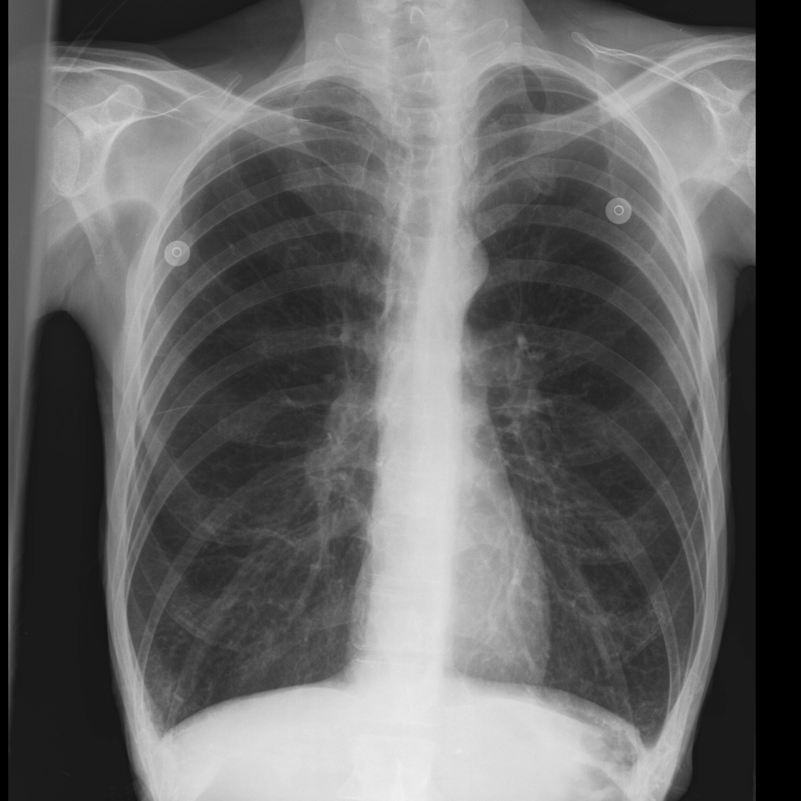
This chest X-ray shows hyperinflation of the lungs with flattening of the diaphragms.
Diagnosis
An infective exacerbation of COPD (IECOPD).
This patient has known COPD and an acute exacerbation of dyspnoea and cough. Considering the acute onset, production of green sputum, and fever, this is the most likely diagnosis.
The correct answer is Haemophilus influenza.
Around 50-70% of infective exacerbations of COPD are caused by bacterial pathogens, with the most common including Haemophilus influenzae, Streptococcus pneumoniae, and Moraxella catarrhalis.
Spirometry report
After looking through the patient’s notes, you find a spirometry report from 6 months ago.
- VC: 3.08 (88% predicted value)
- FEV1: 0.99 (43% predicted value)
- FEV1/FVC: 0.32
This spirometry results shows a severe obstructive lung disease.
FVC, or forced vital capacity, is the total volume of air that the patient can forcibly exhale in one breath. This is >80% predicted so can be considered normal.
FEV1, or forced expiratory volume in 1 second, is the volume of air exhaled in the first second after deep inspiration and forced expiration. This is much less than 80% predicted so is abnormal.
The FEV1/FVC ratio is <0.7 in obstructive lung disease, as although the patient’s lungs can exhale a normal amount of air, an obstruction prevents it from doing so quickly (within the first second).
The GOLD classification system is used to assess the severity of COPD.
Management
An acutely unwell patient should be assessed with an ABCDE approach. Given the likely diagnosis of IECOPD, the following steps should be taken:
- Short-acting beta-agonists (e.g. salbutamol): use an increased dose from the patient’s normal dose, and use a nebulised form for those with moderate-severe exacerbations.
- Corticosteroids: a 5-day course of 30mg prednisolone is preferred, although 100mg IV hydrocortisone may be required in patients who cannot tolerate oral delivery.
- Oxygen: titrate oxygen supply using Venturi masks to target 88-92% saturation.
- Antibiotics: the antibiotic choice will depend on local guidance, however, some example regimes are:
- Amoxicillin 500 mg three times a day for 5 days OR
- Doxycycline 200 mg on the first day, then 100mg once a day for 5 days in total OR
- Clarithromycin 500 mg twice a day for 5 days.
Prognosis
Smoking cessation is the most important lifestyle intervention for COPD.
Discussions regarding smoking cessation can take the ‘5 A’s’ approach.
- Ask: ask and record the patient’s smoking status
- Advise: explain the risks of smoking, its long-term effects, and what support is available
- Assess: assess the patient’s views of cessation and level of motivation
- Assist: plan an approach to cessation and discuss pharmacological and non-pharmacological therapies
- Arrange: arrange a follow-up appointment to re-assess
Editor
Dr Jess Speller
References
- Fletcher W. Peripheral cyanosis. License: [CC BY-SA]
- Case courtesy of Frank Gaillard, Radiopaedia.org, rID: 10550. License: [CC BY-NC-SA]
- NICE guideline. Chronic obstructive pulmonary disease in over 16s: diagnosis and management. Available from: [LINK].
- Global Initiative for Chronic Obstructive Lung Disease. Global Strategy for the Diagnosis, Management and Prevention of COPD. Published in 2020. Available from: [LINK]
- Spirometry in Practice: A Practical Guide to Using Spirometry in Primary Care 2nd Ed (2005). British Thoracic Society COPD Consortium. Available from: [LINK]
