

- 📖 Geeky Medics OSCE Book
- ⚡ Geeky Medics Bundles
- ✨ 1300+ OSCE Stations
- ✅ OSCE Checklist PDF Booklet
- 🧠 UKMLA AKT Question Bank
- 💊 PSA Question Bank
- 💉 Clinical Skills App
- 🗂️ Flashcard Collections | OSCE, Medicine, Surgery, Anatomy
- 💬 SCA Cases for MRCGP
To be the first to know about our latest videos subscribe to our YouTube channel 🙌
Introduction
Rhabdomyolysis is a clinical syndrome characterised by the breakdown of skeletal muscle cells (myocytes) and the subsequent release of intracellular contents into the circulation.1 This can result in acute kidney injury (AKI) and life-threatening electrolyte disturbances.
Aetiology
A variety of factors can trigger skeletal muscle breakdown.2
Traumatic factors include:
- Crush injuries
- Prolonged immobilisation
Non-traumatic factors include:
- Extreme exertion: including prolonged seizures (status epilepticus)
- Surgery: due to prolonged immobilisation or direct muscle ischemia
- Drugs: statins, antipsychotic medications, illicit drugs
- Infections
- Inherited disorders: muscular dystrophy
Pathophysiology
Regardless of the underlying cause, myocyte cell death (necrosis) releases previously intracellular material into the bloodstream.
Key intracellular components released include creatine kinase (CK), myoglobin, urate and electrolytes (potassium, phosphate).
Release of myoglobin into the bloodstream is particularly important, as this is the mechanism by which acute kidney injury (AKI) can develop. The kidneys filter myoglobin. However, the heme pigment can precipitate out of the glomerular filtrate within the renal tubules, causing tubular obstruction.3
When the kidney’s filtering system is blocked, AKI will develop. This can worsen electrolyte imbalances as the homeostatic functions of the kidney are impaired.
Risk factors
Risk factors for rhabdomyolysis include:4
- Male sex
- Body mass index (BMI) >40
- Chronic use of lipid-lowering medications (e.g. statins)
Clinical features
History
The history of rhabdomyolysis is variable and non-specific. Typical symptoms may include:
- Malaise
- Myalgias
- Muscle weakness
It is important to identify any potential triggers of rhabdomyolysis, including:
- Fall with a ‘long lie’ (common in elderly populations)
- Extreme recent exercise
- Initiation of new medications / illicit drugs
Clinical examination
There are few specific signs of rhabdomyolysis. There may be muscular soreness, muscle oedema, or direct evidence of trauma.
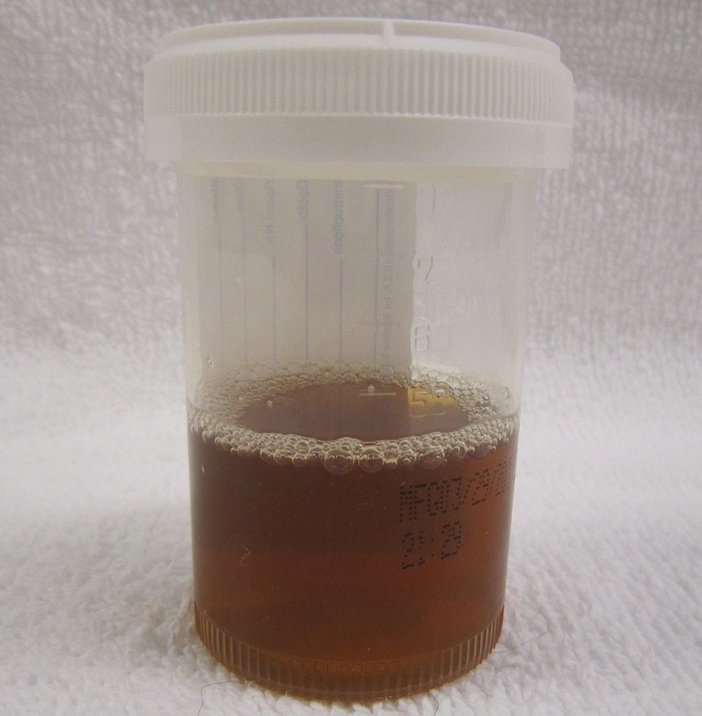
Patients may develop ‘tea-coloured urine’ caused by myoglobin in the urine (myoglobinuria).
Investigations
Bedside investigations
Relevant bedside investigations include:
- Urinalysis: myoglobin in the urine produces a false positive result for haematuria. Although the dipstick will show haematuria, no red blood cells will be present on high-powered microscopy when the laboratory examines the urine.
- ECG: to assess for signs of life-threatening complications, such as hyperkalaemia.
- Blood gas (ABG/VBG): metabolic acidosis with a raised anion gap.
Laboratory investigations
Relevant laboratory investigations include:
- Serum creatine kinase: essential for diagnosing rhabdomyolysis, usually >5x normal limits.5
- U&E: assess for the presence of AKI and identify hyperkalaemia.
- Liver function tests: elevated transaminases (AST/ALT) are a non-specific marker of muscle injury.
- Bone profile: hyperphosphatemia and hypocalcemia are often seen (hypocalcemia develops as myocyte necrosis is associated with significant calcium influx to the cell).
Management
Initial management of rhabdomyolysis involves rehydration with intravenous fluids to prevent acute kidney injury and close monitoring of electrolytes.
Intravenous fluid rehydration
IV fluid resuscitation is the most important aspect of management, aiming to reduce the rate of myoglobin precipitation and kidney toxicity.5
Prompt administration of IV fluid alone is often the only specific management required, with the rate adjusted to maintain high urine output.
Management of electrolyte disturbances
The most acute, life-threatening complication of rhabdomyolysis is hyperkalaemia.
Initial acute management of hyperkalaemia involves:
- Stabilising the myocardium (intravenous calcium)
- Shifting potassium intracellularly (insulin-glucose infusion)
Drugs which can increase serum potassium should be withheld. For more information, see the Geeky Medics guide to managing hyperkalaemia.
Hyperphosphataemia and hypocalcemia usually will not require specific treatment and will self-correct as the CK level falls.
Other management
In severe cases, specialist treatments include IV sodium bicarbonate to alkalinise the urinary filtrate and reduce the rate of myoglobin precipitation.6 Occasionally, patients with severe acute kidney injury require emergency renal replacement therapy.
Complications
Complications of rhabdomyolysis include:
- Acute kidney injury (may require renal replacement therapy if severe)
- Electrolyte disturbances
- Disseminated intravascular coagulation
Key points
- Rhabdomyolysis is a clinical syndrome characterised by the breakdown of skeletal muscle cells and the release of cell contents such as myoglobin into circulation.
- The kidneys filter myoglobin, however it can precipitate out of the filtrate in the tubules to cause significant renal damage.
- Symptoms are usually vague, with myalgia and ‘tea-coloured’ urine classical features.
- Serum creatine kinase (CK) levels are markedly elevated (> 5x times), and myoglobin in the urine will cause the urine dipstick to be falsely positive for the presence of blood.
- Rehydration with intravenous fluids is the mainstay of treatment, alongside monitoring and treating complications such as hyperkalemia.
Editor
Dr Chris Jefferies
References
- Patient.info Professional. Rhabdomyolysis and Myoglobinuria. September 2021. Available from: [LINK]
- Torres PA, Helmstetter JA, Kaye AM, et al; Rhabdomyolysis: pathogenesis, diagnosis, and treatment. Spring 2015. Available from [LINK]
- UpToDate. Clinical features and diagnosis of heme pigment-induced acute kidney injury. November 2021. Available from [LINK].
- UpToDate. Rhabdomyolysis: Epidemiology and etiology. January 2023. Available from [LINK].
- BMJ Best Practice. Rhabdomyolysis. November 2022. Available from: [LINK].
- UpToDate. Prevention and treatment of heme pigment-induced acute kidney injury (including rhabdomyolysis). January 2023. Available from: [LINK].
