
- 📖 Geeky Medics OSCE Book
- ⚡ Geeky Medics Bundles
- ✨ 1300+ OSCE Stations
- ✅ OSCE Checklist PDF Booklet
- 🧠 UKMLA AKT Question Bank
- 💊 PSA Question Bank
- 💉 Clinical Skills App
- 🗂️ Flashcard Collections | OSCE, Medicine, Surgery, Anatomy
- 💬 SCA Cases for MRCGP
To be the first to know about our latest videos subscribe to our YouTube channel 🙌
Introduction
The heart has an intricate electrical system responsible for ensuring the most efficient sequence of cardiac contraction. Any change in this electrical conduction system can affect cardiac output.
This article will focus on the physiology, pathophysiology, and clinical consequences of damage to the bundle branches, including left bundle branch block (LBBB) and right bundle branch block (RBBB).
The cardiac conduction system
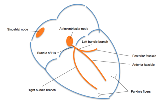
Electrical impulses travel through the heart via a specific conduction pathway. The sinoatrial node (SAN) acts as the initial pacemaker before the impulse spreads throughout the atria and towards the atrioventricular node (AVN).
The depolarisation wave travels through the heart’s septum via the Bundle of His and Purkinje fibres. These are organised into the left and right bundle branches.
The right bundle branch depolarises the right ventricle, and the left bundle branch depolarises the left ventricle simultaneously. The septum is depolarised by the left bundle branch, resulting in the septum being depolarised from left to right.
For more information, see the Geeky Medics guide to the heart’s conduction system.
ECG basics
The ECG is a graphical representation of the net direction of electrical depolarisation in the heart at any one time.3
Different leads look at the heart from different angles (the most important to know for understanding bundle branch blocks is that V1 views the heart from the right and V6 from the left).
An upwards spike means the net depolarisation is heading towards that lead. A downward spike means the net depolarisation is heading away from that lead.
There is a greater muscle mass on the left side of the heart compared to the right, so depolarisation within the left ventricles has a greater impact on the ECG trace.
The right and left ventricles should depolarise simultaneously, producing one uniform R wave.
Ventricular depolarisation using normal pathways is complete within 120ms. Depolarisation takes longer when these pathways are disrupted or changed in any way, causing broad QRS complexes. A broad QRS complex always indicates abnormal ventricular depolarisation.3
For more information on ECG interpretation, see the Geeky Medics guides to reading an ECG and understanding an ECG.
Table 1. Components of an ECG trace.
| ECG component | Definition |
| P wave | Atrial depolarisation |
| PR interval | Conduction through the AVN to the ventricles |
| QRS complex | Ventricular depolarisation |
| Q wave | The first downward deflection |
| R wave | Any upwards deflection |
| S wave | Any downward deflection after an R wave |
| T wave | Ventricular repolarisation |
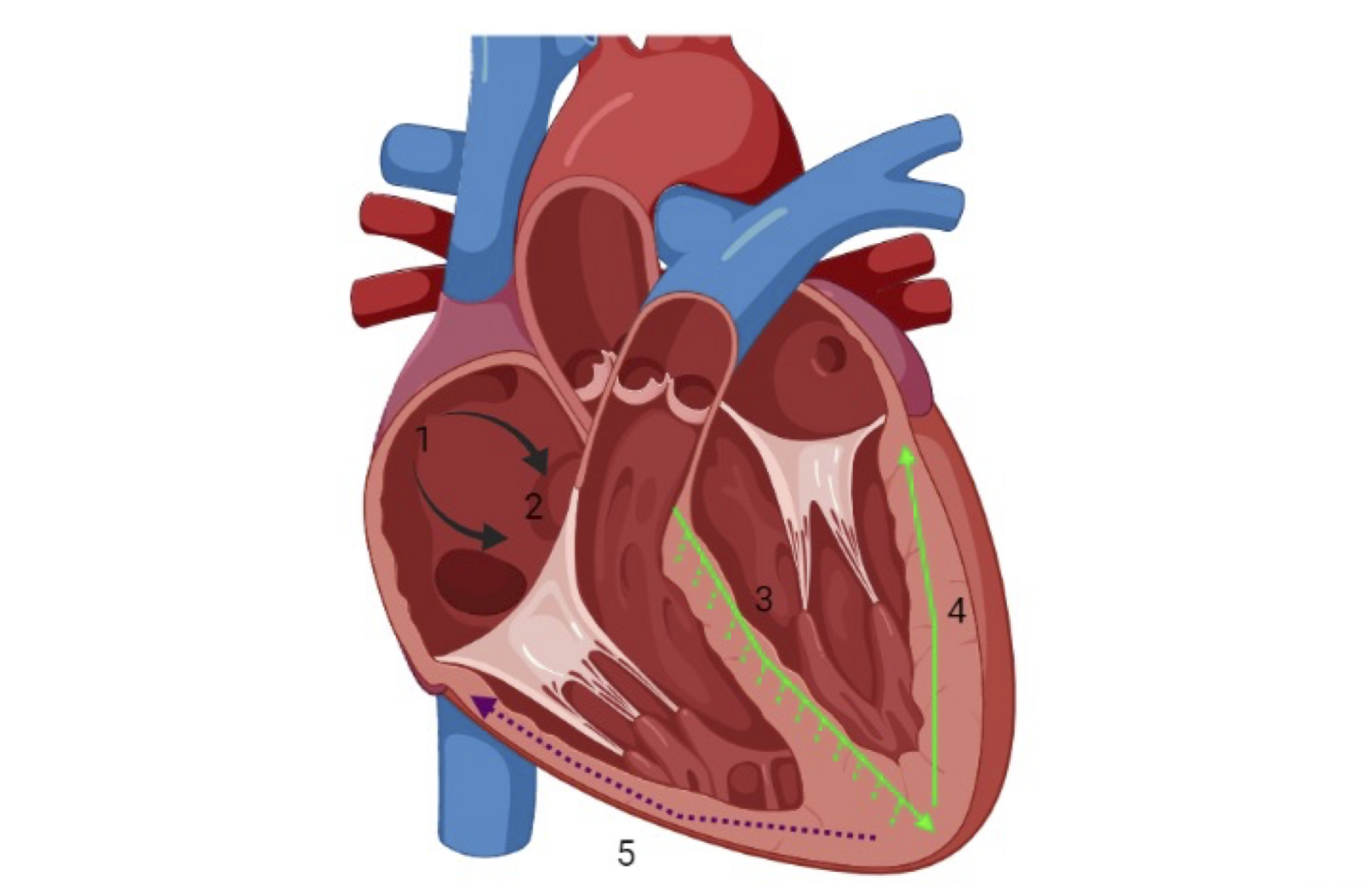
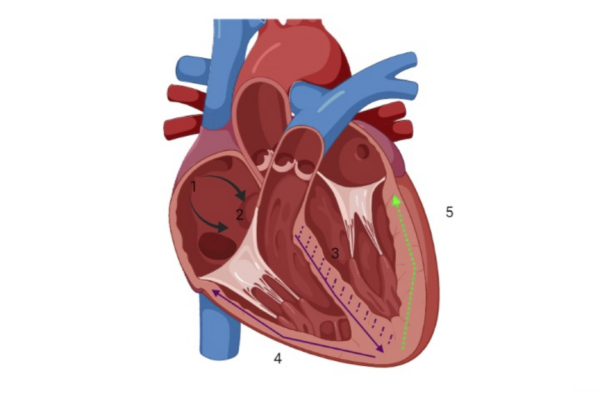
In normal cardiac conduction (Figure 1):
- The sino-atrial node acts as the initial pacemaker
- Depolarisation reaches the atrioventricular node
- Impulses travel simultaneously down the bundle of His via the left and right bundle branches. The septum is depolarised from the left.
- Both the left and right ventricular walls are depolarised simultaneously.


The main feature of bundle branch blocks is the broadening of QRS complexes. It is important to ensure other causes of broad complexes are excluded. For more information, see the Geeky Medics guide to atrioventricular blocks.
As the problem is below the atria, the P waves and the PR intervals are normal.
Right bundle branch block (RBBB)
Diagnostic criteria
The diagnostic criteria for RBBB are:2
- Broad QRS complex: >120 ms (3 small squares)
- RSR’ pattern in V1-V3: an initial small upward deflection (R wave), a larger downward deflection (S wave), then another large upward deflection (a second R wave, which is indicated as R’)
- Wide, slurred S wave in lateral leads: I, aVL, V5-V6
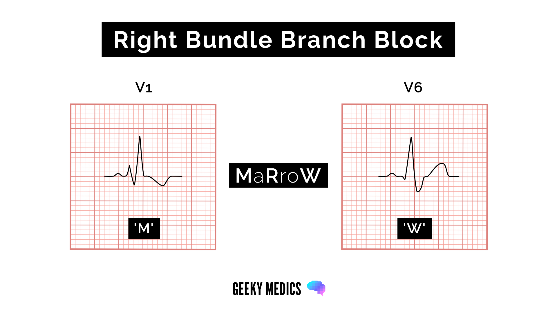
WiLLiaM MaRRoW mnemonic
The WiLLiaM MaRRoW mnemonic can be used to quickly recognise left and right bundle branch blocks by looking at V1 and V6:
- WiLLiaM refers to the ECG appearance of left bundle branch block (see next section)
- MaRRoW refers to the ECG appearance of right bundle branch block
The middle letters of the names help you remember which bundle branch block each name is referring two (two Ls in WiLLiaM = left bundle branch block, two Rs in MaRRoW = right bundle branch block).
Each name’s first and last letter helps you recognise the ECG features of the associated bundle branch block.
To recognise right bundle branch block, we use the name MaRRoW and look at the first and last letters:
- M: complexes in V1 resemble the letter M: initial small upward deflection (r wave), a larger downward deflection (S wave), then another large upward deflection (second R wave)
- W: complexes in V6 resemble a W: initial small downward deflection (Q wave), then a larger upward deflection (R wave), and then a wide downward deflection (S wave)
Pathophysiology
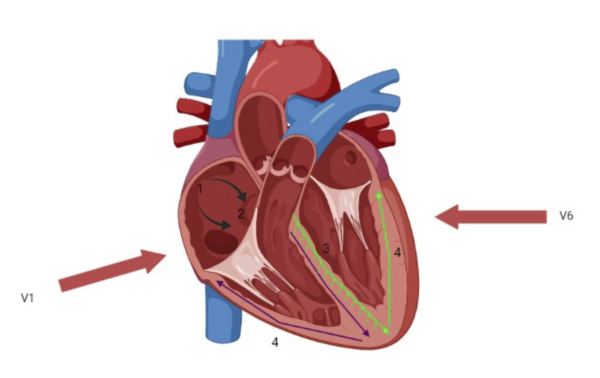
In right bundle branch block (Figure 3):
- The sino-atrial node acts as the initial pacemaker
- Depolarisation reaches the atrioventricular node
- Depolarisation through the bundle of His occurs only via the left bundle branch. The left branch still depolarises the septum as normal.
- The left ventricular wall depolarises as normal.
- The right ventricular walls are eventually depolarised by the left bundle branch, this occurs by a slower, less efficient pathway.

Clinical relevance
RBBB can be either physiological or the result of damage to the right bundle branch. Causes of damage include underlying lung pathology (COPD, pulmonary emboli, cor pulmonale), primary heart muscle disease (ARVC), congenital heart disease (e.g. ASD), ischaemic heart disease and primary degeneration of the right bundle.
Left bundle branch block (LBBB)
Diagnostic criteria
The diagnostic criteria for LBBB are:
- Broad QRS complex: >120 ms (3 small squares)
- Dominant S wave in V1
- Broad, monophasic R wave in lateral leads: I, aVL, V5-V6
- Absence of Q waves in lateral leads
- Prolonged R wave >60ms in leads V5-V6
WiLLiaM MaRRoW mnemonic
As discussed above, the WiLLiaM MaRRoW mnemonic can be used to quickly recognise left and right bundle branch blocks by looking at V1 and V6.
To recognise left bundle branch block, we use the name WiLLiaM and look at the first and last letters:
- W: complexes in V1 resemble the letter W: deep downward deflection (dominant S wave), which may be notched
- M: complexes in V6 resemble the letter M: broad, notched or ‘M’ shaped R wave in V6

Pathophysiology
When viewed from the right-hand side (V1), net depolarisation travels away (towards the left), resulting in negative ECG deflections. The first downward deflection represents the right ventricle, and the slightly delayed 2nd downward deflection corresponds to the depolarisation of the left ventricle.
When viewed from the left-hand side (V6), where the net depolarisation is travelling towards the detector, deflections are positive on the ECG. Again, there will be two peaks (RR) due to the delay in left ventricular depolarisation.1
In left bundle branch block (Figure 4):
- The sino-atrial node acts as the initial pacemaker
- Depolarisation reaches the atrioventricular node
- Depolarisation down the bundle of His occurs only via the right bundle branch. The septum is abnormally depolarised from right to left.
- The right ventricular wall is depolarised as normal.
- The left ventricular walls are eventually depolarised by the right bundle branch, this occurs by a slower, less efficient pathway.

Clinical relevance
LBBB is always pathological. Left bundle branch block may be due to conduction system degeneration or myocardial pathologies such as ischaemic heart disease, cardiomyopathy and valvular heart disease.
LBBB may also occur after cardiac procedures, which damage the left bundle branch or His bundle. A STEMI presenting as chest pain with LBBB is exceedingly rare.
Branches of the left bundle branch
Due to the relatively greater mass of the left ventricle, disruptions in the depolarisation of the left ventricular muscle can cause cardiac axis changes. The left bundle branch splits into anterior and posterior fascicles.
LBBB = Left anterior fasicular block (LAFB) + Left posterior fasicular block (LPFB)
Each branch of the left bundle branch may be damaged in isolation. Anterior fascicle block, which is much more common, causes left axis deviation. Posterior fascicle block may cause right axis deviation. However, the posterior fascicle does much less work than the anterior fascicle, so it can be blocked without any obvious ECG changes.
The right ventricular muscle does not have enough mass to significantly deviate the cardiac axis.
Other types of block
Bifascicular block involves both right bundle branch block and the blockade of one of the fascicles of the left bundle branch.
Trifascicular block is present when a 3rd-degree heart block exists alongside bifascicular block.
Key points
- Consider bundle branch block in an ECG trace with broad complexes.
- A broad complex tachycardia requires differentiation between ventricular tachycardia or sinus tachycardia with concurrent bundle branch block.
- Left bundle branch block is always pathological and indicates significant damage to the cardiac conduction system.
- When assessing whether a broad QRS complex is LBBB or RBBB, the appearances of V1 and V6 are often enough to provide the answer using the WiLliaM and MaRroW technique.
Reviewers
Dr Matt Jackson
Consultant Cardiologist
Dr Ben Marrow
Cardiology Registrar
Editor
Dr Chris Jefferies
References
- Burns, R. B. (2021, December 10). Left Bundle Branch Block. Available from: [LINK]
- Buttner, E. B. (2021, March 5). Right bundle branch block. Available from: [LINK]
- Hampton, J. (2013). ECG Made Easy.
