

- 📖 Geeky Medics OSCE Book
- ⚡ Geeky Medics Bundles
- ✨ 1300+ OSCE Stations
- ✅ OSCE Checklist PDF Booklet
- 🧠 UKMLA AKT Question Bank
- 💊 PSA Question Bank
- 💉 Clinical Skills App
- 🗂️ Flashcard Collections | OSCE, Medicine, Surgery, Anatomy
- 💬 SCA Cases for MRCGP
To be the first to know about our latest videos subscribe to our YouTube channel 🙌
This guide discusses how to approach performing a focused deep vein thrombosis (DVT) examination on a patient in an OSCE setting.
In OSCE scenarios, you may be asked to perform a focused examination to determine the presence (or absence) of a certain condition. It is important to be able to confidently elicit the main diagnostic signs of the condition. In order to do this, you need to be comfortable with the relevant basic system examination (i.e. for an acromegaly examination you need to be able to perform assessments such as a full examination of the peripheral vascular system).
Background
A deep vein thrombosis (DVT) refers to the formation of a thrombus within the deep venous system, most often occurring in the veins of the leg but also in the pelvis and arm.
If this thrombus becomes dislodged, it becomes an embolus and can travel to the lungs causing a pulmonary embolism (PE). PEs and DVTs are both types of venous thromboembolism (VTE).
Pathophysiology and risk factors
Factors that contribute to the development of VTE can be summarised using Virchow’s triad, which consists of:
Hypercoagulability (a state in which blood coagulates quicker than usual). Examples include:
- Inherited thrombophilia (e.g. Factor V Leiden)
- Pregnancy and oestrogen therapy
- Malignancy
- Infection and inflammation
- Dehydration
- Nephrotic syndrome
Stasis (a state in which blood flows slowly or becomes turbulent). Examples include:
- Immobility (hospitalised patients, long flights)
- Varicose veins
- Obesity
Endothelial injury (a state in which there has been damage to the vascular wall). Examples include:
- Physical trauma (including surgery)
- Foreign devices (e.g. stents)
- Hypertension
- Bacterial infection
Clinical features of a DVT
Typical clinical features of a DVT include:
- Oedema
- Pain (often cramping, may progress over several days)
- Erythema & warmth
- Peripheral venous distention
These signs are most often unilateral unless the DVT has occurred on both sides.
Red flags for pulmonary embolism
A pulmonary embolism is a life-threatening complication of DVTs. The following clinical features are red flags for the development of a PE:
- Sudden-onset shortness of breath
- Tachycardia
- Haemoptysis
- Chest pain (usually pleuritic)
Introduction
Wash your hands and don PPE if required.
Introduce yourself including your name and role.
Confirm the patient’s name and date of birth.
Briefly explain the examination using patient-friendly language. Establish any questions or concerns. Offer a chaperone if appropriate.
Gain consent to proceed with the examination and establish that the examination can be stopped at any point.
Adjust the head of the bed to a 45° angle and ask the patient to lay on the bed.
Adequately expose the patient’s lower limbs.
Ask the patient if they have any pain before proceeding with the clinical examination.
General inspection
Risk factors
Perform a brief general inspection of the patient, looking for risk factors for venous thromboembolism such as:1,2
- Age: patients aged over 50 are at an increased risk
- Sex: incidence is similar between men and women, however, females have a higher risk during childbearing years and males have a comparatively higher risk after the age of 45
- Obesity: is associated with decreased fibrinolytic activity
- Pregnancy: is associated with up to a 5x increased risk, especially in the 3rd trimester
- Immobility: leads to venous stasis
- Trauma: direct damage to the endothelial vascular lining can trigger thrombosis, particularly if paired with immobility
- Medication: oral contraceptive pill, anticoagulants, chemotherapy
Clinical signs
Look for obvious clinical signs suggestive of venous thromboembolism including:
- Unilateral leg or arm swelling, pain or erythema (deep vein thrombosis)
- Shortness of breath (pulmonary embolism)
Objects and equipment
Look for objects or equipment on or around the patient that may provide useful insights into their medical history and current clinical status:
- Medical equipment: note any oxygen delivery devices and ECG leads.
- Mobility aids: items such as walking aids give an indication of the patient’s current mobility status.
- Vital signs: charts on which vital signs are recorded will give an indication of the patient’s current clinical status and how their physiological parameters have changed over time.
- Prescriptions: prescribing charts or personal prescriptions can provide useful information about the patient’s recent medications.
Pulse and blood pressure
Radial pulse
Palpate the patient’s radial pulse, located at the radial side of the wrist, with the tips of your index and middle fingers aligned longitudinally over the course of the artery.
Once you have located the radial pulse, assess the rate and rhythm:
- Tachycardia (red flag for pulmonary embolism)
- Thready pulse (may indicate dehydration, a risk factor for venous thromboembolism)
- Bounding pulse (may indicate sepsis, a risk factor for venous thromboembolism)
Blood pressure
Offer to measure the patient’s blood pressure (see our blood pressure guide for more details).
A comprehensive blood pressure assessment should also include lying and standing blood pressure.
In an OSCE station, you are unlikely to have to carry out a thorough blood pressure assessment due to time restraints, however, you should demonstrate that you have an awareness of what this would involve.
Hypertension can be a response to significant pain caused by PE or DVT.
Hypotension may be a late and very concerning sign of a large or bilateral PE and should prompt consideration of thrombolysis. It can also be an indicator of hypovolaemia secondary to dehydration.
Upper limbs
Inspection
Inspect the upper limbs for signs of a DVT. Unilateral engorgement of peripheral veins is a particularly useful clinical sign in the upper limbs.
Ask the patient about any localised neck or shoulder pain.
Neck and chest
Inspection
The most important risk factor for upper limb DVT is the presence of a foreign body in the deep venous system.3
On inspection, look for:
- Central venous catheters
- Cardiac pacemakers: usually marked by a scar inferior to the left clavicle and palpable on the thoracic wall
- PICC lines: especially the triple lumen subtype
Infection and introduction of chemotherapeutic agents through these devices increases the risk of upper limb DVT.
Superior vena cava (SVC) syndrome can be a result of malignant disease in the mediastinum obstructing the SVC and resulting in engorgement of the upper extremity and neck veins. Malignancy is an important risk factor for upper limb DVTs.
Pemberton’s sign
Pemberton’s sign may be elicited in patients with SVC syndrome:
- Ask the patient to raise their arms as high as possible
- Observe for one minute
A positive sign is characterised by facial plethora and shortness of breath due to reduced venous return.
Thoracic outlet syndrome is a disorder in which the structures of the thoracic outlet (including veins supplying the upper limb and neck) become compressed. This compression results in damage to the vascular walls and causes thrombosis. Causes of thoracic outlet syndrome include trauma, cervical ribs and hypertrophic neck muscles.
Leg inspection
Ask the patient to expose their legs fully (if not already exposed).
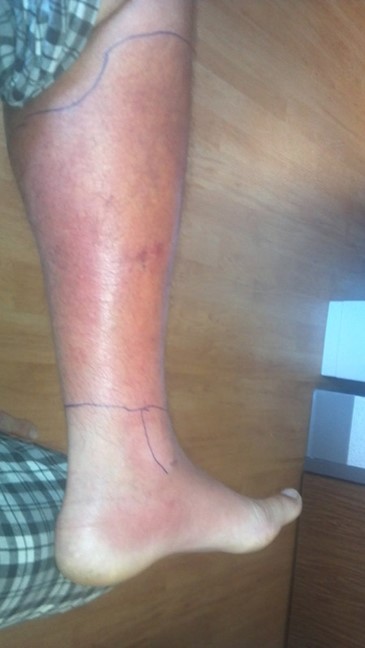
Inspect and compare the legs looking for
- Erythema: due to the pooling of blood in the lower extremity
- Oedema: caused by the blockage of venous return and therefore fluid build-up due to increased hydrostatic pressure, forcing fluid out of the vessels
- Distended peripheral veins: like in most other places in the body, there are collateral ways for blood to flow if major veins are blocked. Peripheral veins act as a diversion around the blockage and may therefore appear distended.
- Varicose veins: although varicose veins are not directly related to DVTs they may indicate venous insufficiency (a risk factor for DVT)
- Ulcers: these also indicate venous insufficiency

Palpation
When performing palpation, both legs should be compared as DVTs are most likely to occur only on one side.
Temperature
Check the temperature of both legs by placing your hand along at least three sites moving proximally or distally.
Warmth is a common sign of DVT, but also of infection/inflammation (e.g cellulitis).
Pitting oedema
Palpate the patient’s legs checking for pitting oedema. Press your thumb (as firmly as comfortable for the patient) along bony prominences, moving from the medial malleolus proximally along the tibia (until no oedema is present).
Pitting oedema is observable as a marked imprint where pressure was applied, but it may not be very visible in less severe cases. Gently feel over the pressure points for a slight dip.
Unilateral oedema should raise suspicion of a DVT. If the oedema is bilateral, alternative causes should be considered (e.g. heart failure).
Tenderness
Gently squeeze the calves moving proximally. Verbally confirm whether there is any pain and watch the patient’s facial expression for any signs of discomfort.
Measure
Using measuring tape, measure the circumference of each calf 10cm below the tibial tuberosity. If the difference between the two is greater than 3cm, this is a clinically significant finding.
Calculating the Wells score
If a DVT is suspected, a two-level Wells score for DVT is recommended. Remember, some patients can present asymptomatically, so it is important to consider the history as well.5,6
Table 1. The two-level Wells score for DVT
| Clinical feature | Points |
| Active cancer (currently receiving treatment or treatment within 6 months or palliative) | 1 |
| Paralysis, paresis or recent plaster immobilisation | 1 |
| Recently bedridden (3 days or more), or major surgery within the last 3 months | 1 |
| Localised tenderness along the distribution of the deep venous system | 1 |
| Entire leg swollen | 1 |
| Calf swelling at least 3cm larger than the asymptomatic side | 1 |
| Pitting oedema in the symptomatic leg | 1 |
| Collateral superficial veins (non-varicose) | 1 |
| Previously documented DVT | 1 |
| An alternative diagnosis is at least as likely as DVT | -2 |
* if both legs are symptomatic the more symptomatic leg should be used
Remember the rule of 3s:
- Bedridden for 3 days
- Surgery in the last 3 months
- Leg 3cm larger
Interpretation of the Wells score for DVT
Interpretation of the score:3
- DVT likely: ≥2 points
- DVT unlikely: <2 points
DVT likely (≥2 points)
If a DVT is likely, a proximal leg vein doppler ultrasound should be requested with the results available within four hours:
- If the ultrasound is positive: treat the DVT with an anticoagulant.
- If the ultrasound can’t be done within 4 hours: D-dimer and offer an interim anticoagulant until results are available.
- If the ultrasound is negative: check the D-dimer. If it is positive, repeat the scan in 6-8 days (stop interim coagulation if started). If it is negative, another diagnosis should be considered and no anticoagulation given.
DVT unlikely (<2 points)
If DVT is unlikely, offer a D-dimer test with the results available within four hours:
- If the D-dimer is positive: offer a proximal leg vein doppler ultrasound or provide interim coagulation if ultrasound results cannot be obtained within four hours. If the ultrasound is positive treat the DVT with an anticoagulant.
- If the D-dimer results can’t be obtained within four hours: offer interim anticoagulation until results are available.
- If the D-Dimer is negative: another diagnosis should be considered and no anticoagulation given.
Interpretation of the D-dimer test
The interpretation of a D-Dimer is only helpful if you know the Wells score (pre-test probability).
D-dimer is sensitive but not specific. Many patients may have an elevated D-dimer for various reasons unrelated to venous thromboembolism.7
Differential diagnoses
It is important to consider other differential diagnoses in the context of a suspected DVT. A DVT is usually unilateral. Bilateral peripheral pitting oedema should raise the suspicion for an alternative diagnosis (e.g. heart failure, liver failure or renal failure), however, these conditions can co-exist with a DVT.
Acute limb ischaemia
Acute limb ischaemia occurs secondary to occlusion of the leg arteries. Like a DVT the symptoms are unilateral. A history of peripheral arterial disease can be a helpful clue.
Remember the 6Ps of limb ischaemia: pain, pallor, paralysis, paraesthesia, perishingly cold and pulseless.8
On examination, a pulseless cool limb should point towards arterial rather than venous disease. For more information, see the Geeky Medics guides to acute limb ischaemia and peripheral vascular examination.
Compartment syndrome
Compartment syndrome occurs when there is a pressure build-up in an anatomical compartment of the body that is surrounded by fascia. When the pressure rises, it can stop blood flow to the affected region and is therefore a medical emergency.
The most common acute cause of compartment syndrome is trauma causing tissue swelling or bleeding into a compartment. Compartment syndrome is a surgical emergency and requires immediate orthopaedic review.9
Cellulitis
Cellulitis is one of the most common and important mimics of DVTs. It is caused by bacterial infection of the dermis and subcutaneous tissue.
Erythema, swelling pain and warmth are common features and cellulitis usually only affects one limb, usually a leg.
As opposed to DVTs, cellulitis can cause systemic symptoms such as fever, chills and nausea. On examination, there is often a visible skin break where infectious organisms may have entered, and the skin may have developed blisters. Additionally, the erythematous changes are usually well demarcated. The spread of the infection is often monitored by marking edges with a pen.
Diabetic and immunocompromised patients as well as those with venous insufficiency are more at risk of developing cellulitis.10
Ruptured Baker’s cyst
A Baker’s cyst or popliteal cyst is caused by a cystic swelling of the gastrocnemio-semimembranosus bursa. It presents as a palpable swelling in the popliteal fossa, often accompanied by stiffness and pain. Sometimes these cysts can cause leg oedema due to compression of the popliteal vessels.
The rupture of a Baker’s cyst can present with an audible ‘pop’ and associated pain redness and warmth. Ultrasonography is the key tool to distinguish Baker’s cysts from DVTs.12
A ‘crescent sign’, which is the appearance of bruising and swelling below the ankle, specifically along the medial malleolus, may develop secondary to the rupture and can be an important finding, especially in late presentations.13
To complete the examination…
Explain to the patient that the examination is now finished.
Thank the patient for their time.
Dispose of PPE appropriately and wash your hands.
Summarise your findings.
Example summary
“Today I performed a focused DVT examination on a 59-year-old gentleman. On general inspection, the patient appeared comfortable at rest, and there were no objects or obvious risk factors associated with VTE.”
“On peripheral examination of the hands, there were no obvious identifiable risk factors of thromboembolic disease and no acute signs of a pulmonary embolism.”
“Gross inspection of the legs revealed no obvious erythema, swelling or signs of venous insufficiency.”
“Palpation of the legs revealed no warmth or tenderness along the distribution of the deep veins. The calf circumference was normal and equal.”
“In summary, there are no clinical findings indicating a DVT.”
“For completeness, I would like to take a full history and calculate a Wells score according to clinical suspicion and perform a venous Doppler or D-dimer test guided by the results.”
Further assessments and investigations
- Cardiovascular examination including measuring blood pressure: to identify cardiovascular involvement including hypotension.
- Ankle-brachial pressure index (ABPI) measurement: to assess arterial perfusion.
- Peripheral arterial examination: to assess for evidence of arterial disease.
- Peripheral vascular examination: to assess for evidence of venous disease.
Reviewer
Dr Leonidas Zachariades
Consultant in Acute Medicine
Editor
Dr Chris Jefferies
References
- Heit J A., Spencer F A., White R H. The epidemiology of venous thromboembolism. 2016. Available from: [LINK]
- Motykie, G D et al. A Guide to Venous Thromboembolism Risk Factor Assessment. 2000. Available from: [LINK]
- Heil J et al. Deep Vein Thrombosis of the Upper Extremity: A Systematic Review. 2017. Available from: [LINK]
- James Heilman MD. Lower Limb DVT. Licence: [CC BY-SA 4.0]
- NiCE Guidance. Venous thromboembolic diseases: diagnosis, management and thrombophilia testing. Published 26 March 2020. Available from: [LINK]
- Wells P et al. Evaluation of D-Dimer in the Diagnosis of Suspected Deep-Vein Thrombosis. 2003. Available from: [LINK]
- Linkins L A, Takach Lapner S. Review of D-dimer testing: Good, Bad, and Ugly. May 2017. Available from: [LINK]
- NICE Clinical Knowledge Summaries. Peripheral Arterial Disease – Features of acute limb ischaemia. Revised in July 2022. Available from: [LINK]
- NHS. Compartment Syndrome. Revised in September 2019. Available from: [LINK]
- NICE Clinical Knowledge Summaries. Acute Cellulitis – Diagnosis. Revised in January 202. Available from: [LINK]
- Pshawnoah. Cellulitis marked with pen. Licence: [CC BY-SA 3.0]
- NICE Clinical Knowledge Summaries. Baker’s cyst – Diagnosis. Revised in May 2020. Available from: [LINK]
- Mizumoto, J. The crescent sign of ruptured baker’s cyst. May 2019. Available from: [LINK]
