

- 📖 Geeky Medics OSCE Book
- ⚡ Geeky Medics Bundles
- ✨ 1300+ OSCE Stations
- ✅ OSCE Checklist PDF Booklet
- 🧠 UKMLA AKT Question Bank
- 💊 PSA Question Bank
- 💉 Clinical Skills App
- 🗂️ Flashcard Collections | OSCE, Medicine, Surgery, Anatomy
- 💬 SCA Cases for MRCGP
To be the first to know about our latest videos subscribe to our YouTube channel 🙌
Introduction
A 21-year-old man is brought to emergency department by their friend following a head injury. Work through the case to reach a diagnosis.
UK Medical Licensing Assessment (UKMLA)
This clinical case maps to the following UKMLA presentations:
- Decreased/loss of consciousness
- Head injury
History
Presenting complaint
“I was playing hockey earlier and think I must have fainted or something”
History of presenting complaint
Can you remember what happened before you lost consciousness?
“I remember I was playing hockey but can’t really remember anything after that. What day is it?”
Do you remember anything about what happened when you lost consciousness?
“My friend told me I was on the floor and was unconscious for at least a minute but I’m not sure. Where am I now?”
What happened after you came round?
“I remember waking up on the side of the pitch with loads of people around me. They tried to call an ambulance but I didn’t feel too bad so my friend drove me here.”
How do you feel now?
“My head is painful and I feel a bit dizzy but other than that I’m okay. I have been sick 2 or 3 times since but I went out last night so I’m not too worried.”
Collateral history is vital particularly when there is impaired or loss of consciousness. You could speak to the friend who saw him on the floor. Key questions for this patient include exactly what happened before he collapsed (as he has retrograde amnesia) and what happened while he was unconscious:
“It was around 3 hours ago. He got hit in the side of the head with the hockey ball. It was an accident, it just flew up in the air and hit him. He collapsed on the floor and his whole body shook a couple of times. His face went really pale and he wasn’t responding to us. I checked his pulse and that seemed ok. He woke up after about 2 minutes and was really confused for around 5 minutes. He’s better now but I don’t think he’s back to normal.”
NICE has produced guidelines on imaging following head injury. The criteria includes:
- GCS <15 over 2 hours after injury
- GCS <13 on initial ED assessment
- Post-traumatic seizure
- Suspected skull fracture
- Signs of basal skull fracture
- Focal neurological deficit
- More than 1 episode of vomiting
In addition, there are other factors that, in combination suggest the need for investigation:
Loss of consciousness and/or amnesia plus any of:
- Age over 65
- High energy mechanism of injury
- History of bleeding disorders
- More than 30 minutes of retrograde amnesia prior to the injury
Clinical examination
- General examination
- Cranial nerve examination
- Upper and lower limb neurological examination
- Glasgow Coma Scale (GCS)
Examination findings
- Tender bruising over the right frontotemporal region.
- No signs of basal skull fracture.
- No cervical pain, tenderness, or restricted movement.
- No other obvious signs of external injury.
- Cranial nerve examination normal
- Upper and lower limb neurological examinations normal
- GCS 14/15: E4V4M6
Investigations
A CT head is appropriate in this case, as the patient has 2 NICE ‘red flags’ indicating the need for imaging post-head injury (GCS <15 more than 2 hours after injury, and vomiting more than once). This should ideally be performed within 1 hour.
Diagnosis
From outside to inside, bleeding may be:
- In the extradural space
- In the subdural space: either acute, chronic, or acute-on-chronic
- In the subarachnoid space: may be traumatic or non-traumatic
- Intraparenchymal: may be traumatic or spontaneous
- Intraventricular: usually related to intraparenchymal or subarachnoid bleeding, but occasionally primary intraventricular haemorrhage can occur
CT head
A CT head is performed for this patient, and a slice from this is shown below.
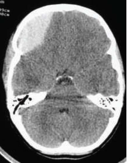
This CT head shows a right-sided convex hyperdensity in keeping with an extradural haematoma. It is causing mass effect on the frontal lobe and a shift of the midline structures to the left.
Extradural haematomas are classically due to bleeding from the middle meningeal artery, which can be damaged by trauma, particularly over the pterion.
Management
This patient will require craniotomy and evacuation of the haematoma. This is usually the operation of choice for evacuation of extradural or acute subdural hematoma.
Outcome
The patient begins to deteriorate in the emergency department. His Glasgow Coma Scale (GCS) score drops to 9 (E2 V2 M5), and one of his pupils becomes fixed and dilated.
The patient’s intracranial pressure (ICP) is rising rapidly. He needs emergency surgical intervention to relieve this pressure. Mannitol is an osmotic diuretic which can be used as a temporising measure to lower ICP. It is given intravenously and tends to cause diuresis, so also requires the insertion of a urinary catheter.
Editor
Dr Jess Speller
References
- The Royal Children’s Hospital Melbourne. Head Injury. CT image of right extradural haematoma. Available from: [LINK]
- National Institute for Health and Care Excellence (NICE). Head injury: assessment and early management. CG 176, published Jan 2014. Available from: [LINK]
