- 📖 Geeky Medics OSCE Book
- ⚡ Geeky Medics Bundles
- ✨ 1300+ OSCE Stations
- ✅ OSCE Checklist PDF Booklet
- 🧠 UKMLA AKT Question Bank
- 💊 PSA Question Bank
- 💉 Clinical Skills App
- 🗂️ Flashcard Collections | OSCE, Medicine, Surgery, Anatomy
- 💬 SCA Cases for MRCGP
To be the first to know about our latest videos subscribe to our YouTube channel 🙌
Examining the neurological system is different in young children compared with older children and adults. The components of the complete exam are extensive and usually cannot be performed in a classical fashion. This approach may be carried out on a cooperative school-aged child – but always be mindful of keeping the examination fun.
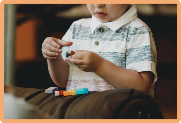
Observation is key. Make the most of every opportunity to examine the child. See how they play, taking into account handedness and motor deficits. These observations, especially in younger children, will ultimately give you the best insight into their daily functioning and paint a broad picture of their neurological function.
Some tips include:
- Using items such as a tennis ball, small toys (including a toy car), bells, bubbles and an object that will attract the child’s attention (like a pinwheel).
- Be mindful to postpone uncomfortable tasks until the end, such as head circumference, fundoscopy and sensory testing.
Introduction
Wash your hands and don PPE if appropriate.
Introduce yourself to the parents and the child, including your name and role.
Confirm the child’s name and date of birth.
Briefly explain what the examination will involve using patient-friendly language: “Today I’d like to perform a neurological examination, which will involve me testing the nerves that supply different parts of the body.”
Gain consent from the parents/carers and/or child before proceeding: “Are you happy for me to carry out the examination?”
General inspection
With toddlers – the initial phase of observation is best done with the child in the parent’s lap. Through minimising apprehension, assessment of higher cortical function, muscle tone and tendon reflexes becomes easier.
Higher cortical functions
Observe the child during play:
- Attention span
- Gross and fine motor coordination
- Problem-solving abilities
Observe for age-appropriate milestones (see our guide on developmental milestones).
Cranial nerves
Testing in infants is often by observation for specific movements and responses, which is ultimately less reliable. In older children, it may be possible to formally assess at least some cranial nerves, however, this very much depends on the exact age of the child, their current state and the environment. We have provided a guide to each of the cranial nerves below, however, it is unlikely you will be able to carry out a complete neurological assessment in one sitting with most children.
Olfactory nerve (I)
The olfactory nerve is responsible for the sense of smell.
Assessment
- Test the ability to detect a smell with the child’s eyes closed (i.e. chocolate).
- Olfaction is not assessed in small children or infants.
- Olfaction can be impaired after closed head injury and in infants with arhinencephaly-holoprosencephaly.
Optic nerve (II)
The optic nerve is responsible for vision and afferent pupillary light reflexes.
Testing visual acuity
- Infant: observe the infant reach for objects of varying size.
- >6 months old: observe reaching for scraps of paper less than 5mm in size when placed on a dark background.
- Older children: standard recognition of letters, numbers or shapes on a Snellen chart (see our guide to visual assessment).
Visual fields
- Introduce objects into the peripheral field of vision as the child focuses on an object held directly in front of them.
- Note if the child becomes aware of the peripheral object (e.g. turning head towards it).
Pupillary reflexes
To best see pupillary reflexes the room should be dimly lit.
Direct pupillary reflex (afferent CN II, efferent CN III):
- Shine a light into the pupil and observe constriction of that pupil.
- Sluggish reaction or lack of constriction may suggest pathology (optic nerve or brainstem lesion).
Consensual pupillary reflex:
- Again shine a light into the pupil, but this time observe the contralateral pupil.
- A normal consensual response involves the contralateral pupil constricting.
- Lack of a normal consensual response may suggest damage to one or both optic nerves or damage to the Edinger-Westphal nucleus.
Fundoscopy
Fundoscopy is often difficult and requires patience. If in doubt, it is often best to arrange for a specialist to do this examination using equipment designed specifically for children.
Assess the fundal reflex:
-
Look through the ophthalmoscope, shining the light towards the child’s eye at a distance of approximately one arm’s length.
-
Observe for a reddish/orange reflection in each pupil, caused by light reflecting back from the vascularised retina.
-
A white fundal reflex (leukocoria) may indicate the presence of cataract, or in rare circumstances retinoblastoma.
Assess the fundus:
1. If you are assessing the child’s right eye, you should hold the ophthalmoscope in your right hand and vice versa. Place the hand not holding the ophthalmoscope onto the child’s forehead to prevent accidental collision between yours and the child’s face.
2. Approaching from a 10-15 degree angle slightly temporal to the child, move closer whilst maintaining the fundal reflex.
3. Begin by identifying a blood vessel and then follow the branching of this blood vessel towards the optic disc (the branches point like arrows towards the optic disc).
4. Once you identify the optic disc assess its characteristics including the contour, colour and cup (“3Cs”):
- Contour: the borders of the optic disc should be clear and well defined. If the borders appear blurred it may suggest the presence of optic disc swelling (papilloedema) secondary to raised intracranial pressure.
- Colour: a healthy optic disc should look like an orange-pink doughnut with a pale centre. The orange-pink colour represents well-perfused neuro-retinal tissue. A pale optic disc suggests the presence of optic atrophy which can occur as a result of optic neuritis, advanced glaucoma and ischaemic vascular events.
- Cup: the cup is the pale centre of the orange-pink doughnut mentioned previously. The pale colour of the cup is due to the absence of neuroretinal tissue. The vertical size of the cup can be estimated in relation to the optic disc as a whole, known as the “cup-to-disc ratio“. A cup-to-disc ratio of 0.3 (i.e. the cup occupies one-third of the height of the optic disc) is generally considered normal. An increased cup-to-disc ratio suggests a reduced volume of healthy neuro-retinal tissue, which can occur in glaucoma.
5. Methodically assess each quadrant of the retina and the associated vascular arcades in a clockwise or anticlockwise fashion looking for evidence of pathology:
- Superior temporal (ST)
- Superior nasal (SN)
- Inferior nasal (IN)
- Inferior temporal (IT)
Oculomotor, trochlear and abducens nerves (III, IV, VI)
The oculomotor (CN III), trochlear (CN IV) and abducens (CN VI) nerves transmit motor information to the extraocular muscles to control eye movement and eyelid function. The oculomotor nerve also carries parasympathetic fibres responsible for pupillary constriction.
Inspect for ptosis
- Look for evidence of eyelid asymmetry suggestive of ptosis.
- Causes include sympathetic paralysis from lesions of cranial nerve III, Horner’s syndrome, myopathies, myasthenia gravis and structural eye lesions (e.g. neurofibroma).
Assess extraocular eye movements
- Test horizontal, vertical and oblique planes of eye movement by drawing an imaginary “H” with a brightly coloured soft toy or light and asking the child to follow it.
Oculomotor, trochlear and abducens nerve palsy
Damage to any of the three cranial nerves innervating the extraocular muscles can result in paralysis of the corresponding muscles.Oculomotor nerve palsy (CN III)
The oculomotor nerve supplies all extraocular muscles except the superior oblique (CNIV) and the lateral rectus (CNVI). Oculomotor palsy (a.k.a. ‘third nerve palsy’), therefore, results in the unopposed action of both the lateral rectus and superior oblique muscles, which pull the eye inferolaterally. As a result, children typically present with a ‘down and out’ appearance of the affected eye. Oculomotor nerve palsy can also cause ptosis (due to a loss of innervation to levator palpebrae superioris) as well as mydriasis (pupillary dilation) due to the loss of parasympathetic fibres responsible for innervating to the sphincter pupillae muscle.
Trochlear nerve palsy (CN IV)
The only muscle the trochlear nerve innervates is the superior oblique muscle. As a result, trochlear nerve palsy (‘fourth nerve palsy’) typically results in vertical diplopia when looking inferiorly, due to loss of the superior oblique’s action of pulling the eye downwards. Children often try to compensate for this by tilting their head forwards and tucking their chin in, which minimises vertical diplopia. Trochlear nerve palsy also causes torsional diplopia (as the superior oblique muscle assists with intorsion of the eye as the head tilts). To compensate for this, children with trochlear nerve palsy tilt their head to the opposite side, in order to fuse the two images together.
Abducens nerve palsy (CN VI)
The abducens nerve (CN VI) innervates the lateral rectus muscle. Abducens nerve palsy (‘sixth nerve palsy’) results in unopposed adduction of the eye (by the medial rectus muscle), resulting in a convergent squint. Children typically present with horizontal diplopia which is worsened when they attempt to look towards the affected side.
Gaze abnormalities associated with paediatric neurological disease:
- Opsoclonus: chaotic bursts of eye movements, often associated with myoclonus caused by neuroblastoma.
- Up gaze paresis: associated with Parinaud syndrome.
- Impaired downwards gaze: associated with Niemann Pick Type C disease.
- Oculomotor apraxia: delayed initiation of eye movement and jerky pursuit movements. Associated with Joubert syndrome and oculomotor apraxia-ataxia syndrome.
Trigeminal nerve (V)
The trigeminal nerve (CN V) transmits both sensory information about facial sensation and motor information to the muscles of mastication.
The trigeminal nerve has three sub-divisions, each of which has its own broad set of functions (not all are covered below):
- Ophthalmic (V1): carries sensory information from the scalp and forehead, nose, upper eyelid as well as the conjunctiva and cornea of the eye.
- Maxillary (V2): carries sensory information from the lower eyelid, cheek, nares, upper lip, upper teeth and gums.
- Mandibular (V3): carries sensory information from the chin, jaw, lower lip, mouth, lower teeth and gums. It also carries motor information to the muscles of mastication (masseter, temporal muscle and the medial/lateral pterygoids) as well as the tensor tympani, tensor veli palatini, mylohyoid and digastric muscles.
Assess sensory function
- Assess response to light touch over the three sub-divisions of the trigeminal nerve using a piece of cotton wool.
- In a baby, the presence of the rooting reflex confirms intact facial sensation.
Assess motor function
- Ask the child to open their mouth against resistance.
- Jaw jerk reflex (tests sensory and motor function) – very rarely performed.
Facial nerve (VII)
The facial nerve provides motor innervation to the muscles of facial expression and is also involved in taste sensation.
Inspection
- Inspect the child’s face for asymmetry.
- Compare the nasolabial folds to identify subtle asymmetry.
Assess motor function
- It is difficult to formally assess the power of the facial muscles, particularly in children.
- Instead, observe their facial expressions for any asymmetry (e.g. when smiling, crying etc).
- In older children, you may be able to ask them to copy your facial expressions (e.g. blowing out your cheeks, showing teeth, screwing up eyes, wrinkling forehead).
Vestibulocochlear nerve (VIII)
The vestibulocochlear nerve is responsible for balance and hearing.
Assessment
- Infants: make a soft sound close to the ear (i.e. rustling of paper). The child should show an ‘alerting response’.
- >5-6 months: the child should be able to localise the sound to a specific quadrant.
- School-age children: softly whisper a number approximately 30 cm from the ear. Rinne and Weber’s can also be used.
- Vestibular function: poor head control, truncal unsteadiness, gait ataxia, nausea, vomiting and horizontal nystagmus may indicate vestibular system dysfunction.
- See our guide to hearing assessment for more details.
Glossopharyngeal and vagus nerve (IX, X)
The glossopharyngeal (IX) and vagus (X) nerves have various functions including:
- IX: sensation from the soft palate and taste.
- X: palatal movement, vocal cord control and cough.
Assessment
Observe the child swallowing:
- Observe the child drinking or eating.
- Dysfunctional swallowing may present with salivary drooling, pooling of saliva and coughing during feeding.
Observe the movement of the soft palate:
- Observe the uvula and ask the child to say “Ahh” (if possible).
- Unilateral CNX lesions result in deviation of the uvula to the side contralateral to the lesion.
Listen to the child’s voice:
- Hoarseness may be due to unilateral dysfunction of the recurrent laryngeal nerve (X).
- Bilateral dysfunction results in a bovine cough.
Gag reflex
- Assesses both the afferent pathway of CN IX and efferent pathway of CN X.
- You should have an awareness of this test, however, it is not performed in routine clinical practice.
Accessory nerve (XI)
The accessory nerve provides motor innervation to the trapezius and sternocleidomastoid muscles, which assist with head-turning and shoulder shrugging.
Assessment
Test elevation of the shoulders:
- If the child is old enough, ask them to scrunch their shoulders up towards their ears (demonstrate for them).
Test turning the neck against resistance:
- If the child is old enough, ask them to look over their shoulder whilst you observe the sternocleidomastoid muscle.
- Small child: when supine, gently push the head laterally while supporting the shoulder.
Hypoglossal nerve (XII)
The hypoglossal nerve is responsible for the movement of the tongue.
Assessment
- Inspect the tongue when inside the mouth for fasciculations.
- Ask the child to stick out their tongue: a unilateral CNXII lesion results in deviation of the tongue to the affected side.
- Check whether the tongue can be equally protruded on both sides.
Upper and lower limb examination
This portion of the examination requires an assessment of muscle tone, strength, reflexes and sensation of the upper and lower limbs.
Upper motor neuron (UMN) lesions
Lesions result in loss of muscle strength and dexterity distal to the injury, hypertonia and hyperreflexia.
Due to the corticospinal tract crossing at the pyramidal decussation, UMN lesions will present with contralateral deficits for lesions above the pyramids and ipsilateral defects for lesions of the spinal cord.
Spinal cord lesions will also present with LMN findings at the level of the injury due to damage to the ventral root or ventral nerve at that level.
Lower motor neuron (LMN) lesions
Lesions result in muscle fasciculations and atrophy, loss of strength, decreased tone and absent deep tendon reflexes.
Inspection
Begin by inspecting the limbs for symmetry, muscle bulk and posture.
Look for any evidence of abnormalities:
- Asymmetry at rest in infants: may suggest hemiparesis.
- Opisthotonus: persistent arching of the neck and trunk due to bilateral cerebral cortical dysfunction.
- Abducted hips or ‘frog-legged’ posture: hypotonia
- Making a fist with the hand or holding the thumb adducted across the palm during quiet wakefulness: suggestive of corticospinal tract involvement.
- Tremor: rhythmic, fine amplitude flexion-extension movements of the distal extremity.
- Myoclonus: quick, non-stereotyped jerks around a segment of the body.
- Athetosis: slow, sinuous movement of the distal extremity with pronation of the distal extremity.
- Chorea: rapid, quasi-purposive, non-stereotyped movements of a segment of the body that is generally proximal.
- Tics: highly stereotyped and repetitive movements.
- Muscle atrophy: may be segmental or generalised and can be associated with neuropathy, myopathy or disuse atrophy.
- Pseudohypertrophy: bulky appearance of a muscle with associated weakness (e.g. Duchenne muscular dystrophy).
- Fasciculations: ripple-like movements of the muscles that accompany the degeneration of anterior horn cells.
- Stereotyped hand wringing movements and bruxism: may be seen in Rett syndrome.
Gait
Observe the child walking (if able): pay attention to their posture, arm swing, stride length, speed, symmetry, balance and any abnormal movements.
Gait abnormalities
- Ataxic gait: broad-based, unsteady and associated with either cerebellar pathology or sensory ataxia (e.g. vestibular or proprioceptive dysfunction). In the context of proprioceptive sensory ataxia, children typically watch their feet intently to compensate for the proprioceptive loss. If a cerebellar lesion is present the child may veer to the side of the lesion.
- High-stepping gait: can be unilateral or bilateral and is typically caused by foot drop (weakness of ankle dorsiflexion). The child also won’t be able to walk on their heel(s).
- Waddling gait: shoulders sway from side to side, legs lifted off ground with the aid of tilting the trunk. Waddling gait is commonly caused by proximal lower limb weakness (e.g. myopathy).
- Hemiparetic gait: one leg held stiffly and swings round in an arc with each stride (circumduction). This type of gait is commonly associated with individuals who have had a stroke.
- Spastic paraparesis: similar to hemiparetic gait but bilateral, with both legs stiff and circumducting. The child’s feet may be inverted and “scissor”. This type of gait is typically associated with hereditary spastic paraplegia.
Tone
Muscle tone is assessed by passively taking the limb through a range of motion – including the shoulder, elbow and wrist bilaterally in the upper limb and the hip, knee and ankle bilaterally in the lower limb.
Spasticity
Spasticity is “velocity-dependent”, meaning the faster you move the limb, the worse it is. There is typically increased tone in the initial part of the movement which then suddenly reduces past a certain point (known as “clasp knife spasticity”). Spasticity is also typically accompanied by weakness.
Spasticity is associated with pyramidal tract lesions (e.g. cerebral palsy).
Rigidity
Rigidity is “velocity independent” meaning it feels the same if you move the limb rapidly or slowly.
Rigidity is associated with extrapyramidal tract lesions.
Clonus
Clonus is a series of involuntary rhythmic muscular contractions and relaxations that is associated with upper motor neuron lesions of the descending motor pathways (e.g. cerebral palsy).
1. Position the child’s leg so that the knee and ankle are slightly flexed, supporting the leg with your hand under their knee, so they can relax.
2. Rapidly dorsiflex and partially evert the foot to stretch the gastrocnemius muscle.
3. Keep the foot in this position and observe for clonus. Clonus is felt as rhythmic beats of dorsiflexion and plantarflexion. If more than 5 beats of clonus are present, this would be classed as an abnormal finding.
Further upper and lower limb assessment
The assessment of muscle power in young children is less formal and involves comparing the strength of their natural movements between sides.
The assessment of reflexes will vary depending on the age of the child. See our NIPE guide for details on how primitive reflexes are assessed in infants.
The formal assessment of sensation is often not possible in young children and gross assessment is used instead.
See our upper and lower limb neurological examination guides for more details on the formal assessment of power, reflexes and sensation.
Cerebellar examination
Cerebellar function should be assessed as part of a complete neurological examination.
If the child is unable to follow instructions – noting how a child reaches for and manipulates toys can be used as a crude assessment of coordination.
See our cerebellar examination guide for more information (adapting it as appropriate to the age of the child).
Neurological examination of the infant and primitive reflexes
See our guide to the newborn infant physical examination (NIPE), which covers basic neurological assessment of the infant, including primitive reflexes.
Cognitive assessment
Young children
Cognitive assessment in young children typically focuses on whether they are currently meeting the various milestones that would be expected of a child that age (e.g. communication abilities).
Parents should be asked about how the child is progressing in relation to their milestones. A history of loss or plateauing of developmental milestones is a red flag that should be investigated in greater detail.
See our developmental milestones guide for more details.
Children aged >7 years
The mini-mental state examination (MMSE) may be used, with modifications available for children of different ages/stages (e.g. MMSPE for preschool-aged children).
To complete the examination…
Explain to the child and parents that the examination is now finished.
Ensure the child is re-dressed after the examination.
Thank the child and parents for their time.
Explain your findings to the parents.
Ask if the parents and child (if appropriate) have any questions.
Dispose of PPE appropriately and wash your hands.
Summarise your findings to the examiner.
Further assessments and investigations
Suggest further assessments and investigations to the examiner:
- Skin assessment: both the skin and the nervous system develop from ectoderm during embryogenesis, so dermatological findings can sometimes relate to underlying neurological disease (e.g. Café au lait spots in neurofibromatosis and Ash-leaf spots in tuberous sclerosis).
- Assessment of the back: scoliosis or a patch of hair which may indicate an undetected vertebral anomaly (e.g. spina bifida).
- Cardiovascular examination: important if considering causes of loss of consciousness or a thromboembolic source of stroke.
- Abdominal examination: important if considering metabolic diseases (e.g. hepatomegaly in glycogen storage disorders).
- Vital signs
- Measure and plot height and weight on a growth chart.
- Lumbar puncture (e.g. meningitis, encephalitis)
- Neuroimaging (e.g. CT/MRI)
- EEG: if investigating possible seizures.
- Nerve conduction studies (e.g. visual and somatosensory evoked potentials).
Reviewer
Dr Sunil Bhopal
Senior Paediatric Registrar
References
Text references
- Acedillo, R (2011). Pediatric Neurological Exam Checklist. Learnpediatrics.com Narration. The University of British Columbia. [LINK](Accessed 20 Mar 2019)
- Bishop & Statham (2011). Neurology Examination. Learnpediatrics.com Narration. The University of British Columbia. [LINK](Accessed 20 Mar 2019)
- Hills, W. Pediatric and Infant Neurologic Examination. OHSU. (Accessed 20 Mar 2019)
- Kotagal (2019). Detailed neurologic assessment of infants and children. Nordli Jr. (ed). UpToDate. Waltham MA. [LINK](Accessed 20 Mar 2019).
- Lissauer, T., Clayden, G., & Craft, A. (2012). Illustrated textbook of paediatrics. Edinburgh: Mosby.
- Miin Lee (2014). Neurological Examination Guide. MRCPCH Clinical Revision. Trainees Committee, London School of Paediatrics. [LINK](Accessed 20 Mar 2019)
- Snowdon, D (2006). Neurological examination.
- Tasker, R. C., McClure, R. J. & Acerini, C. L. (2013). Oxford handbook of paediatrics. Oxford: Oxford University Press.
Image references
- Photo by Caleb Woods on Unsplash
- Photo by Christian Bowen on Unsplash


